Review
. 2017 Jul 1;41(4):512-537.
doi: 10.1093/femsre/fux008.

The hidden life of integrative and conjugative elements

Affiliations
Review

The hidden life of integrative and conjugative elements

François Delavat et al. FEMS Microbiol Rev. .

Abstract

Integrative and conjugative elements (ICEs) are widespread mobile DNA that transmit both vertically, in a host-integrated state, and horizontally, through excision and transfer to new recipients. Different families of ICEs have been discovered with more or less restricted host ranges, which operate by similar mechanisms but differ in regulatory networks, evolutionary origin and the types of variable genes they contribute to the host. Based on reviewing recent experimental data, we propose a general model of ICE life style that explains the transition between vertical and horizontal transmission as a result of a bistable decision in the ICE-host partnership. In the large majority of cells, the ICE remains silent and integrated, but hidden at low to very low frequencies in the population specialized host cells appear in which the ICE starts its process of horizontal transmission. This bistable process leads to host cell differentiation, ICE excision and transfer, when suitable recipients are present. The ratio of ICE bistability (i.e. ratio of horizontal to vertical transmission) is the outcome of a balance between fitness costs imposed by the ICE horizontal transmission process on the host cell, and selection for ICE distribution (i.e. ICE 'fitness'). From this emerges a picture of ICEs as elements that have adapted to a mostly confined life style within their host, but with a very effective and dynamic transfer from a subpopulation of dedicated cells.

Keywords: bistability; cellular differentiation; fitness cost; horizontal gene transfer.


Figures

Figure 1.
Figure 1.
Generalized conceptual model of transfer of ICEs. (A) The ICE is integrated in the host chromosome (brown bars) but can excise by site-specific recombination (red cross) between the attachment ends (attL and attR), a process mediated by an ICE-specific integrase. The excised ICE molecule (1) undergoes single-strand cleavage at the origin of transfer (oriT), unwinds and reconstitutes by rolling-circle replication as a result of TraI relaxase activity (2). One single-stranded copy covered by single-strand DNA binding protein (Ssb) passes through a type IV conjugative channel (pink and grey membrane structure) or other into a recipient cell. (3) The double-stranded DNA is reconstituted and site specifically recombines with the recipient's attB attchment site to become re-integrated (4). (B) In certain cases, ICEs can mobilize other integrated elements (IME), which can excise by themselves (magenta cross), may have their own TraI relaxase, but rely on the transfer system of the ICE or even of a conjugative plasmid (C). After transfer, the IME can reintegrate into the recipient chromosome by site-specific recombination at its attIME site.
Figure 2.
Figure 2.
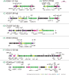
Genetic organization of ICE models with relevant gene names. (AG) ICEBs1 of B. subtilis, Tn916 of E. faecalis DS16, ICESXT of V. cholerae, CTnDOT of B. thetaiotaomicron, ICESt3 of S. thermophilus, ICEclc of P. knackmussii B13 and ICEMlSymR7A of M. loti R7A. Coding sequences of the ‘core’ ICE genes (i.e. important for its life style) are represented as thick arrows filled with different colors depending on (deduced or experimentally demonstrated) functions according to the color-scale below (mob, DNA mobilization; mpf, mating pair formation complex; reg, regulation; oriT, origin of transfer [blue ellipses]; chrom, chromosomal genes). Variable gene regions are omitted for clarity; their positions are indicated by non-connecting horizontal lines. Crucial promoters experimentally characterized are shown with bent arrows or with names (e.g. Pint in ICEclc). Plus signs on ICESXT indicate SetCD-regulated promoters. ICE ends, attR and attL, are indicated as vertical black lines. All ICEs are depicted to the same scale, and their lengths are shown within brackets. Note the double gene assignments in the Tn916 system (e.g. orf24-22 is tecLKJ, orf20 is tecH).
Figure 3.
Figure 3.
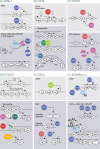
Schematic outlines of the main regulatory networks controlling ICE transmission in the ICEBs1 (A), Tn916 (B), ICESXT (C), CTnDOT (D), ICEclc (E) and ICEMlSymR7A (F) models. Depicted are the integrated, silent (upper panels) and the activated state, leading to horizontal transmission (lower panels, dark background). Hooked arrows indicate promoters, other black arrows point to activation and blocked lines to repression; waved arrows (A) point to protein interactions. Open arrows indicate relevant genes; waved lines represent mRNAs, colored circles or hexagones point to key proteins in the network. Protein degradation is symbolized by broken circles, whereas protein phosphorylation is indicated by an encircled P. EXPO, exponential phase; STAT, stationary phase; AI, N-homoserinelactone autoinducer. (A) In short, the ICEBs1 balance is controlled by ImmR, which prevents transcription of the xis excisionase gene. When ImmR is degraded or overruled, xis is transcribed, leading to ICEBs1 excision. (B) Orf9 is the major repressor blocking Tn916 transmission. In the presence of tetracycline, an antisense mRNA prevents orf9 mRNA translation (red hybrid). In the absence of Orf9, the cascade of orf7/8 transcription can start, leading to xis, int and tra gene expression. The ICESXT element is controlled by a double-negative feedback loop implicating CroS and SetR (C). Under SOS response, SetR is preferentially degraded, liberating transcription of setCD for the master regulators, which then further activate the ICE excision and transfer system. (D) A small RNA named rteR and an RNA stem-loop structure are the major inhibitors for CTnDOT transmission. The presence of tetracycline liberates the inhibitory stem-loop, leading to transcription of rteA and rteB that elicit the activation cascade. (E) ICEclc is only activated in stationary-phase cells. Activation is dependent on the TciR transcription regulator, which stimulates an as yet unknown bistability generator, whose activation is transmitted to the downstream tra and int genes. (F) Two proteins QseC and QseM control ICEMlSymR7Atransmission. QseM inhibits downstream transfer factors, but is itself transcriptionally repressed by high intracellular amounts of QseC, upon which TraR, AI and FseA can elicit the transfer. For more details and references, see the main text.
Figure 4.
Figure 4.
Concept of bistability in ICE horizontal transmission. ICE regulatory architectures contain a bistability generator (e.g. double inhibitory cyan/magenta loop as shown), which permits at low frequency in a clonal population (A) to activate the ICE horizontal transmission process (B, green gene arrow, individual green cell). In most other cells, the ICE will remain in its OFF state. At this point, the ICE is still in its integrated state (represented by the black bar inside the cells). Growth phase or environmental conditions can change the frequency of the bistable decision outcome, as illustrated in the details of Fig. 3. (C) Activation of the ICE will continue as (bistable) propagation of the horizontal transmission pathway in the same individual cells (e.g. activation of tra transfer, int integrase and xis excisionase genes), resulting in ICE excision and transfer (represented by the black circle and small black stick in the green cell), when suitable donors are present. This may require specific mechanisms to ensure preventing downstream pathway expression in non-active cells (shielding), and a faithful progression of the different steps of the transmission cascade in activated cells (fidelity). Finally, the ICE bistable horizontal transmission pathway has to end (D), either by death of the donor cell, by ICE reintegration, loss or some other mechanism.
Figure 5.
Figure 5.
Methods to detect ICE bistability and transfer. (A) Fusing ICE-core promoters (in this example Pint and PinR from ICEclc) to fluorescent reporter genes (like egfp or mcherry) allows observing ICE bistable gene expression (Minoia et al.; Reinhard and van der Meer 2013). The example schematically shows cells carrying ICEclc and additional single copy Pint- and PinR-reporter fusions, leading to the appearance of a subpopulation of fluorescent cells in stationary (STAT) phase. The subpopulation size can be deduced from quantile–quantile plots of observed versus expected fluorescence distribution (Reinhard and van der Meer 2013), or from scatter plots of dual fluorescent marker expression among individual cells (lower panel). (B) ICEclc transfer can be followed at single cell level by fusing the egfp gene downstream of the intB13 integrase gene (Delavat et al.2016). Donor cells (d) activating the ICEclc transfer competence (tc) program express eGFP through the Pint promoter, but become brighter fluorescent upon ICEclc excision (tc+exc) as a result of the Pcirc promoter being transcriptionally fused upstream of Pint. Silent donor cells (d, non-tc) are barely visible in background. ICEclc transfer (lower panel) can be detected from donor cells with an excised ICEclc (d, tc+exc) to recipient cells (r) expressing a different fluorescent protein (e.g. mCherry), as a result of combined colors (r+ICE). For further details, see the main text.
Figure 6.
Figure 6.
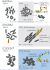
Inferred ICE–host cell adaptations selected for optimal ICE transmission. (A) ICEclc in P. knackmussii or P. putida remains integrated in exponentially growing cells (EXPO, white bar inside black cells), but is activated in ∼3% of individual cells during stationary phase (STAT, green cells). Such cells become transfer competent (tc), but only excise ICEclc once they are provided with new nutrients (EXPO, white circles inside green cells). tc cells can divide a few times to produce a microcolony that improves the transfer probability, but individual cells in such microcolony show highly variable ICE and cell fates (illustrated here as lysing cells [with holes], ICE replication, single or multiple ICE transfer [small sticks pointing out from cells], or ICE loss). Eventually, tc cells perish and are overgrown by the non-tc cells, and the cycle repeats itself in a next stationary phase (Reinhard et al.; Delavat et al.2016). ICEclc transferred to a new recipient (r, light brown cell) may integrate depending on the availability and sequence match with the chromosomal attachment site (i.e. integration efficiency). (B) ICEBs1 in B. subtilis remains integrated in exponentially growing cells (EXPO) and when the density of ICE-free cells (r, light brown) is low. Stationary phase (STAT) conditions and high density of ICE-free cells, or occurrence of DNA damage, stimulate the process of ICEBs1 horizontal transmission from donors (tc, green cell) to ICE-free cells. ICEBs1 replicates in the donor to avoid loss upon donor cell division. Optimal conjugation further requires specific hydrolysis of the recipient cell wall and efficient ICEBs1 replication from its single-stranded origin (sso) aids in maintenance in the recipient. Transfer can directly continue in cell chains (r to r). Wild-type ICEBs1 transfer rates estimated to between 10–7 and 3×10⁻⁵ per colony-forming cell (A. Grossman, personal communication). (C) ICESXT transfer in V. cholerae cells is stimulated ca 100-fold by DNA damage induced SOS response from a background level of around 2×10⁻⁷ per cell (Waldor, Tschäpe and Mekalanos ; Beaber, Hochhut and Waldor 2004). ICESXT replication and partitioning ensure proper segregation among dividing donor cells with excised ICE (tc, green cell; excised ICE as circles), but TA systems may specifically inhibit any ICE-free daughter cell (punctured green cell in illustration). Successful conjugation and integration in a recipient (r, light brown cell) further depends on the recipient's SOS response, exclusion mechanisms or defense systems such as restriction-modification (R/M). Note that cases (B) and (C) are inferred from bistability assumptions but have no current support from single cell observations. For further details and references, see the main text.

Similar articles

Cited by

References

    1. Ackermann M, Stecher B, Freed NE et al. . Self-destructive cooperation mediated by phenotypic noise. Nature 2008;454:987–90. - PubMed
    1. Ambroset C, Coluzzi C, Guedon G et al. . New insights into the classification and integration specificity of Streptococcus Integrative Conjugative Elements through extensive genome exploration. Front Microbiol 2015;6:1483. - PMC - PubMed
    1. Arkin A, Ross J, McAdams HH. Stochastic kinetic analysis of developmental pathway bifurcation in phage lambda-infected Escherichia coli cells. Genetics 1998;149:1633–48. - PMC - PubMed
    1. Auchtung JM, Aleksanyan N, Bulku A et al. . Biology of ICEBs1, an integrative and conjugative element in Bacillus subtilis. Plasmid 2016;86:14–25. - PubMed
    1. Auchtung JM, Lee CA, Garrison KL et al. . Identification and characterization of the immunity repressor (ImmR) that controls the mobile genetic element ICEBs1 of Bacillus subtilis. Mol Microbiol 2007;64:1515–28. - PMC - PubMed

Publication types

MeSH terms

Substances