

The plantar calcaneal spur: a review of anatomy, histology, etiology and key associations
- PMID: 28369929
- PMCID: PMC5442149
- DOI: 10.1111/joa.12607
The plantar calcaneal spur: a review of anatomy, histology, etiology and key associations
Abstract
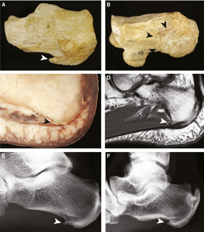
The plantar calcaneal spur (PCS) is a bony outgrowth from the calcaneal tuberosity and has been studied using various methods including cadavers, radiography, histology and surgery. However, there are currently a number of discrepancies in the literature regarding the anatomical relations, histological descriptions and clinical associations of PCS. Historically, authors have described the intrinsic muscles of the foot and/or the plantar fascia as attaching to the PCS. In this article we review the relationship between the PCS and surrounding soft tissues as well as examining the histology of the PCS. We identify a number of key associations with PCS, including age, weight, gender, arthritides, plantar fasciitis and foot position; these factors may function as risk factors in PCS formation. The etiology of these spurs is a contentious issue and it has been explained through a number of theories including the degenerative, inflammatory, traction, repetitive trauma, bone-formers and vertical compression theories. We review these and finish by looking clinically at the evidence that PCS causes heel pain.
Keywords: bony outgrowth; calcaneal tuberosity; plantar calcaneal spur.
© 2017 Anatomical Society.
Figures
References
-
- Abreu MR, Chung CB, Mendes L, et al. (2003) Plantar calcaneal enthesophytes: new observations regarding sites of origin based on radiographic, MR imaging, anatomic, and paleopathologic analysis. Skeletal Radiol 32, 13–21. - PubMed
-
- Ali M, Chen T, Crues J (2006) MRI of the foot. 12, 10.
-
- Alshami AM, Souvlis T, Coppieters MW (2008) A review of plantar heel pain of neural origin: differential diagnosis and management. Man Ther 13, 103–111. - PubMed
-
- Barrett SL, Day SV, Pignetti TT, et al. (1995) Endoscopic heel anatomy: analysis of 200 fresh frozen specimens. J Foot Ankle Surg 34, 51–56. - PubMed
-
- Banadda BM, Gona O, Vaz R, Ndlovu DM (1992) Calcaneal spurs in a black African population. Foot Ankle Int 13, 352–354. - PubMed
Publication types
MeSH terms
LinkOut - more resources
Full Text Sources
Other Literature Sources
