. 2017 Jul 1;595(13):4417-4430.
doi: 10.1113/JP274049. Epub 2017 May 26.

A calcium-dependent pathway underlies activity-dependent plasticity of electrical synapses in the thalamic reticular nucleus

Affiliations

A calcium-dependent pathway underlies activity-dependent plasticity of electrical synapses in the thalamic reticular nucleus

Jessica Sevetson et al. J Physiol. .

Abstract

Key points: Electrical synapses are modified by various forms of activity, including paired activity in coupled neurons and tetanization of the input to coupled neurons. We show that plasticity of electrical synapses that results from paired spiking activity in coupled neurons depends on calcium influx and calcium-initiated signalling pathways. Plasticity that results from tetanization of input fibres does not depend on calcium influx or dynamics. These results imply that electrically coupled neurons have distinct sets of mechanisms for adjusting coupling according to the specific type of activity they experience.

Abstract: Recent results have demonstrated modification of electrical synapse strength by varied forms of neuronal activity. However, the mechanisms underlying plasticity induction in central mammalian neurons are unclear. Here we show that the two established inductors of plasticity at electrical synapses in the thalamic reticular nucleus - paired burst spiking in coupled neurons, and mGluR-dependent tetanization of synaptic input - are separate pathways that converge at a common downstream endpoint. Using occlusion experiments and pharmacology in patched pairs of coupled neurons in vitro, we show that burst-induced depression depends on calcium entry via voltage-gated channels, is blocked by BAPTA chelation, and recruits intracellular calcium release on its way to activation of phosphatase activity. In contrast, mGluR-dependent plasticity is independent of calcium entry or calcium dynamics. Together, these results show that the spiking-initiated mechanisms underlying electrical synapse plasticity are similar to those that induce plasticity at chemical synapses, and offer the possibility that calcium-regulated mechanisms may also lead to alternate outcomes, such as potentiation. Because these mechanistic elements are widely found in mature neurons, we expect them to apply broadly to electrical synapses across the brain, acting as the crucial link between neuronal activity and electrical synapse strength.

Keywords: burst firing; calcium; depression; electrical synapse; plasticity.


Figures

Figure 1
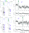
Figure 1. Paired bursting and ACPD each occlude further LTD induction
A, example recordings from paired whole‐cell recordings of electrically coupled neurons. In this pair, the coupling coeffient (cc) was initially 0.13. Scale bar 100 ms, 5 mV (blue), 1.67 mV (green). Following paired bursting in both cells, cc decreased to 0.097, and was unchanged after subsequent bath application of ACPD. B and C, coupling conductance (G C) and cc, respectively, for the cohort of pairs used in this experiment, shown in control, after paired bursting (diamonds), and after subsequent ACPD application (purple). D, LTD was induced by paired bursting in two coupled TRN neurons. Between 0 and 15 min, ΔG C (black) was −11.6 ± 1.0%, P t = 0.003, P s = 0.008; and Δcc (grey) was −11.8 ± 1.3%, P t = 0.002, P s = 0.008 from pre‐stimulus control (n = 8 pairs). ACPD was subsequently bath‐applied for 4 min, resulting in no further changes in electrical synapse strength (P t = 0.58, P s = 0.95 for G C, P t = 0.66 and P s = 0.74 for cc). E, input resistance (R in) over the experiment. F, in a pair with initial cc of 0.19, ACPD application decreased cc to 0.15 and was unchanged by subsequent paired bursting activity in both cells. Scale bar 100 ms, 5 mV (blue), 2.5 mV (green). G and H, G C and cc, respectively, for the cohort of pairs used in this experiment, shown in control, after ACPD application (purple), and after subsequent paired bursting (diamonds). I, LTD was induced by bath‐applied ACPD (50 μm) in two coupled TRN neurons. Between 0 and 20 min, ΔG C was −8.6 ± 0.9%, P t = 0.0001, P s = 0.008; and Δcc was −8.2 ± 0.9%, P t = 0.01, P s = 0.02 from pre‐stimulus control (n = 8 pairs). Paired bursting was then induced, resulting in no further changes in electrical synapse strength (P t = 0.22, P s = 0.15 for G C, P t = 0.35 and P s = 0.55 for cc for the post‐ACPD and post‐burst comparisons). J, input resistance (R in) over the experiment.
Figure 2
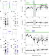
Figure 2. T‐type calcium channels are necessary for burst‐induced but not ACPD‐induced LTD
A, example recordings from a pair before and after paired bursting in TTA‐A2. Scale bar 100 ms, 5 mV (green), 2.5 mV (blue). B and C, coupling conductance (G C) and coupling coeffient (cc), respectively, for the cohort of pairs used in this experiment, shown in control and after paired bursting (diamonds). D, paired bursting in two coupled TRN neurons did not result in LTD induction in the presence of the T‐channel antagonist TTA‐A2 (ΔG C = −1.2 ± 1.0%, P t = 0.9, P s = 0.77; Δcc = −1.4 ± 1.5%, P t = 0.8, P s = 1; n = 10 pairs). E, input resistance (R in) over the experiment. F, example recordings from a pair before and after ACPD application in TTA‐A2. Scale bar 100 ms, 5 mV (green), 2.5 mV (blue). G and H, G C and cc, respectively, for the cohort of pairs used in this experiment, shown in control and after ACPD (purple). I, ACPD application resulted in LTD induction in the presence of the T‐channel antagonist TTA‐A2 (ΔG C = −12.0 ± 1.5%, P t = 0.002, P s = 0.008; Δcc = −11.0 ± 1.5%, P t = 0.001, P s = 0.008; n = 8 pairs). J, input resistance (R in) over the experiment.
Figure 3
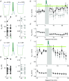
Figure 3. Calcium influx through high voltage‐activated channels is not necessary for burst‐induced LTD
A, example recordings from a pair before and after paired bursting in nimodipine. Scale bar 100 ms, 5 mV. B and C, coupling conductance (G C) and coupling coeffient (cc), respectively, for the cohort of pairs used in this experiment, shown in control and after paired bursting (diamonds). D, paired bursting induced decreased LTD in two coupled TRN neurons in the presence of the L‐type channel antagonist nimodipine (ΔG C = −6.2 ± 0.8%, P t = 0.047, P s = 0.07; Δcc = −7.6 ± 0.7%, P t = 0.012, P s = 0.07; n = 11 pairs) E, input resistance (R in) over the experiment.
Figure 4
Figure 4. Calcium chelation prevents burst‐induced but not ACPD‐induced LTD
A, example recordings from a pair before and after paired bursting with BAPTA in the intracellular solution. Scale bar 100 ms, 5 mV (green), 2.5 mV (blue). B and C, coupling conductance (G C) and coupling coeffient (cc), respectively, for the cohort of pairs used in this experiment, shown in control and after paired bursting (diamonds). D, paired bursting failed to induce LTD in coupled TRN neurons, both with BAPTA chelating calcium in the intracellular solution. After paired bursting, ΔG C = 1.67 ± 1.2%, P t = 0.28, P s = 0.47 and Δcc = −0.34 ± 0.9%, P t = 0.71, P s = 0.85, n = 12 pairs) from control values. E, input resistance (R in) over the experiment. F, example recordings from a pair before and after ACPD application with BAPTA. Scale bar 100 ms, 5 mV (green), 2.5 mV (blue). G and H, G C and cc, respectively, for the cohort of pairs used in this experiment, shown in control and after ACPD (purple). I, ACPD application induced LTD in coupled TRN neurons, both with BAPTA to chelate calcium in the intracellular solution. After paired bursting, ΔG C was −12.0 ± 1.2%, P t = 0.026, P s = 0.015 and Δcc = −8.9 ± 1.2%, P t = 4.4 × 10−6, P s = 0.014; n = 8 pairs) relative to control values. J, input resistance (R in) over the experiment.
Figure 5
Figure 5. Ryanodine receptor‐mediated intracellular calcium release is necessary for burst‐induced LTD
A, example recordings from a pair before and after paired bursting in caffeine. Scale bar 100 ms, 5 mV (green), 2.5 mV (blue). B and C, coupling conductance (G C) and coupling coeffient (cc), respectively, for the cohort of pairs used in this experiment, shown in control and after paired bursting (diamonds). D, paired bursting was induced in two coupled TRN neurons in the presence of the ryanodine receptor antagonist caffeine (ΔG C = 6.7 ± 1.4%, P t = 0.31, P s = 0.41; ΔR in = −11.7 ± 1.6% P = 0.007; Δcc = −5.7 ± 1.0%, P t = 0.09, P s = 0.31; n = 11 pairs). E, input resistance (R in) over the experiment. F, example recordings from a pair before and after paired bursting in ryanodine. Scale bar 100 ms, 5 mV (green), 2.5 mV (blue). G and H, G C and cc, respectively, for the cohort of pairs used in this experiment, shown in control and after paired bursting (diamonds). I, paired bursting was induced in two coupled TRN neurons in the presence of the ryanodine receptor antagonist ryanodine (for t < 15 min, ΔG C = 0.88 ± 1.8%, P t = 0.6, P s = 0.6; Δcc = −2.1 ± 1.9%, P t = 0.07, P s = 0.32; n = 11 pairs). F, input resistance (R in) over the experiment.
Figure 6
Figure 6. IP3‐mediated intracellular calcium release is not necessary for burst‐induced LTD
A, example recordings from a pair before and after paired bursting in xestospongin. Scale bar 100 ms, 5 mV. B and C, coupling conductance (G C) and coupling coeffient (cc), respectively, for the cohort of pairs used in this experiment, shown in control and after paired bursting (diamonds). D, paired bursting was induced in two coupled TRN neurons in the presence of the IP3 antagonist xestospongin (ΔG C = −6.0 ± 1.1%, P t = 0.16, P s = 0.09; Δcc = −12.4 ± 1.1%, P t = 0.093, P s = 0.098; n = 9 pairs). E, input resistance (R in) over the experiment.
Figure 7
Figure 7. Burst‐induced LTD requires calcineurin activation
A, example recordings from a pair before and after paired bursting in FK‐506. Scale bar 100 ms, 5 mV (green), 2.5 mV (blue). B and C, coupling conductance (G C) and coupling coeffient (cc), respectively, for the cohort of pairs used in this experiment, shown in control and after paired bursting (diamonds). D, paired bursting was induced in two coupled TRN neurons in the presence of the calcineurin antagonist FK‐506, resulting in ΔG C = −4.1 ± 1.0%, P t = 0.91, P s = 0.37; Δcc = −3.1 ± 0.9%, P t = 0.28, P s = 0.41 relative to control values (n = 9 pairs). E, input resistance (R in) over the experiment. F, example recordings from a pair before and after paired bursting with cyclosprin A in the internal solution. Scale bar 100 ms, 5 mV (green), 3 mV (blue). G and H, G C and cc, respectively, for the cohort of pairs used in this experiment, shown in control and after paired bursting (diamonds). I, paired bursting was induced in two coupled TRN neurons in the presence of the calcineurin inhibitor cyclosporin A (ΔG C = 0.04 ± 1.5%, P t = 0.36, P s = 0.81; Δcc = −2.5 ± 1.6%, P t = 0.38, P s = 0.58; n = 7 pairs). F, input resistance (R in) over the experiment.
Figure 8
Figure 8. Proposed core mechanistic pathway for activity‐dependent plasticity of electrical synapses
For paired bursting, calcium enters through voltage‐gated channels (T). Influx of calcium causes calcium‐induced calcium release from internal stores (ER), and activates calcineurin (PP2B), resulting in a dephosphorylation of the connexin36 protein (GJ). Separately, tetanic input activates metabotropic glutamate receptors (mGluR), and a chain eventually resuting in PKA activation also results in phosphorylation changes of the connexin36 protein (Wang et al. 2015).

Comment in

References

    1. Alev C, Urschel S, Sonntag S, Zoidl G, Fort AG, Hoher T, Matsubara M, Willecke K, Spray DC & Dermietzel R (2008). The neuronal connexin36 interacts with and is phosphorylated by CaMKII in a way similar to CaMKII interaction with glutamate receptors. Proc Natl Acad Sci USA 105, 20964–20969. - PMC - PubMed
    1. Apostolides PF & Trussell LO (2013). Regulation of interneuron excitability by gap junction coupling with principal cells. Nat Neurosci 16, 1764–1772. - PMC - PubMed
    1. Apostolides PF & Trussell LO (2014). Control of interneuron firing by subthreshold synaptic potentials in principal cells of the dorsal cochlear nucleus. Neuron 83, 324–330. - PMC - PubMed
    1. Beierlein M, Gibson JR & Connors BW (2003). Two dynamically distinct inhibitory networks in layer 4 of the neocortex. J Neurophysiol 90, 2987–3000. - PubMed
    1. Burr GS, Mitchell CK, Keflemariam YJ, Heidelberger R & O'Brien J (2005). Calcium‐dependent binding of calmodulin to neuronal gap junction proteins. Biochem Biophys Res Commun 335, 1191–1198. - PMC - PubMed

Publication types

LinkOut - more resources