Review
. 2017 Sep;38(17):2100-2114.
doi: 10.1002/elps.201700042. Epub 2017 May 17.

Characterization of isomeric glycan structures by LC-MS/MS

Affiliations
Review

Characterization of isomeric glycan structures by LC-MS/MS

Lucas Veillon et al. Electrophoresis. 2017 Sep.

Abstract

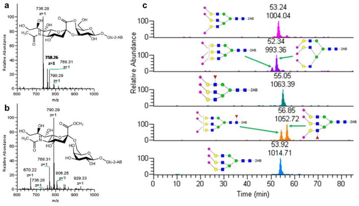
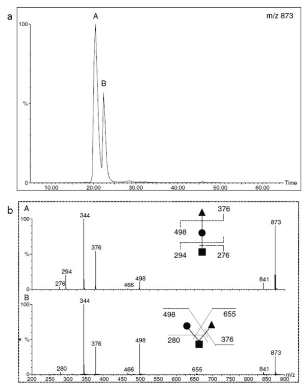
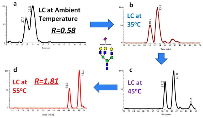
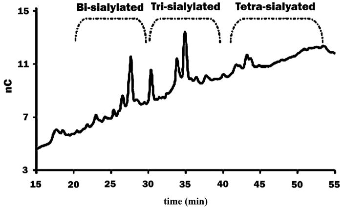
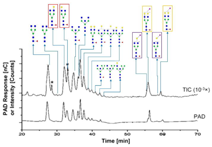
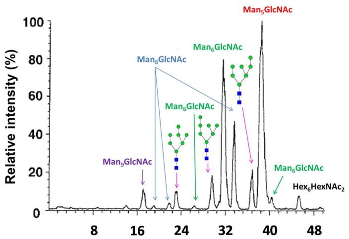
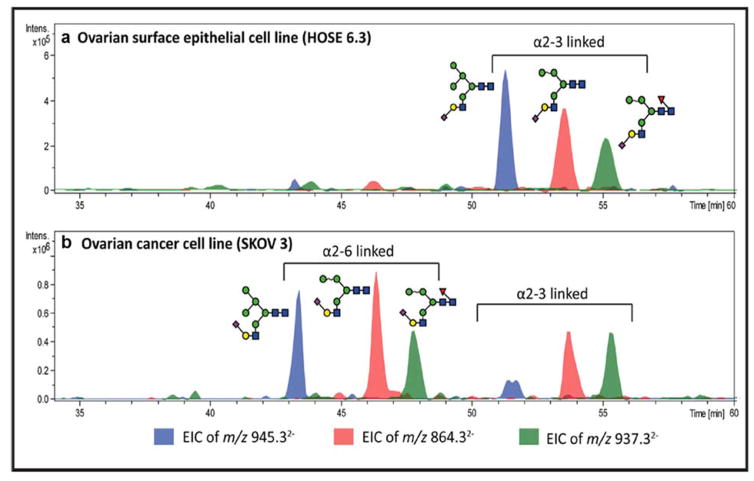
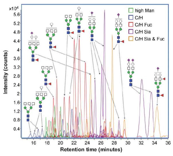
The characterization of glycosylation is critical for obtaining a comprehensive view of the regulation and functions of glycoproteins of interest. Due to the complex nature of oligosaccharides, stemming from variable compositions and linkages, and ion suppression effects, the chromatographic separation of glycans, including isomeric structures, is necessary for exhaustive characterization by MS. This review introduces the fundamental principles underlying the techniques in LC utilized by modern day glycomics researchers. Recent advances in porous graphitized carbon, reverse phase, ion exchange, and hydrophilic interaction LC utilized in conjunction with MS, for the characterization of protein glycosylation, are described with an emphasis on methods capable of resolving isomeric glycan structures.

Keywords: Glycan analysis; Isomeric separation; LC-MS/MS; Liquid chromatography; Mass spectrometry.


Conflict of interest statement

Conflict of interest statement

The authors declare no conflict of interest.

Figures

Figure 1
Figure 1
Scheme of (a) α-2,3 and (b) α-2,6 linked sialic acid molecular structures after DMT/MM derivatization. (c) EIC of the identified compositions in haptoglobin trisialylated N-glycans after DMT-MM/methanol derivatization. Reprinted and modified from [53], with permission.
Figure 2
Figure 2
(a) RPLC separation of permethylated isomeric O-glycan trisaccharides. (b) MS/MS spectra of isomers in peaks A and B (upper panel). Reprinted from [90], with permission.
Figure 3
Figure 3
EICs of tri-antennary mono-sialylated glycan derived from pooled human blood serum eluted by a gradient of 2% ACN, 98% H2O and 100% ACN (each containing 0.1% formic acid) at (a) ambient temperature, (b) 35 °C, (c) 45 °C, (d) 55 °C. Symbols in accordance with the Consortium for Functional Glycomics (CFG) notation. [71].
Figure 4
Figure 4
Separation of glycans from AGP by HPAEC. Glycans can be separated by the number of sialic acid residues in their structure. Reprinted and modified from [95], with permission.
Figure 5
Figure 5
Isomeric separation performed by HPAEC. HPAEC-PAD chromatograms and total ion current chromatograms from positive-mode HPAEC-MS (intensity attenuation: 10−3×) demonstrating the isomeric separation of PNGase F-released N-glycans from a purified human IgG (h-IgG, 99%). Six branch isomers are highlighted by frames with different colors. The same frame color denotes the isomers from the same glycan composition. Symbols in accordance with CFG notation. A spike peak is indicated by an asterisk. Reprinted and modified from [33].
Figure 6
Figure 6
Base peak chromatogram of RNase B oligosaccharide alditols. Reprinted from [120], with permission.
Figure 7
Figure 7
EIC of 3 hybrid monosialylated structures in (a) an ovarian surface epithelial cell line and (b) an ovarian cancer cell line. The sialic acid linkage isomers were separated with α-2,6 linked structures eluting earlier than α-2,3 linked species. Reprinted and modified from [124].
Figure 8
Figure 8
ECC of identified glycans from a representative serum sample. Different charge states and adducts were all taken into account. Reprinted and modified from [125], with permission.
Figure 9
Figure 9
(a) EIC of permethylated Man-7 glycans at [M+2Na]2+ m/z 1013.49. Although the 3 peaks were separated, the resolution was below 1. Reprinted from [131], with permission. (b) EIC of permethylated Man-7 glycans at [M+2H]2+ m/z 991.52. Man7 isomers were base line separated. Reprinted from [132], with permission.
Figure 10
Figure 10
Extracted ion chromatogram (EIC) of permethylated Hex4HexNAc4Fuc1 glycans at m/z 1017.5413 (a), Hex5HexNAc4Fuc1NeuAc1 glycans at m/z 867.1213 (b) derived from human blood serum, and Hex5HexNAc4Fuc1NeuAc1 glycans at m/z 867.1213 (c) from human milk. Reprinted and modified from [23], with permission.

References

    1. Taylor ME, Drickamer K. Current opinion in structural biology. 2014;28:14–22. - PMC - PubMed
    1. Ohtsubo K, Marth JD. Cell. 2006;126:855–867. - PubMed
    1. Yoshida-Moriguchi T, Yu L, Stalnaker SH, Davis S, Kunz S, Madson M, Oldstone MB, Schachter H, Wells L, Campbell KP. Science (New York, NY) 2010;327:88–92. - PMC - PubMed
    1. Phillips ML, Nudelman E, Gaeta FC, Perez M, Singhal AK, Hakomori S, Paulson JC. Science (New York, NY) 1990;250:1130–1132. - PubMed
    1. Sperandio M, Gleissner CA, Ley K. Immunological reviews. 2009;230:97–113. - PMC - PubMed

Publication types

MeSH terms

Substances

LinkOut - more resources