. 2017 Jul;105(1):1-24.
doi: 10.1111/mmi.13682. Epub 2017 Apr 24.

cpsA regulates mycotoxin production, morphogenesis and cell wall biosynthesis in the fungus Aspergillus nidulans

Affiliations

cpsA regulates mycotoxin production, morphogenesis and cell wall biosynthesis in the fungus Aspergillus nidulans

Xuehuan Feng et al. Mol Microbiol. 2017 Jul.

Abstract

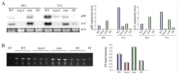
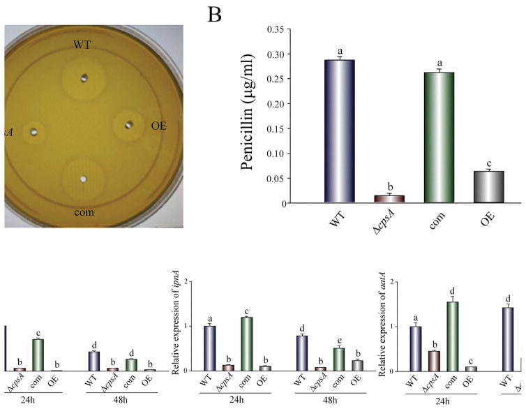
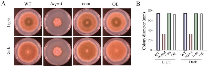
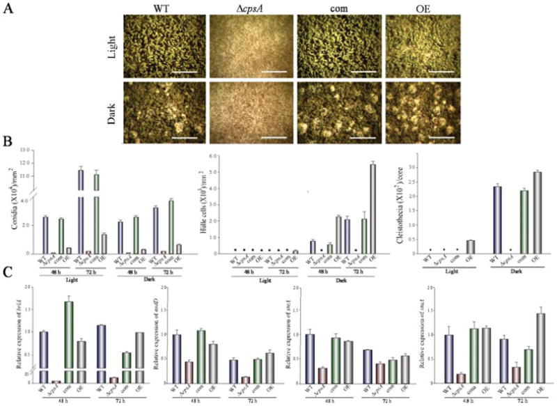
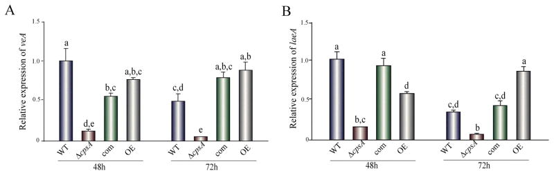
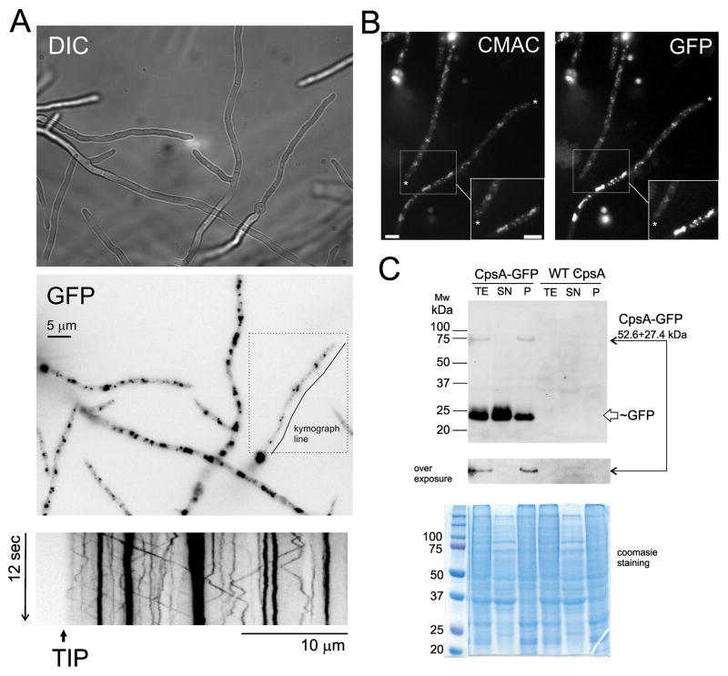
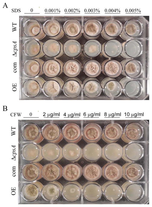
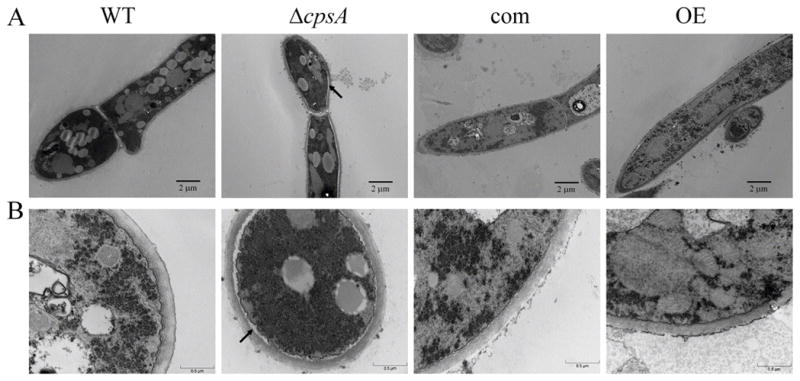
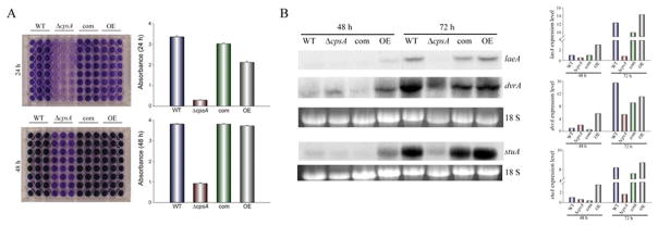
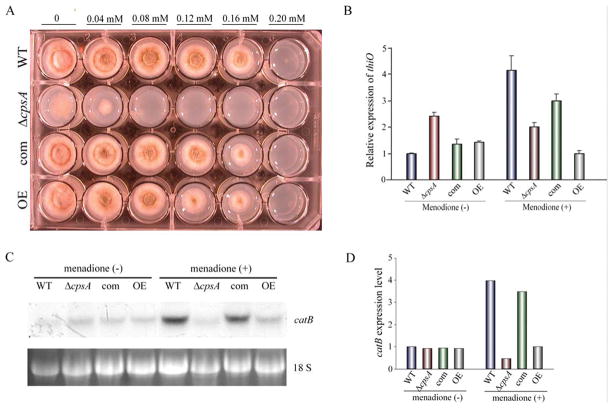
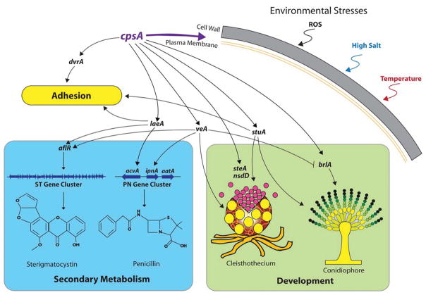
The model fungus Aspergillus nidulans synthesizes numerous secondary metabolites, including sterigmatocystin (ST). The production of this toxin is positively controlled by the global regulator veA. In the absence of veA (ΔveA), ST biosynthesis is blocked. Previously, we performed random mutagenesis in a ΔveA strain and identified revertant mutants able to synthesize ST, among them RM1. Complementation of RM1 with a genomic library revealed that the mutation occurred in a gene designated as cpsA. While in the ΔveA genetic background cpsA deletion restores ST production, in a veA wild-type background absence of cpsA reduces and delays ST biosynthesis decreasing the expression of ST genes. Furthermore, cpsA is also necessary for the production of other secondary metabolites, including penicillin, affecting the expression of PN genes. In addition, cpsA is necessary for normal asexual and sexual development. Chemical and microscopy analyses revealed that CpsA is found in cytoplasmic vesicles and it is required for normal cell wall composition and integrity, affecting adhesion capacity and oxidative stress sensitivity. The conservation of cpsA in Ascomycetes suggests that cpsA homologs might have similar roles in other fungal species.


Figures

Figure 1
Figure 1. TLC analysis of NOR production in GMM cultures
(A) The strains ΔveA ΔstcE (RDAEp206), veA1 ΔstcE (RAV1p), cpsA− ΔveA ΔstcE (RM1p), cpsAcpsA+ ΔveA ΔstcE (RM1p-com), cpsAveA1 ΔstcE (RM1p-R2), ΔcpsA ΔveA ΔstcE (TDAEΔcpsA), ΔcpsA cpsA+ ΔveA ΔstcE (TDAEΔcpsA-com) (Table S2), were top-agar inoculated and incubated at 37 °C in the dark for 5 days. NOR production was analyzed by TLC. (B) Intensity of NOR bands. Densitometry was carried out with GelQuant.NET software. NOR band intensity was normalized using the intensity corresponding to RDAEp206 as 1. The letters above the bars represent significantly different values (P ≤0.05, Tukey test).
Figure 2
Figure 2. cpsA is necessary for normal expression of ST genes and mycotoxin production in A. nidulans strains with a veA+ wild-type background
(A) Wild type (TRV50.2) (WT) veA+ control, ΔcpsA (TRVpΔcpsA), complementation (TRVpΔcpsA-com)(com), and overexpression (OEpcpsA)(OE) strains were inoculated in liquid GMM. Mycelia were collected after 48 h and 72 h of culture in a shaker incubator at 250 rpm at 37 °C. Expression of aflR and stcU was analyzed by Northern blot. rRNA serves as loading control. Asterisk indicates not detected. Densitometry of the Northern blot results is shown. (B) TLC analysis of ST production in 72 h cultures described in (A), densitometry of ST band intensity is shown.
Figure 3
Figure 3. cpsA affects penicillin production
A) Extracts from wild-type (WT) veA+ control, ΔcpsA, complementation (com) and overexpression (OE) strains were analyzed for penicillin content as described in Materials and Methods section. (B) Quantification of penicillin content in the analyzed extracts. Values are means of three replicates. (C) qRT-PCR gene expression analysis by of acvA, ipnA and aatA from mycelial samples collected after 24 h and 48 h of incubation in PN inducing medium. The relative expression was calculated using the 2−ΔΔCT method, as described by Livak and Schmittgen (2001). Values were normalized to the expression levels in the wild type, considered 1. Error bars represent standard errors. The letters above the bars represent significantly different values (P ≤0.05).
Figure 4
Figure 4. cpsA positively affects A. nidulans colony growth
(A) Photographs of wild-type (WT) veA+ control, ΔcpsA, complementation (com) and overexpression (OE) strains point-inoculated on GMM plates and incubated at 37 °C in either light or dark for 6 days. (B) Colony growth measured as colony diameter. Standard error is shown. Means are average of three replicates.
Figure 5
Figure 5. cpsA is necessary for proper asexual and sexual development
(A) Micrographs of wild-type (WT) veA+ control, ΔcpsA, complementation (com) and overexpression (OE) strains point-inoculated GMM cultures incubated at 37 °C in either light or dark for 6 days. Samples were observed two centimeters from the inoculation point. Images were captured with an upright Leica MZ75 stereomicroscope. Scale bar corresponds to 200 μm. (B) Quantitative analysis of production of conidia, Hülle cells and cleistothecia from top-agar inoculated GMM cultures incubated at 37 °C in either light or dark for 48 h and 72 h, in the case of conidia and Hülle cells, and 6 days in the case of cleistothecia. Asterisk indicates not detected. The experiment included three replicates. (C) qRT-PCR gene expression analysis of brlA, nsdD, steA and stuA from mycelial samples from 48 h and 72 h GMM liquid stationary cultures. The relative expression was calculated using the 2−ΔΔCT method, as described by Livak and Schmittgen (2001). Values were normalized to the expression levels in the wild type, considered 1. Error bars represent standard errors.
Figure 6
Figure 6. Expression of veA and laeA is positively affected by cpsA
qRT-PCR gene expression analysis of veA and laeA from liquid stationary GMM cultures 48 h and 72 h after incubation in the dark at 37 °C. The relative expression was calculated using the 2−ΔΔCT method, as described by Livak and Schmittgen (2001). Levels in the wild type, considered 1. Error bars represent standard errors. The letters above the bars represent significantly different values (P ≤0.05).
Figure 7
Figure 7. Localization of CpsA protein
(A) Cellular localization of CpsA-GFP fusion protein expressed by its endogenous promoter. Cells were grown for 16 h at 25 °C. CpsA accumulates at small vesicles resembling vacuoles and endosomes. The kymograph obtained from a stack of 90 images for 12 seconds (200 ms exposure each) along the line shown in GFP inset. Dashed line square indicates the limits of Movie 1. Kymograph shows the presence of static and motile vesicles migrating in an anterograde (to the tip) and retrograde (from tip to base of cells) modes. (B) CMAC staining confirm the presence of CpsA-GFP in small vacuoles. Details of the aggregates are shown at the bottom of the images. Asterisks indicate cell tips. Scale bar represents 5 micrometers. (C) Detection of CpsA-GFP fusion using GFP antibodies. TE indicates a total protein extract using an alkaline lysis protocol. Soluble protein from A50 extraction protocol is shown in line SN. Insoluble protein in A50 treatment was analyzed in line P. An overexposure of the filter on the CpsA-GFP fusion region is shown. In both protocols a band corresponding to GFP is visualized as indication of protein degradation in vacuoles or MVs (multivesicular bodies)/lysosomes.
Figure 8
Figure 8. Absence of or overexpression of cpsA increases sensitivity to SDS and CFW
(A) Wild-type (WT) veA+ control, ΔcpsA, complementation (com) and overexpression (OE) strains were point inoculated on GMM containing SDS (A) and CFW (B) at different concentrations. Cultures were incubated at 37 °C in the dark for 3 days.
Figure 9
Figure 9. cpsA is required for proper attachment of the cell wall to the plasma membrane
Transmission electron microscopy images of wild-type (WT) veA+ control, ΔcpsA, complementation (com) and overexpression (OE) strains. The strains were grown in liquid stationary GMM cultures at 37 °C for 48 h. Mycelia were fixed and stained with 2% uranyl acetate and 0.2% lead citrate. The arrows show the cell structure alteration with detachment of plasma membrane and cell wall. Micrographs were acquired with different magnifications represented by panels in (A) and (B).
Figure 10
Figure 10. cpsA is involved in adhesion to surfaces
(A) Conidia of wild-type (WT) veA+ control, ΔcpsA, complementation (com) and overexpression (OE) strains were inoculated in liquid GMM (105 spores/ml), and incubated at 37 °C for 24 h and 48 h as stationary cultures. Mycelium was stained with Crystal violet as described in Materials and Methods section. The 48 h samples in panel A were dilluted 2-fold for measurement at 560 nm. (B) Expression analysis of laeA, dvrA, and stuA by Northern blot. The strains were inoculated (105 spores/ml) in Petri dishes containing 20 ml of liquid GMM, and incubated at 37 °C for 48 h and 72 h as stationary cultures. rRNA serves as loading control. Densitometry of the Northern blot results is shown.
Figure 11
Figure 11. cpsA is necessary in resistance to oxidative stress in A. nidulans
(A) Wild-type (WT) veA+ control, ΔcpsA, complementation (com) and overexpression (OE) strains were point-inoculated on GMM containing different concentrations of menadione and incubated at 37 °C in dark for 3 days. Expression analysis of thiO by qRT-PCR (B) and catB by Northern blot (C and D). The strains were incubated in liquid GMM at 250 rpm at 37°C in the dark for 48 h. After that, mycelia were transferred to GMM with or without menadione (0.08 mM). rRNA serves as loading control. Densitometry of the Northern blot results is shown.
Figure 12
Figure 12. Model of the role of cpsA in A. nidulans
cpsA is necessary for proper attachment of the cell wall to the plasma membrane, and the normal cell wall composition and integrity that protect fungal cells from environmental stresses. All the genes in this model are downregulated in the absence of cpsA. The cpsA gene positively influences the expression of the master regulators veA, laeA and stuA, and is required for normal sexual and asexual development, secondary metabolism and adhesion capacity.

References

    1. Adams TH, Boylan MT, Timberlake WE. brlA is necessary and sufficient to direct conidiophore development in Aspergillus nidulans. Cell. 1988;54:353–362. - PubMed
    1. Adams TH, Yu JH. Coordinate control of secondary metabolite production and asexual sporulation in Aspergillus nidulans. Curr Opin Microbiol. 1998;1:674–677. - PubMed
    1. Adrio JL, Demain AL. Fungal biotechnology. Int Microbiol. 2003;6:191–199. - PubMed
    1. Ahmed YL, Gerke J, Park HS, Bayram Ö, Neumann P, Ni M, et al. The velvet family of fungal regulators contains a DNA-binding domain structurally similar to NF-κB. PLoS Biol. 2013;11(12):e1001750. - PMC - PubMed
    1. Amare MG, Keller NP. Molecular mechanisms of Aspergillus flavus secondary metabolism and development. Fungal Genet Biol. 2014;66:11–18. - PubMed

LinkOut - more resources