Review
. 2017 Apr;25(2):177-191.
doi: 10.1111/wrr.12516. Epub 2017 Apr 27.

Regeneration of injured skin and peripheral nerves requires control of wound contraction, not scar formation

Affiliations
Review

Regeneration of injured skin and peripheral nerves requires control of wound contraction, not scar formation

Ioannis V Yannas et al. Wound Repair Regen. 2017 Apr.

Abstract

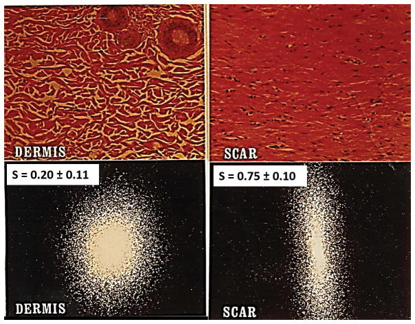
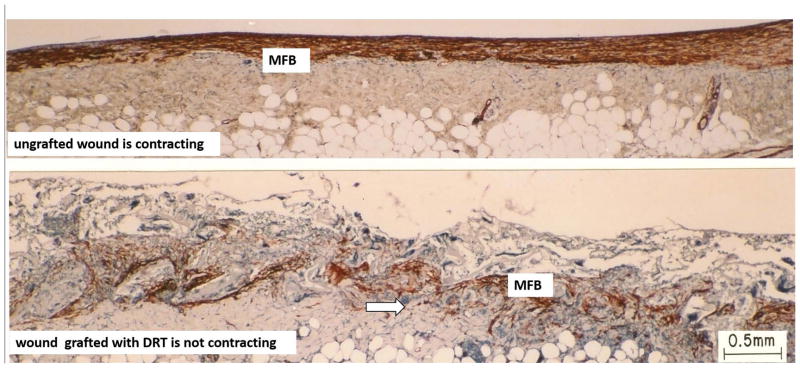
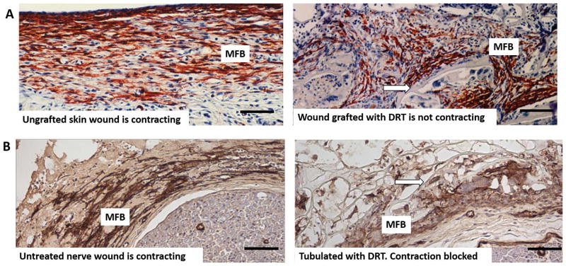
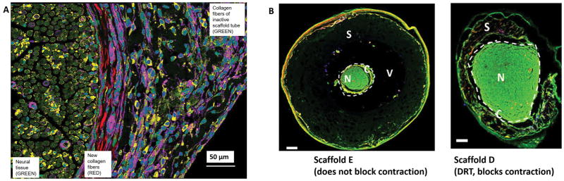
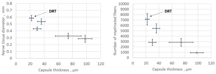
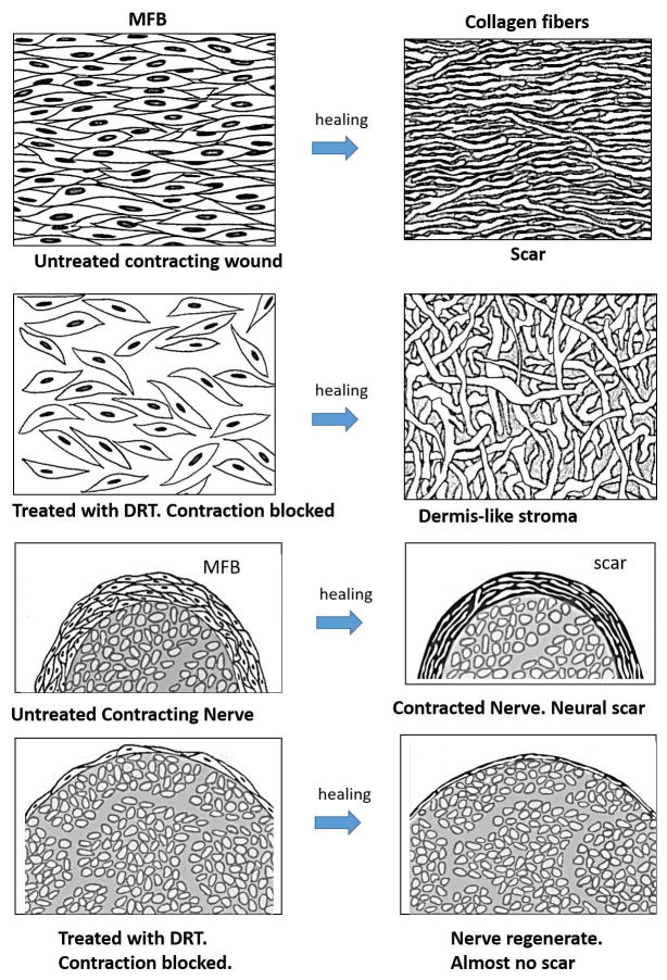
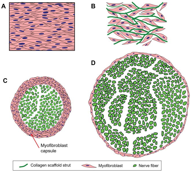
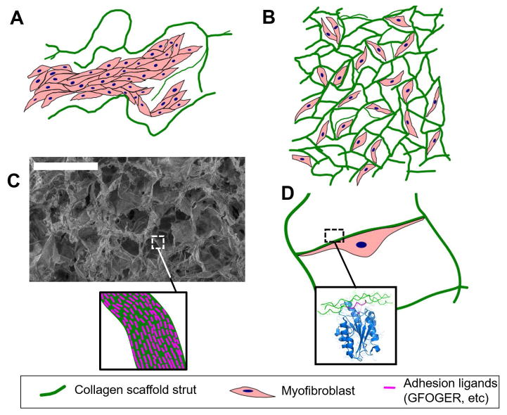
We review the mounting evidence that regeneration is induced in wounds in skin and peripheral nerves by a simple modification of the wound healing process. Here, the process of induced regeneration is compared to the other two well-known processes by which wounds close, i.e., contraction and scar formation. Direct evidence supports the hypothesis that the mechanical force of contraction (planar in skin wounds, circumferential in nerve wounds) is the driver guiding the orientation of assemblies of myofibroblasts (MFB) and collagen fibers during scar formation in untreated wounds. We conclude that scar formation depends critically on wound contraction and is, therefore, a healing process secondary to contraction. Wound contraction and regeneration did not coincide during healing in a number of experimental models of spontaneous (untreated) regeneration described in the literature. Furthermore, in other studies in which an efficient contraction-blocker, a collagen scaffold named dermis regeneration template (DRT), and variants of it, were grafted on skin wounds or peripheral nerve wounds, regeneration was systematically observed in the absence of contraction. We conclude that contraction and regeneration are mutually antagonistic processes. A dramatic change in the phenotype of MFB was observed when the contraction-blocking scaffold DRT was used to treat wounds in skin and peripheral nerves. The phenotype change was directly observed as drastic reduction in MFB density, dispersion of MFB assemblies and loss of alignment of the long MFB axes. These observations were explained by the evidence of a surface-biological interaction of MFB with the scaffold, specifically involving binding of MFB integrins α1 β1 and α2 β1 to ligands GFOGER and GLOGER naturally present on the surface of the collagen scaffold. In summary, we show that regeneration of wounded skin and peripheral nerves in the adult mammal can be induced simply by appropriate control of wound contraction, rather than of scar formation.


Conflict of interest statement

Conflict statement

I.V. Yannas has participated in the founding of Integra LifeSciences, Plainsboro, NJ. He currently has no financial connection with the company and owns no stock of Integra LifeSciences.

Figures

Figure 1
Figure 1
Quantitative distinction between scar and physiologic dermis in guinea pig skin using laser light scattering from histological tissue sections. Representative scattering patterns for dermis and scar can be analyzed to compute the orientation index, S, which varies from 0 (perfectly random alignment) to 1 (perfect alignment). Top: Histologic views from the reticular region of normal dermis (left) and from scar (right). Bottom left: Laser scattering patterns from dermis, showing S = 0.20 ± 0.11, indicative of a largely random orientation pattern with a small component of alignment (in the epidermal plane). Bottom right: Scar shows S = 0.75 ± 0.10, indicating high, but not perfect, orientation. (Adapted from (30)).
Figure 2
Figure 2. Skin wounds
Sharply contrasting behavior during healing of two full-thickness skin wounds in the guinea pig. Histology sections were stained with antibody to α-smooth muscle actin. Top: Ungrafted wound is contracting vigorously on day 10. Dense assemblies of highly oriented contractile cells (myofibroblasts, MFB; red brown) populate the wound. Bottom: Wound grafted with a collagen scaffold, the dermis regeneration template (DRT), is not contracting on day 11. Grafting with DRT (bottom) resulted in significant reduction in MFB, dispersion of MFB assemblies and randomization of alignment of MFB axes. These changes describe a dramatic change in MFB phenotype and hypothetically account for the observed cancellation of the macroscopic contractile force in the wound. Red brown, MFB. Arrows: Scaffold struts. Scale bar: 0.5 mm. (adapted from (73)).
Figure 3
Figure 3
A: Skin wounds. High magnification view of guinea pig skin wounds from Fig. 2, prepared by full-thickness excision, shows changes in myofibroblast phenotype following grafting with the dermis regeneration template (DRT). Immunohistochemical localization of α-SMA corresponds to the myofibroblast phenotype (MFB, red brown). Left: Untreated skin wound is contracting (10 days). MFB are dense, assembled closely and their long axes are oriented in the plane of the wound. Right: Skin wound grafted with DRT is not contracting (11 days). Compared to untreated wound (left), MFB show lower density, dispersed cell assemblies and lack of alignment of cell axes. Arrow: scaffold strut. Scale bars: 100 μm (adapted from (73)). B: Peripheral nerve wounds. High magnification view of two peripheral nerve wounds observed at 14 days after complete transection of the rat sciatic nerve. Immunohistochemical localization of α-SMA (indicative of myofibroblast phenotype) in the contractile cell capsule surrounding the nerve stumps (MFB, brown). Left: Untreated (untubulated) wound. MFB are dense, assembled closely and their long axes are mostly oriented circumferentially around the neural tissue (bottom right). Right: Wound tubulated with DRT. Compared to untreated nerve (left), MFB show lower density, dispersed cell assemblies and lack of circumferential orientation of cell axes around the neural tissue (bottom right). (adapted from (9)). Arrow: scaffold strut. Scale bars: 100 μm
Figure 4
Figure 4
A: Peripheral nerve wounds. Circumferential arrangement of collagen fibers around neural tissue during healing of the completely transected rat sciatic nerve. Observed by high resolution spectral multi-photon imaging. The transected nerve was tubulated with the regeneratively inactive scaffold A, member of a collagen scaffold library described in the text. The initial gap length was 15 mm. The nerve regenerate was observed at 9 weeks post injury and at 1.5 mm away from the proximal stump. The left section of the photo shows the newly-regenerated neural tissue (green); the central section shows newly synthesized collagen fibers surrounding the perimeter of neural tissue (red); the right section shows the semi degraded remnants of the porous inactive collagen scaffold (purple-green). Scale bar: 50 μm (adapted from (74)). B: Peripheral nerve wounds. Two nerve cross sections, illustrating the effect of two collagen scaffolds, members of a in internally controlled scaffold library, on the extent of contraction of the regenerating nerve diameter at 9 weeks. The rat sciatic nerve was completely transected and the initial gap length was 15 mm. The scaffold library comprised members that differed in degradation half-life (described in detail in Harley et al., 2004; Soller et al., 2012). Left: Tubulated with Scaffold E, with long degradation half-life, which did not interfere with normal contraction of the nerve diameter following transection. Neural tissue (N, green) shows a small diameter, is surrounded by a contractile cell capsule (C) and serum (V). The nerve does not make close contact with the largely undegraded scaffold (S). Right: Nerve was tubulated with Scaffold D, similar in structure to DRT, which blocks contraction of the nerve diameter. Scaffold D had a relatively short degradation half-life (see also text). N, Neural tissue (N, green) shows a large diameter and is surrounded by a contractile cell capsule (C). S, partly degraded scaffold. Fluorescent imaging. Scale bars: 200 μm. (adapted from (37)).
Figure 5
Figure 5. Peripheral nerve wounds
The quantitative effect of thickness of the contractile cell capsule (MFB capsule) on the properties of the regenerating nerve at 9 weeks post injury. The rat sciatic nerve was transected and the stumps were inserted inside five collagen tubes with closely matched but nonidentical scaffold structures, differing in half-life for degradation. The nerve stumps were originally separated by a gap length of 15 ± 1 mm. Data were obtained with the regenerated nerve that formed at the midpoint of the original stump separation. The contractile cell capsule stained positively for the myofibroblast phenotype and surrounded circumferentially the regenerating nerve, as illustrated in Fig. 3B (left) and Fig. 4A. Left: An inverse relationship was observed between the thickness of the contractile cell capsule and the diameter of the regenerating nerve. The nerve tissue diameter was estimated as the square root of the total myelinated area. Right: The number of myelinated axons decreased sharply with increase in thickness of the contractile cell capsule surrounding the regenerating rat sciatic nerve. DRT, indicates location of DRT (dermis regeneration template) scaffolds which were associated with maximum regenerative activity (adapted from (9)).
Figure 6
Figure 6
Schematic illustration of the deformation theory of scar formation in full-thickness skin wounds and in a fully transected peripheral nerves. Row 1: wound untreated with the dermis regeneration template (DRT) contracts normally and heals with scar formation. A dense field of myofibroblasts (MFB), comprising cells that are closely assembled and highly aligned (based on data in Fig. 3A, top left), induces strong contraction and synthesizes collagen fibers that have the same high alignment as the long axes of cells, resulting in skin scar (based on data in Fig. 1 right). Row 2: in a skin wound treated (grafted) with DRT, the MFB density is attenuated, cell assembly is dispersed and alignment of cells is nearly random, leading to synthesis of collagen fibers that are randomly aligned, resembling the normal dermis (based on data in Fig. 3A, top right). Row 3: nerve untreated (untubulated) with DRT contracts normally in the presence of a thick myofibroblast (MFB) layer (based on data in Fig. 4B, left) and heals with formation of a thick layer of circumferential scar (see text for documentation; also see Fig. 7 in (35)). The nerve diameter is relatively small, the myofibroblast layer surrounding the nerve is thick, and the resulting scar layer is also thick. Row 4: in a nerve tubulated with DRT scaffold contraction is blocked and the transected nerve heals with formation of a nearly normal nerve trunk (based on data in Fig. 4B, right). The nerve diameter is large, the myofibroblast layer surrounding it is very thin and the resulting scar layer is also very thin. Schematic myofibroblast layer thickness (capsule thickness) was based on data in Fig. 5; nerve diameters were based on photos in Figs. 4B and 5; neural scar layer thickness in untubulated and tubulated nerve trunks was based on photos in Fig. 7 of (35) (graphic by A. Maragh).
Figure 7
Figure 7
Large-scale organized structures of contractile myofibroblasts (MFB) are the cellular origins of wound contraction and scar formation in skin and peripheral nerves. A: In ungrafted skin wounds, large numbers of MFB form a thick cellular capsule. The long axes of MFB are oriented parallel to the plane of the epidermis. Elementary forces applied by each MFB sum up to a significant resulting force that contracts the injury site. Collagen fibers synthesized by MFB are also oriented along the same direction, leading to scar synthesis. B: In skin wounds grafted with DRT, MFB migrate inside the scaffold, bind on its surface and become randomly oriented. The resulting macroscopic contraction force is much smaller compared to the ungrafted wound. C: In ungrafted transected peripheral nerves, large numbers of MFB form a thick cellular capsule in the outer surface of the nerve regenerate. These MFB are oriented circumferentially around the nerve perimeter, generating a “pressure cuff” effect that compresses the neural tissue along the radial direction. D: In transected peripheral nerves grafted with porous collagen conduits based on DRT, the MFB capsule is much thinner and the resulting neural tissue synthesized is much larger in mass and axon content.
Figure 8
Figure 8
Extensive cell binding to DRT, a biomaterial rich in adhesion ligands, is observed in biomaterial-induced regeneration, both in skin and peripheral nerves. A: In the case of a scaffold with very large pores (e.g., average pore size 400 μm, which does not induce regeneration), the low specific surface (surface per volume) of the material reduces the amount of adhesion ligands available per cell. In this case MFB bind to other MFB, forming large capsules inside the pores of the biomaterial rather than adhering to its surface. B: In the case of DRT (pore size approximately 100 μm, which induces regeneration), the mean pore diameter corresponds to a large specific surface and extensive cell-scaffold adhesion. MFB form few cell-cell contacts and do not form large capsules. C: The chemical stimuli provided by an active biomaterial to interacting cells depend both on the specific surface (SEM image shows the structure of a collagen scaffold of 90 μm mean pore diameter) and the surface chemistry of the scaffold (depicted in the insert as density of ligands of particular adhesion receptors, e.g. the GFOGER ligand for collagen-binding integrins). D: Illustration of a MFB interacting with the DRT surface. The extensive binding of each MFB on the DRT surface is mediated by binding of MFB adhesion receptors on the ligand-rich scaffold surface. For example, the insert depicts the binding of the I domain of integrin α2 on the GFOGER ligand present in collagen molecules (rendering based on the crystal structure 1DZI).

Similar articles

Cited by

References

    1. Yates CC, Hebda P, Wells A. Skin wound healing and scarring: fetal wounds and regenerative restitution. Birth Defects Res C Embryo Today. 2012;96:325–333. - PMC - PubMed
    1. Kiernan JA. Hypotheses concerned with axonal regeneration in the mammalian nervous system. Biol Rev Camb Philos Soc. 1979;54(2):155–97. - PubMed
    1. Esmaeili M, Berry M, Logan A, Ahmed Z. Decorin treatment of spinal cord injury. Neural Regen Res. 2014;9(18):1653–6. - PMC - PubMed
    1. Vogelaar CF, König B, Krafft S, Estrada V, Brazda N, Ziegler B, et al. Pharmacological Suppression of CNS Scarring by Deferoxamine Reduces Lesion Volume and Increases Regeneration in an In Vitro Model for Astroglial-Fibrotic Scarring and in Rat Spinal Cord Injury In Vivo. PLoS One. 2015;10(7):e0134371. - PMC - PubMed
    1. Yannas IV, Tzeranis DS, So PT. Surface biology of collagen scaffold explains blocking of wound contraction and regeneration of skin and peripheral nerves. Biomed Mater. 2015;11(1):014106. - PMC - PubMed