. 2017 Jun;26(6):1182-1195.
doi: 10.1002/pro.3171. Epub 2017 Apr 16.

Structure of the Y. pseudotuberculosis adhesin InvasinE

Affiliations

Structure of the Y. pseudotuberculosis adhesin InvasinE

Pooja Sadana et al. Protein Sci. 2017 Jun.

Abstract

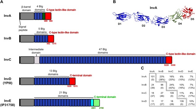
Enteropathogenic Yersinia expresses several invasins that are fundamental virulence factors required for adherence and colonization of tissues in the host. Within the invasin-family of Yersinia adhesins, to date only Invasin has been extensively studied at both structural and functional levels. In this work, we structurally characterize the recently identified inverse autotransporter InvasinE from Yersinia pseudotuberculosis (formerly InvasinD from Yersinia pseudotuberculosis strain IP31758) that belongs to the invasin-family of proteins. The sequence of the C-terminal adhesion domain of InvasinE differs significantly from that of other members of the Yersinia invasin-family and its detailed cellular and molecular function remains elusive. In this work, we present the 1.7 Å crystal structure of the adhesion domain of InvasinE along with two Immunoglobulin-like domains. The structure reveals a rod shaped architecture, confirmed by small angle X-ray scattering in solution. The adhesion domain exhibits strong structural similarities to the C-type lectin-like domain of Yersinia pseudotuberculosis Invasin and enteropathogenic/enterohemorrhagic E. coli Intimin. However, despite the overall structural similarity, the C-type lectin-like domain in InvasinE lacks motifs required for Ca2+ /carbohydrate binding as well as sequence or structural features critical for Tir binding in Intimin and β1 -integrin binding in Invasin, suggesting that InvasinE targets a distinct, yet unidentified molecule on the host-cell surface. Although the biological role and target molecule of InvasinE remain to be elucidated, our structural data provide novel insights into the architecture of invasin-family proteins and a platform for further studies towards unraveling the function of InvasinE in the context of infection and host colonization.

Keywords: C-type lectin-like domain; X-ray crystallography; Yersinia; adhesion; invasion; small angle X-ray scattering.


Figures

Figure 1
Figure 1
Overview of invasin‐family proteins of Y. pseudotuberculosis. (A) Domain organization of the invasin‐family proteins from Y. pseudotuberculosis (modified from9). (The previously identified InvD from strain IP31758 has been renamed to InvE; refer to main text for details.) (B) Cartoon representation of the crystal structure of InvA (PDB ID: 1CWV).7 (C) Matrix table of percentages (dashed line) and number of amino acids (solid line) for the sequence alignment of the C‐terminal domains of InvA, InvB, InvC, InvD and InvE. Top value represents identities and bottom value (in brackets) represents similarities of amino acids. Table has been generated with GeneDoc.32
Figure 2
Figure 2
Domain architecture of Y. pseudotuberculosis InvE and expression/purification of InvE2448. (A) Scheme of the domain organization of Y. pseudotuberculosis InvE with construct boundaries of InvE2448. (B) Elution profile of the size exclusion chromatography run on S200 16/60 column. Absorbance of the protein is measured at 280 nm. (C) Coomassie stained gel of eluted fractions analyzed by SDS‐PAGE. Blue bar represents the corresponding elution fractions in the chromatogram (B).
Figure 3
Figure 3
Overall architecture of Y. pseudotuberculosis InvE2448. (A) Cartoon representation of the crystal structure of InvE2448. InvE2448 consists of BIg20 (dark blue), BIg21 (cyan) and the C‐terminal adhesion domain (with the “adaptor ring” (V2661‐A2677) in orange, the “wedge module” (P2690‐M2712) in magenta and the CTLD in green (E2678‐R2689 and F2713‐L2795), including the LLR in red). (B) Rigid body fitting of the crystal structure into the ab initio determined SAXS envelope. The image in this panel has been rotated 180° relative to panel (A). (C) A representative portion of the electron density map (2F oF c contoured at 1 σ). (D) Fit of the rigid body model (red) with the experimental SAXS data (blue). Quality of the fit is expressed in terms of chi value. An interactive view is available in the electronic version of the article.
Figure 4
Figure 4
Overall structure of InvE BIg20 and BIg21 domains. (A) Topology diagram of the BIg20 (dark blue) and BIg21 (cyan) domains. (B) Cartoon representation of the BIg20/21 domains of InvE (Color coding as in (A); β‐strands are labeled). (C) Detailed view on the inter‐domain interactions of the BIg20–BIg21 domains, with interacting residues shown a stick‐model. (D) Close‐up view of the hydrophobic core of BIg20/21 and its comparison with human NCAM domain‐1 and InvA‐D1. Human NCAM domain‐114 adopts a classical Ig‐like fold (brown, PDB code: 5AEA) and contains a disulfide bond formed by C22 and C77 while in InvA‐D17 (light pink, PDB code: 1CWV), InvE‐BIg20 (dark blue) and InvE‐BIg21 (cyan), Cysteines are replaced by hydrophobic amino acids. (E) Conformation of the C‐E interstrand loop. Structural overlay of BIg20 (dark blue), BIg21 (cyan) and InvA‐D1 (light pink). The black box denotes the C‐E interstrand loop of BIg21. (F) Structural overlay of BIg20 and BIg21. Superposition of BIg20 (dark blue) and BIg21 (cyan) shown in two orientations (β‐strands are labeled). (G) Close up view of residues responsible for stabilizing the C–E interstrand loop. Hydrogen bonds are shown as black dashed lines.
Figure 5
Figure 5
Overall structure of InvE adhesion domain. (A) Cartoon representation of the InvE‐AD [color‐coding as in Fig. 3(A)]. Parts of the connected BIg21 are shown in gray. Secondary structure elements are labeled according to the nomenclature for the canonical CTLD‐fold.10 Labels in brackets correspond to β‐strands present in the canonical CTLD‐fold, which are however not corresponding to a β‐strand conformation in InvE‐CTLD (see also main text). Disulfide bond is shown as stick model in blue with sulfur atoms in yellow. (B) Topology of the InvE‐AD. (C) Wall‐eye stereo image of the interaction network mediated by residues in the “adaptor ring.” Water‐mediated contacts and labels/stick‐models for some residues depicted in (D) are omitted for clarity. Hydrogen bonds are shown as black‐dotted line. (D) Schematic view of the interaction network mediated by residues in the “adaptor ring.” Border thickness of residue label boxes in is proportional to the number of residue interactions. (mc = main chain interaction, sc = side chain interaction).
Figure 6
Figure 6
Structural comparison of CTLDs of InvE and InvA. (A) Structural overlay of the CTLD/“wedge module” of InvE (CTLD in green, LLR in red, “wedge module” in magenta, disulfide bond as stick model in blue/yellow) and the CTLD of Invasin (InvA) (in yellow). (B) Polar residues of InvA‐CTLD involved in the interaction with β1‐integrins are shown as stick‐model. (C) Hydrophobic residues in InvE‐CTLD (green), at the position equivalent to the β1‐integrin binding site of InvA‐CTLD. The “wedge module” in InvE is shown in magenta. (D) Structure‐based alignment of the CTLD/“wedge module” of InvE and the CTLD of InvA using the DALI server.19 Small and capital letters correspond to structurally nonaligned and aligned residues, respectively. Gaps in the sequences are depicted by a dash, residues conserved in the two sequences are linked with a vertical bar, the conserved Cysteines forming the disulfide‐bridge are shown in red. For InvE, secondary structure elements are shown in the respective color code as used in Figure 3(A). Secondary structure elements were labeled according to the canonical CTLD nomenclature.
Figure 7
Figure 7
Structural comparison of CTLDs of InvE and Intimin. (A) Structural overlay of the CTLD/“wedge module” of InvE (CTLD in green, LLR in red, “wedge module” in magenta, disulfide bond as stick model in blue/yellow) and the CTLD of Intimin (light blue) in complex with Tir (pink). (B) Intimin‐Tir complex with the Intimin‐CTLD (light blue) shown as surface and Tir (pink) as cartoon model. (C) InvE‐CTLD/“wedge module” (green/red/magenta) shown as surface and Tir (pink) as cartoon model. In the InvE‐Tir complex model, the InvE‐CTLD clashes with the β‐hairpin element in Tir. (D) Structure‐based alignment of the CTLD/“wedge module” of InvE and the CTLD of Intimin using the DALI server.19 Small and capital letters correspond to structurally non‐aligned and aligned residues, respectively. Gaps in the sequences are depicted by a dash, residues conserved in the two sequences are linked with a vertical bar, the conserved Cysteines forming the disulfide‐bridge are shown in red. For InvE, secondary structure elements are shown in the respective color code as used in Figure 3(A). Secondary structure elements were labeled according to the canonical CTLD nomenclature.
Figure 8
Figure 8
Orientation of InvE AD as compared to InvA and Intimin. (A) Structural overlay of InvE [BIg20/BIg21 in gray, InvE‐AD in color code as used in Fig. 3(A) with InvA (D3/D4/InvA AD, in yellow)]. (B) Structural overlay of InvE [BIg20/BIg21 in gray, InvE‐AD in color code as used in Fig. 3(A) with Intimin (D1/D2/Intimin AD, in light blue)]. (C) Overlay of InvE, InvA and Intimin in surface representation in two orientations. (D) Left: Schematic representation of InvE embedded in the bacterial outer membrane (OM), reminiscent of the putative biological orientation perpendicular to the bacterial surface. Right: Schematic representation of BIg20, BIg21 and AD with the CTLD, “adaptor ring” and “wedge module.” The long axis of the rod‐shaped molecule is depicted by a black dashed line. In comparison to the orientation in InvE (defined as 0°), the CTLDs of InvA and Intimin are tilted by 65° and 44°, respectively, as shown by the dashed yellow and blue lines. (E) Representation of InvE, InvA and Intimin in a parallel arrangement (in analogy to panel D). The CTLDs are shown as surface, emphasizing the tilt of the CTLDs in regard to the BIg‐stalk, as depicted schematically in D. (F) Left: Schematic representation of the four Cα‐positions (C‐terminal residue of CTLD α1/N‐terminal residue of CTLD α1/BIg strand E/BIg strand G) used for determination of the dihedral angle and rotation of the CTLD in regard to the preceding BIg domain. Right: View along the axis connecting Cα‐positions of the N‐terminal residue in helix α1 and of the residue in strand E. (G) View on top of the CTLDs (corresponding to the right panel in F) with the respective dihedral angles between the CTLD and the preceding BIg domain. Helix α1 is shown in a darker tone (InvE, dark green; InvA, orange; Intimin dark blue) and the N‐ and C‐terminal Cα‐positions in the helix are shown as blue and red sphere, respectively. The position of Cα‐residue in BIg strand G is shown as black sphere, the position of Cα‐residue in BIg strand E is in line with the N‐terminal residue of helix α1 and thus not visible in this view. Dihedral angles were determined using the following Cα‐positions: InvE M2721/Q2715/V2631/N2660; InvA C907/S900/I858/P884; Intimin N862/Y853/L814/I836 (C‐terminal residue of CTLD α1/N‐terminal residue of CTLD α1/BIg strand E/BIg strand G).

Similar articles

Cited by

References

    1. Bottone EJ (1999) Yersinia enterocolitica: overview and epidemiologic correlates. Microbes Infect 1:323–333. - PubMed
    1. Isberg RR, Voorhis DL, Falkow S (1987) Identification of invasin: a protein that allows enteric bacteria to penetrate cultured mammalian cells. Cell 50:769–778. - PubMed
    1. Reis RSD, Horn F (2010) Enteropathogenic Escherichia coli, Samonella, Shigella and Yersinia: cellular aspects of host‐bacteria interactions in enteric diseases. Gut Pathog 2:8. - PMC - PubMed
    1. Chauhan N, Wrobel A, Skurnik M, Leo JC (2016) Yersinia adhesins: an arsenal for infection. Proteomics Clin Appl. 10:949–963. - PubMed
    1. Leo JC, Oberhettinger P, Schütz M, Linke D (2015) The inverse autotransporter family: intimin, invasin and related proteins. Int J Med Microbiol 305:276–282. - PubMed

Publication types

MeSH terms

LinkOut - more resources