. 2017 Sep;11(9-10):1600190.
doi: 10.1002/prca.201600190. Epub 2017 May 12.

Complementary proteomic approaches reveal mitochondrial dysfunction, immune and inflammatory dysregulation in a mouse model of Gulf War Illness

Affiliations

Complementary proteomic approaches reveal mitochondrial dysfunction, immune and inflammatory dysregulation in a mouse model of Gulf War Illness

Zuchra Zakirova et al. Proteomics Clin Appl. 2017 Sep.

Abstract

Purpose: Long-term consequences of combined pyridostigmine bromide (PB) and permethrin (PER) exposure in C57BL6/J mice using a well-characterized mouse model of exposure to these Gulf War (GW) agents were explored at the protein level.

Experimental design: We used orthogonal proteomic approaches to identify pathways that are chronically impacted in the mouse CNS due to semiacute GW agent exposure early in life. These analyses were performed on soluble and membrane-bound protein fractions from brain samples using two orthogonal isotopic labeling LC-MS/MS proteomic approaches-stable isotope dimethyl labeling and iTRAQ.

Results: The use of these approaches allowed for greater coverage of proteins than was possible by either one alone and revealed both distinct and overlapping datasets. This combined analysis identified changes in several mitochondrial, as well as immune and inflammatory pathways after GW agent exposure.

Conclusions and clinical relevance: The work discussed here provides insight into GW agent exposure dependent mechanisms that adversely affect mitochondrial function and immune and inflammatory regulation. Collectively, our work identified key pathways which were chronically impacted in the mouse CNS following acute GW agent exposure, this may lead to the identification of potential targets for therapeutic intervention in the future. Long-term consequences of combined PB and PER exposure in C57BL6/J mice using a well-characterized mouse model of exposure to these GW agents were explored at the protein level. Expanding on earlier work, we used orthogonal proteomic approaches to identify pathways that are chronically impacted in the mouse CNS due to semiacute GW agent exposure early in life. These analyses were performed on soluble and membrane-bound protein fractions from brain samples using two orthogonal isotopic labeling LC-MS/MS proteomic approaches-stable isotope dimethyl labeling and iTRAQ. The use of these approaches allowed for greater coverage of proteins than was possible by either one alone and revealed both distinct and overlapping datasets. This combined analysis identified changes in several mitochondrial, as well as immune and inflammatory pathways after GW agent exposure. The work discussed here provides insight into GW agent exposure dependent mechanisms that adversely affect mitochondrial function and immune and inflammatory regulation at 5 months postexposure to PB + PER.

Keywords: Gulf War; MS/MS; SIDL; iTRAQ; mitochondrial dysfunction.


Figures

Figure 1
Figure 1
Proteomics workflow. Workflow (A) depicting protein extraction, and schematic of two proteomics experiments conducted using brain homogenates from PB + PER exposed mice and controls using (B) SIDL and (C) iTRAQ methodologies.
Figure 2
Figure 2
Venn‐diagrams. Venn‐diagram depicting (A) the distinct total number of proteins identified by iTRAQ and SIDL, as well as the overlapping number of proteins. Venn‐diagram illustrating (B) the statistically significant number of proteins identified by iTRAQ and SIDL, unique to each proteomic experiment, as well as the overlapping number of proteins.
Figure 3
Figure 3
Heatmap of overlapping proteins identified by iTRAQ and SIDL. Heatmap generated from proteomics data (iTRAQ and SIDL) reflecting statistically significant overlapping proteins, and their fold change ratios (ln) in brains of PB + PER exposed as compared to control mice.
Figure 4
Figure 4
IPA‐based diseases and biofunctions, and canonical pathways. IPA platform analysis mapped top diseases and biofunctions, and top canonical pathways that were associated with CNS changes at 5 months postexposure to GW agent exposure using SIDL and iTRAQ. (A) Top diseases and biofunctions include Neurological Disease, Psychological Disorders, Metabolic Disease and Skeletal and Muscular Disorders, among others. (B) Top canonical pathways associated with energy production such as Mitochondrial Dysfunction, OXPHOS, and the TCA cycle, as well as immune and inflammatory dysregulation such as mTOR signaling and PI3K/AKT signaling were among the top‐ranking pathways that were affected by GW agent exposure. TCA, tricarboxylic acid.
Figure 5
Figure 5
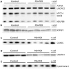
Western blotting analyses using antibodies against mitochondrial proteins. Membrane brain homogenates from control and PB + PER exposed mice were probed with antibodies against mitochondrial proteins. (A) Anti‐OXPHOS cocktail antibody was used to probe membranes using five major proteins (Complex V, ATP5A, 55 kD, Complex III, UQCR2, 48 kD, Complex IV, MTCO1, 40 kD, Complex II, SDHB, 30 kD, and Complex I, NDUFB8, 20 kD) revealed decreased expression in several key proteins such as NDUFB8 (Complex I; t‐test = 3.672, DF = 5, p = 0.014), SDHB (Complex II; t‐test = 3.672, DF = 5, p = 0.014), and MTCO1 (Complex IV; t‐test = 2.856, DF = 5, p = 0.036) in the brains of PB + PER mice as compared to controls. (B) Membranes were probed with anti‐ATP5F1 antibody (Complex V; 29 kD), no statistically significant differences were observed between the two groups. (C) Membranes were probed with an anti‐COX6C antibody (Complex IV, 9 kD), a significant reduction in COX6C expression was revealed in exposed mice as compared to controls (t = 3.472, DF = 4, p = 0.026). (D) Probing with an anti‐UQCRC1 antibody (Complex III, 35 kD) using brain homogenates revealed a significant reduction in exposed mice as compared to controls (t‐test = 3.947, DF = 6, p = 0.008). (E) Probing with anti‐cytochrome c antibody (15 kD) revealed no statistically significant differences between the two groups. All protein expression values were normalized to UQCRC2, as a loading control. ATP5A, ATP Synthase, H+ Transporting, Mitochondrial F1 Complex, Alpha Subunit 1; ATPF1, ATP Synthase Mitochondrial F1 Complex Assembly Factor 1; COX6C, Cytochrome C Oxidase Subunit 6C MTCO1, mitochondrially encoded cytochrome c oxidase I; SDHB, succinate dehydrogenase complex, subunit B; UQCRC1, Ubiquinol‐Cytochrome C Reductase Core Protein I; UQCRC2, Ubiquinol‐Cytochrome C Reductase Core Protein II.
Figure 6
Figure 6
Quantifications of Western blotting data. The histograms represent the average ratio of each representative antibody against UQCRC2 total protein used as a loading control for control and in PB + PER exposed mice. UQCRC2, Ubiquinol‐Cytochrome C Reductase Core Protein II.
Figure 7
Figure 7
Cytokine profiling. Cytokine profile in brain homogenates and plasma of control and PB + PER exposed mice (n = 14/group; N =28), using a Bioplex multiplex system for simultaneous detection of Th17 cytokines. (A and B) Concentrations of cytokines in brain homogenates of control and PB + PER exposed mice, no differences were observed between exposed and control mice. (C and D) Concentrations of cytokines in plasma samples of mice, where IFN‐γ (t‐test = −3.0, DF = 24, p = 0.006), TNF‐α (t‐test = −2.4, DF = 24, p = 0.02), IL‐10 (t‐test = −3.79, DF = 24, p = 0.0009), and IL‐1β (t‐test = −3.85, DF = 24, p = 0.0008) expression was reduced in PB + PER mice as compared to controls. A p‐value lower than 0.05 was found to be statistically significant (*p ≤ 0.05; **p ≤ 0.01; ***p ≤ 0.001). Error bars in the figures present the SEM.

Similar articles

Cited by

References

    1. Wille, T. , Thiermann, H. , Worek, F. , In vitro kinetic interactions of DEET, pyridostigmine and organophosphorus pesticides with human cholinesterases. Chem. Biol. Interact. 2011, 190, 79–83. - PubMed
    1. Binns, J. B. C. , Bloom, E. , Clauw, D. , Gulf war illness and the health of gulf war veterans. Research Advisory Committee on Gulf War Veterans’ Illnesses 2008.
    1. Zakirova, Z. , Crynen, G. , Hassan, S. , Abdullah, L. et al., A chronic longitudinal characterization of neurobehavioral and neuropathological cognitive impairment in a mouse model of Gulf War agent exposure. Front. Integr. Neurosci. 2016, 9, 71–95. - PMC - PubMed
    1. Zakirova, Z. , Tweed, M. , Crynen, G. , Reed, J. et al., Gulf War agent exposure causes impairment of long‐term memory formation and neuropathological changes in a mouse model of Gulf War Illness. PLoS One 2015, 10, e0119579. - PMC - PubMed
    1. Ojo, J. O. , Abdullah, L. , Evans, J. , Reed, J. M. et al., Exposure to an organophosphate pesticide, individually or in combination with other Gulf War agents, impairs synaptic integrity and neuronal differentiation, and is accompanied by subtle microvascular injury in a mouse model of Gulf War agent exposure. Neuropathology 2013, 34, 109–127. - PubMed

Publication types