. 2017 Apr;6(4):1286-1294.
doi: 10.5966/sctm.2015-0363. Epub 2016 Sep 29.

Placental Stromal Cell Therapy for Experimental Autoimmune Encephalomyelitis: The Role of Route of Cell Delivery

Affiliations

Placental Stromal Cell Therapy for Experimental Autoimmune Encephalomyelitis: The Role of Route of Cell Delivery

Ilona Shapira et al. Stem Cells Transl Med. 2017 Apr.

Abstract

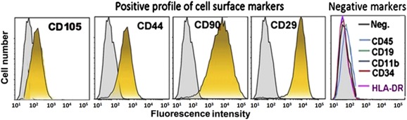
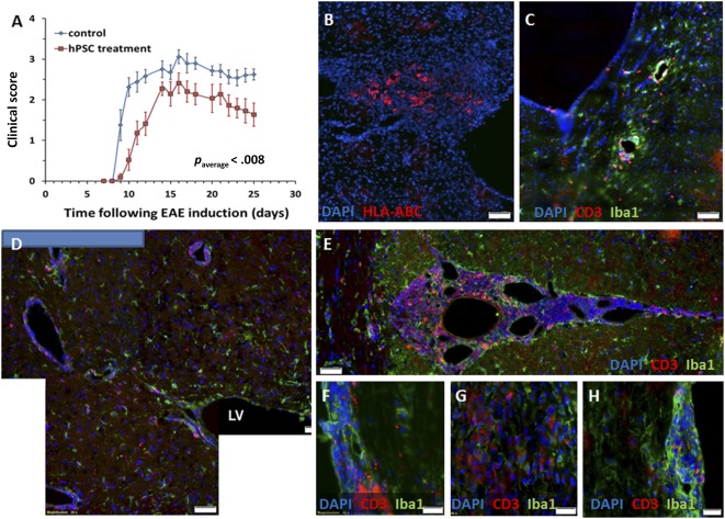
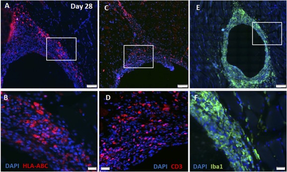
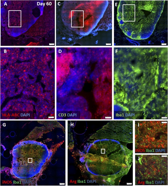
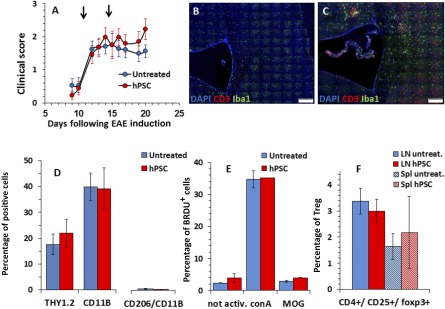
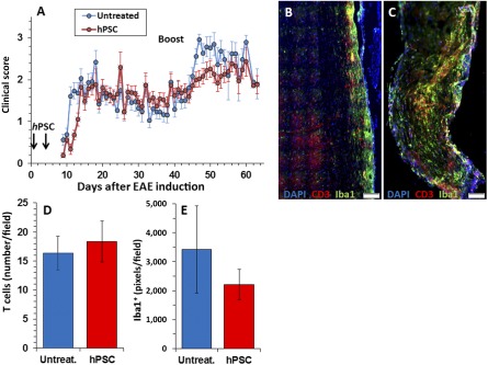
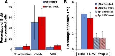
Multiple sclerosis (MS) is an immune-mediated disease of the central nervous system (CNS) with no effective treatment available for the chronic-progressive stage. Cell therapy is a promising therapeutic approach for attenuating the immune-mediated CNS process. Isolated and expanded human placental stromal cells (hPSCs) possess potent immunomodulatory and trophic properties, making them a good candidate for MS therapy. We examined the potential of hPSC therapy in preventing the onset or attenuating the course of established disease in a murine MS model of myelin oligodendrocyte glycoprotein-induced experimental autoimmune encephalomyelitis. We examined the feasibility of hPSC systemic delivery by intramuscular (i.m.) implantation rather than the commonly used intravenous injection, which is dose-limiting and carries the risk of pulmonary obstruction. Our findings showed significant attenuation of the disease only when hPSCs were injected directly to the central nervous system. Intramuscular implanted hPSCs survived at the site of injection for at least 2 months and elicited extensive local immune responses. Intramuscular hPSC implantation before disease onset caused a delay in the appearance of clinical signs and reduced the severity of a relapse induced by repeated challenge with the autoantigen. Intramuscular implantation after disease onset did not affect its course. Thus, pathological analysis of CNS tissue did not show inhibition of neuroinflammation in i.m. hPSC-implanted mice. Moreover, no apparent effect was seen on the proliferative response of peripheral lymph node cells in these animals. We conclude that to maximize their therapeutic potential in MS, hPSCs should be delivered directly to the affected CNS. Stem Cells Translational Medicine 2017;6:1286-1294.

Keywords: Cell therapy; Multiple sclerosis; Placenta; Route of cell delivery.


Figures

Figure 1
Figure 1
Characterization of isolated human placental stromal cells as stromal cells by cell surface marker fluorescence‐activated cell sorting analyses. The cells were negative for markers of hematopoietic or endothelial progenies (CD45, CD19, CD11b, HLA‐DR, and CD34) and positive for typical mesenchymal stromal markers (CD105, CD44, CD29, and CD90). Abbreviations: HLA, human leukocyte antigen; Neg., negative.
Figure 2
Figure 2
Intracranial implantation of hPSCs to EAE mice. hPSCs were implanted in the periventricular white matter of mice on day 7 after EAE induction before the development of clinical signs (n = 12 mice). (A): Significant (p = .001) disease attenuation was observed in the clinical course of hPSC‐implanted mice compared with a control EAE group (n = 11). (B): hPSCs were detected 18 days after implantation by HLA‐ABC immunostaining of the mouse brain sections. (C): EAE mice exhibited a typical inflammatory response in their brains on day 25 after EAE induction. (D, E): A strong inflammatory response was noted in the hPSC‐transplanted mice in proximity to the implantation site and in the brain meninges. Both meningeal and parenchymal spinal cord infiltrates were detected in the EAE mice (F, G), but the hPSC‐implanted mice exhibited mostly meningeal infiltrates (H). Scale bars = 50 µm (B–E) and 20 µm (F–H). Abbreviations: DAPI, 4′,6‐diamidino‐2‐phenylindole; EAE, experimental autoimmune encephalomyelitis; HLA, human leukocyte antigen; hPSC, human placental stromal cell; LV, lateral ventricle.
Figure 3
Figure 3
Intramuscularly implanted human placental stromal cells (hPSCs) survive and elicit an immune response. At 28 days after intramuscular implantation, numerous hPSCs were detected in the recipient muscles by HLA‐ABC immunostaining (A, B). A significant immune response was evident in proximity to the graft, as manifested by extensive infiltration of CD3+ T cells (C, D) and Iba1+ macrophages (E, F). Insets in (A, C, E) are magnified in (B, D, F), respectively; n = 5) in (C). Scale bars = 100 µm (A–C) and 20 µm (D–F). Abbreviations: DAPI, 4′,6‐diamidino‐2‐phenylindole; HLA, human leukocyte antigen.
Figure 4
Figure 4
Long‐term survival of hPSCs in muscles along with an aggravated immune response. At 60 days after intramuscular implantation, numerous hPSCs were still detected in the recipient muscles (n = 20) by HLA‐ABC immunostaining (A, B). A further increase in the severity of the local immune response, with formation of ectopic lymph node‐like structures was evident in proximity to the graft, as shown by immunostaining for CD3+ T cells (C, D) and Iba1+ macrophages (E, F). Characterization of macrophage polarization showed marked domination of classic iNOS+ (M1) macrophages (G, I) versus alternative dispersed Arg+ (M2) macrophages (H, J). Insets in (A, C, E, G, H) are magnified in (B, D, F, I, J), respectively. Scale bars = 500 µm (A, C, E, G, H) and 50 µm (B, D, F, I, J). Abbreviations: Arg, arginase; DAPI, 4′,6‐diamidino‐2‐phenylindole; HLA, human leukocyte antigen; iNOS, inducible nitric oxide synthase.
Figure 5
Figure 5
Intramuscular hPSC injection after the onset of clinical signs did not affect the course of EAE; 2 × 10⁶ of hPSCs were injected i.m. on days 11 and 15 (arrows) after EAE induction. (A): No difference was observed in disease severity between the untreated control and implanted mice (n = 20, each group, A). Immunofluorescent staining of brain section for CD3+ T cells and Iba1+ microglia/macrophages showed a certain increase in inflammatory processes in the brains of implanted mice (C) compared with untreated mice (B). The immune cells were extracted from the brains and spinal cords of i.m. hPSC‐treated and untreated mice (n = 6, per group) and characterized by fluorescence‐activated cell sorting analysis. No difference was found in the fraction or polarization of CD11b+ microglia/macrophages or Thy1.2+ expressing T cells between the two experimental groups (D). To evaluate whether i.m. hPSC delivery induces any systemic immunomodulatory effects, we examined the proliferative response of lymph node cells in vitro on day 22 after EAE induction. ConA induced a similar proliferation of LNCs in the implanted and control groups (n = 6 per group, E). At this time point, myelin oligodendrocyte glycoprotein peptide corresponding to amino acids 35–55 induced only a borderline proliferative response (E). No difference was observed between the fractions of LN‐ and Spl‐derived regulatory T cells in the two groups (F). Scale bars = 100 µm (B, C). Abbreviations: conA, concanavalin A; DAPI, 4′,6‐diamidino‐2‐phenylindole; EAE, experimental autoimmune encephalomyelitis; hPSC, human placental stromal cell; LN, lymph node; LNCs, lymph node cells; MOG, myelin oligodendrocyte glycoprotein; not activ., not activated; Spl, spleen; Treg, regulatory T cells; untreat., untreated.
Figure 6
Figure 6
Intramuscular hPSC injection before EAE induction delayed disease onset and attenuated relapse; 2 × 10⁶ hPSCs were injected i.m. on days 1 and 5 after EAE induction before the onset of clinical signs. The clinical scores of all mice participating in this project (n = 42 implanted and 45 control mice, part of which were sacrificed at different time points for pathological and immunological studies) were analyzed. (A): A mild, but statistically significant, postponement of disease onset by 2 days was observed in i.m. implanted mice (p = .026) followed by a comparable disease course. In the postacute phase, no significant difference was found between the groups. To evaluate whether i.m. hPSC treatment exerted long‐term therapeutic effects, a relapse was induced on day 40. Relapse was observed in 85% of the control versus 54% of the hPSC‐treated mice (p < .05). The intensity of the relapse was 2.32 ± 1 in the control group versus 1.51 ± 0.43 in the hPSC‐injected mice (p < .0005). By day 60, these differences were annulled, and the two experimental groups were identical clinically. Quantification of the inflammatory response in the spinal cord at this time showed no difference in the number of CD3+ T cells, with a mild nonsignificant decline in Iba1+ macrophage/microglia in the hPSC‐treated (n = 16; C–E) versus control (n = 13; B, D, E) mice. Scale bars = 100 µm (B, C). Abbreviations: DAPI, 4′,6‐diamidino‐2‐phenylindole; EAE, experimental autoimmune encephalomyelitis; hPSC, human placental stromal cell; Untreat., untreated.
Figure 7
Figure 7
Intramuscular hPSC implantation before EAE induction did not reduce systemic LNC response. To assess whether hPSC i.m. injection before the onset of clinical signs attenuated the systemic immune response, a proliferative BrdU incorporation assay was conducted on LNCs excised on day 10 after EAE induction. Both ConA and MOG peptide stimulation caused a similar proliferative response in the control and hPSC‐injected mice (A). Also, no difference was detected in the fraction of CD4+, CD25+, Fox3+ regulatory T cells from the lymph nodes and spleen of hPSC‐treated and control groups (n = 6 per group). Abbreviations: BrdU, bromodeoxyuridine; conA, concanavalin A; hPSC, human placental stromal cell; LN, lymph node; MOG, myelin oligodendrocyte glycoprotein; Spl, spleen; Treg, regulatory T cells; treat., treated.

References

    1. Aharonowiz M, Einstein O, Fainstein N et al. Neuroprotective effect of transplanted human embryonic stem cell‐derived neural precursors in an animal model of multiple sclerosis. PLoS One 2008;3:e3145. - PMC - PubMed
    1. Einstein O, Fainstein N, Vaknin I et al. Neural precursors attenuate autoimmune encephalomyelitis by peripheral immunosuppression. Ann Neurol 2007;61:209–218. - PubMed
    1. Einstein O, Grigoriadis N, Mizrachi‐Kol R et al. Transplanted neural precursor cells reduce brain inflammation to attenuate chronic experimental autoimmune encephalomyelitis. Exp Neurol 2006;198:275–284. - PubMed
    1. Zappia E, Casazza S, Pedemonte E et al. Mesenchymal stem cells ameliorate experimental autoimmune encephalomyelitis inducing T‐cell anergy. Blood 2005;106:1755–1761. - PubMed
    1. Pluchino S, Martino G. The therapeutic use of stem cells for myelin repair in autoimmune demyelinating disorders. J Neurol Sci 2005;233:117–119. - PubMed

Publication types

Substances