

Cysteine persulfides and polysulfides produced by exchange reactions with H2S protect SH-SY5Y cells from methylglyoxal-induced toxicity through Nrf2 activation
- PMID: 28371750
- PMCID: PMC5377440
- DOI: 10.1016/j.redox.2017.03.020
Cysteine persulfides and polysulfides produced by exchange reactions with H2S protect SH-SY5Y cells from methylglyoxal-induced toxicity through Nrf2 activation
Abstract
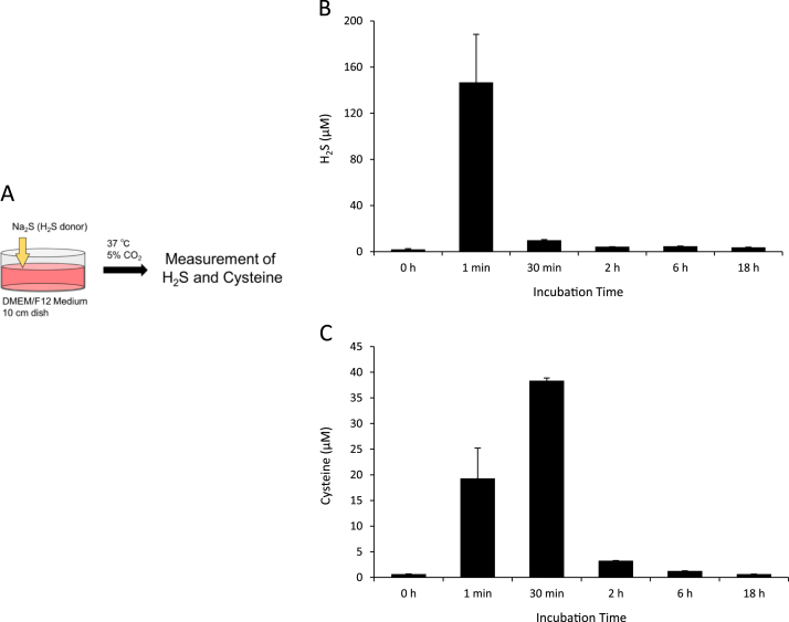
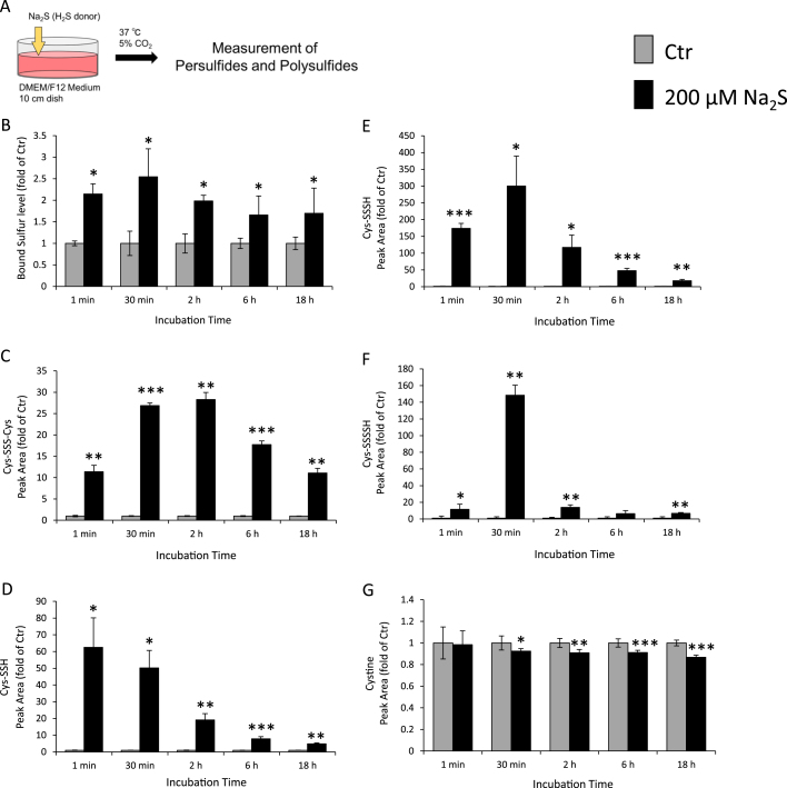
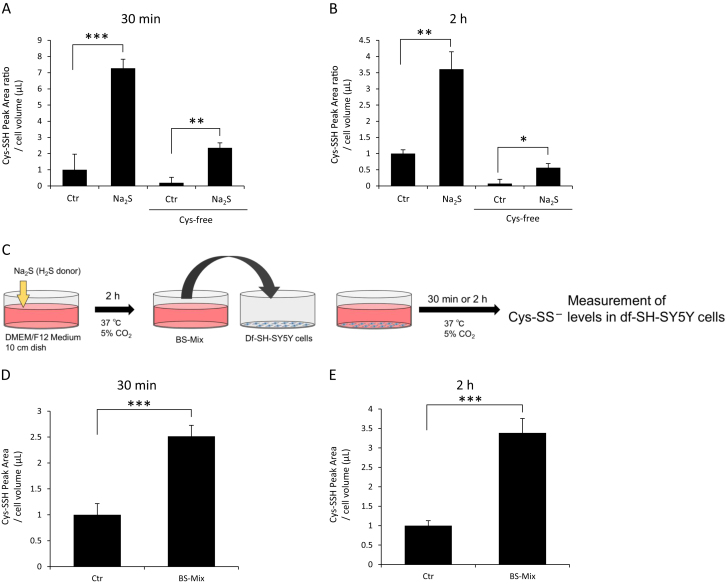
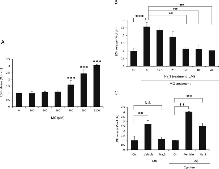
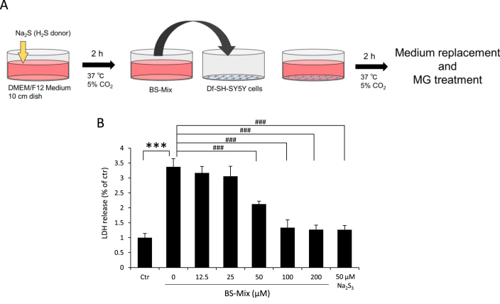
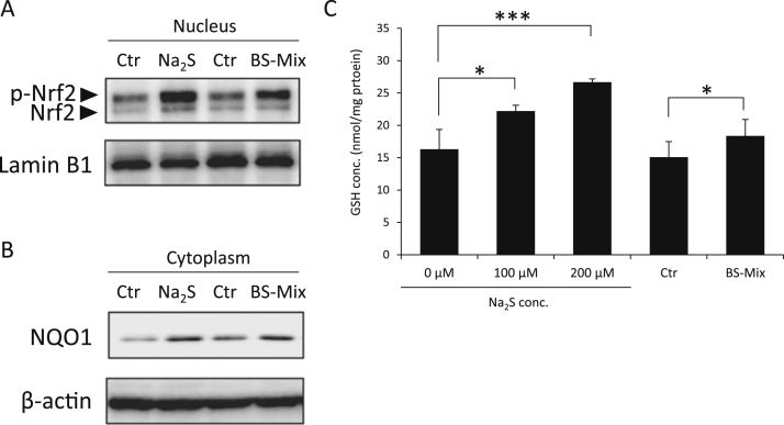
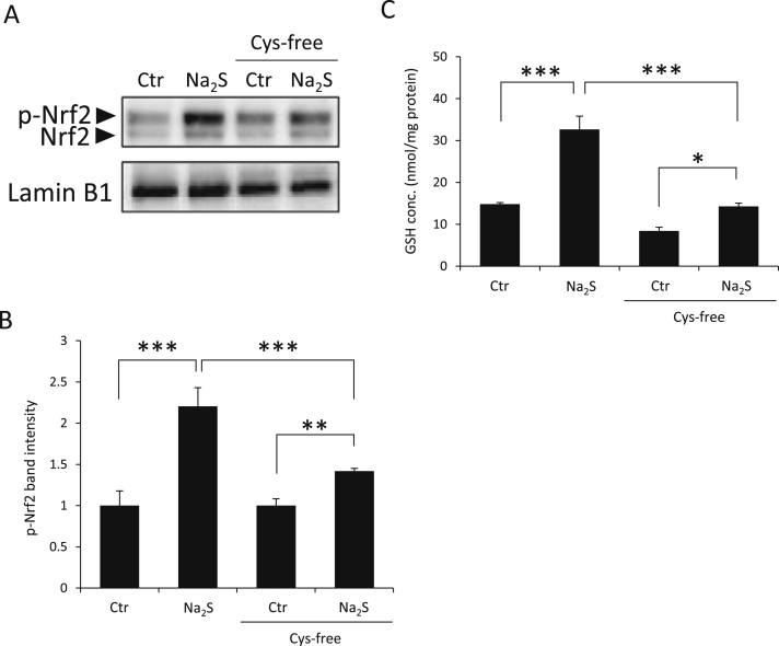
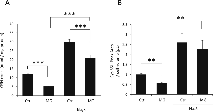
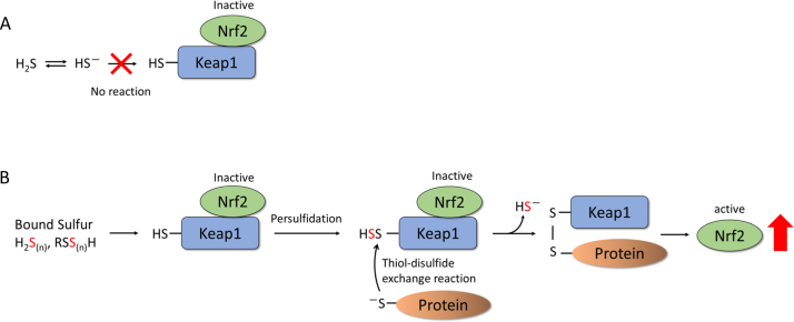
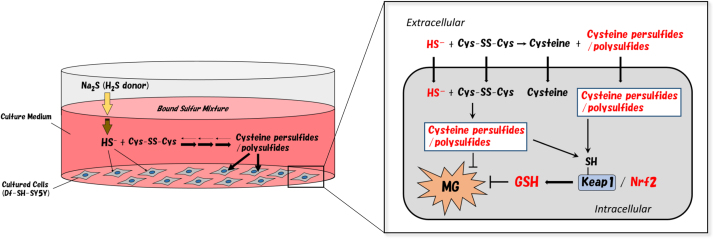
Many physiological functions of hydrogen sulfide (H2S) have been reported in mammalian cells over the last 20 years. These physiological effects have been ascertained through in vitro treatment of cells with Na2S or NaHS, both of which are precursors of H2S. Since H2S exists as HS- in a neutral solution, a disulfide compound such as cystine could react with HS- in culture medium as well as in the cell. This study demonstrated that after the addition of Na2S solution into culture medium, HS- was transiently generated and disappeared immediately through the reaction between HS- and cystine to form cysteine persulfides and polysulfides in the culture medium (bound sulfur mixture: BS-Mix). Furthermore, we found that the addition of Na2S solution resulted in an increase of intracellular cysteine persulfide levels in SH-SY5Y cells. This alteration in intracellular persulfide was also observed in cystine-free medium. Considering this reaction of HS- as a precursor of BS-Mix, we highlighted the cytoprotective effect of Na2S on human neuroblastoma SH-SY5Y cells against methylglyoxal (MG)-induced toxicity. BS-Mix produced with Na2S in cystine-containing medium provided SH-SY5Y cells significant protective effect against MG-induced toxicity. However, the protective effect was attenuated in cystine-free medium. Moreover, we observed that Na2S or BS-Mix activated the Keap1/Nrf2 system and increased glutathione (GSH) levels in the cell. In addition, the activation of Nrf2 is significantly attenuated in cystine-free medium. These results suggested that Na2S protects SH-SY5Y cells from MG cytotoxicity through the activation of Nrf2, mediated by cysteine persulfides and polysulfides that were generated by Na2S addition.
Keywords: Bound sulfur species; Hydrogen sulfide; Methylglyoxal; Nrf2; Persulfide; Polysulfide.
Copyright © 2017 The Authors. Published by Elsevier B.V. All rights reserved.
Figures

Similar articles
-
Polysulfides protect SH-SY5Y cells from methylglyoxal-induced toxicity by suppressing protein carbonylation: A possible physiological scavenger for carbonyl stress in the brain.Neurotoxicology. 2016 Jul;55:13-19. doi: 10.1016/j.neuro.2016.05.003. Epub 2016 May 6. Neurotoxicology. 2016. PMID: 27163164
-
S-Transnitrosation reactions of hydrogen sulfide (H2S/HS-/S2-) with S-nitrosated cysteinyl thiols in phosphate buffer of pH 7.4: Results and review of the literature.Nitric Oxide. 2017 May 1;65:22-36. doi: 10.1016/j.niox.2017.02.001. Epub 2017 Feb 6. Nitric Oxide. 2017. PMID: 28185882
-
Tanshinone I Induces Mitochondrial Protection by a Mechanism Involving the Nrf2/GSH Axis in the Human Neuroblastoma SH-SY5Y Cells Exposed to Methylglyoxal.Neurotox Res. 2019 Oct;36(3):491-502. doi: 10.1007/s12640-019-00091-1. Epub 2019 Jul 29. Neurotox Res. 2019. PMID: 31359290
-
Enzymatic Regulation and Biological Functions of Reactive Cysteine Persulfides and Polysulfides.Biomolecules. 2020 Aug 27;10(9):1245. doi: 10.3390/biom10091245. Biomolecules. 2020. PMID: 32867265 Free PMC article. Review.
-
Signaling by hydrogen sulfide (H2S) and polysulfides (H2Sn) in the central nervous system.Neurochem Int. 2019 Jun;126:118-125. doi: 10.1016/j.neuint.2019.01.027. Epub 2019 Mar 6. Neurochem Int. 2019. PMID: 30849397 Review.
Cited by
-
The hepatic compensatory response to elevated systemic sulfide promotes diabetes.Cell Rep. 2021 Nov 9;37(6):109958. doi: 10.1016/j.celrep.2021.109958. Cell Rep. 2021. PMID: 34758301 Free PMC article.
-
The Human Mercaptopyruvate Sulfurtransferase TUM1 Is Involved in Moco Biosynthesis, Cytosolic tRNA Thiolation and Cellular Bioenergetics in Human Embryonic Kidney Cells.Biomolecules. 2023 Jan 10;13(1):144. doi: 10.3390/biom13010144. Biomolecules. 2023. PMID: 36671528 Free PMC article.
-
Characterization of the Inducible and Slow-Releasing Hydrogen Sulfide and Persulfide Donor P*: Insights into Hydrogen Sulfide Signaling.Antioxidants (Basel). 2021 Jun 29;10(7):1049. doi: 10.3390/antiox10071049. Antioxidants (Basel). 2021. PMID: 34209813 Free PMC article.
-
Expanding the Reactive Sulfur Metabolome: Intracellular and Efflux Measurements of Small Oxoacids of Sulfur (SOS) and H2S in Human Primary Vascular Cell Culture.Molecules. 2021 Nov 26;26(23):7160. doi: 10.3390/molecules26237160. Molecules. 2021. PMID: 34885743 Free PMC article.
-
Effects of Manganese Porphyrins on Cellular Sulfur Metabolism.Molecules. 2020 Feb 22;25(4):980. doi: 10.3390/molecules25040980. Molecules. 2020. PMID: 32098303 Free PMC article.
References
-
- Hosoki R., Matsuki N., Kimura H. The possible role of hydrogen sulfide as an endogenous smooth muscle relaxant in synergy with nitric oxide. Biochem. Biophys. Res. Commun. 1997;237(3):527–531. - PubMed
-
- Shibuya N., Tanaka M., Yoshida M., Ogasawara Y., Togawa T., Ishii K., Kimura H. 3-Mercaptopyruvate sulfurtransferase produces hydrogen sulfide and bound sulfane sulfur in the brain. Antioxid. Redox Signal. 2009;11(4):703–714. - PubMed
-
- Shibuya N., Koike S., Tanaka M., Ishigami-yuasa M., Kimura Y., Ogasawara Y., Fukui K., Nagahara N., Kimura H. A novel pathway for the production of hydrogen sulfide from D-cysteine in mammalian cells. Nat. Commun. 2013;4:1366–1367. - PubMed
MeSH terms
Substances
LinkOut - more resources
Full Text Sources
Other Literature Sources
