. 2017 Sep 1;19(9):1183-1194.
doi: 10.1093/neuonc/nox027.

Telomerase activation in posterior fossa group A ependymomas is associated with dismal prognosis and chromosome 1q gain

Affiliations

Telomerase activation in posterior fossa group A ependymomas is associated with dismal prognosis and chromosome 1q gain

Johannes Gojo et al. Neuro Oncol. .

Abstract

Background: Ependymomas account for up to 10% of childhood CNS tumors and have a high rate of tumor recurrence despite gross total resection. Recently, classification into molecular ependymoma subgroups has been established, but the mechanisms underlying the aggressiveness of certain subtypes remain widely enigmatic. The aim of this study was to dissect the clinical and biological role of telomerase reactivation, a frequent mechanism of cancer cells to evade cellular senescence, in pediatric ependymoma.

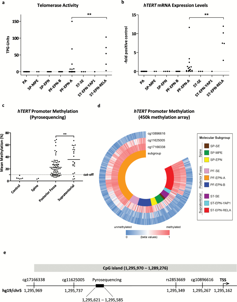
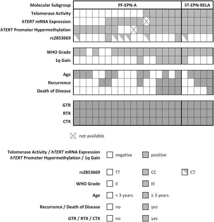
Methods: We determined telomerase enzymatic activity, hTERT mRNA expression, promoter methylation, and the rs2853669 single nucleotide polymorphism located in the hTERT promoter in a well-characterized cohort of pediatric intracranial ependymomas.

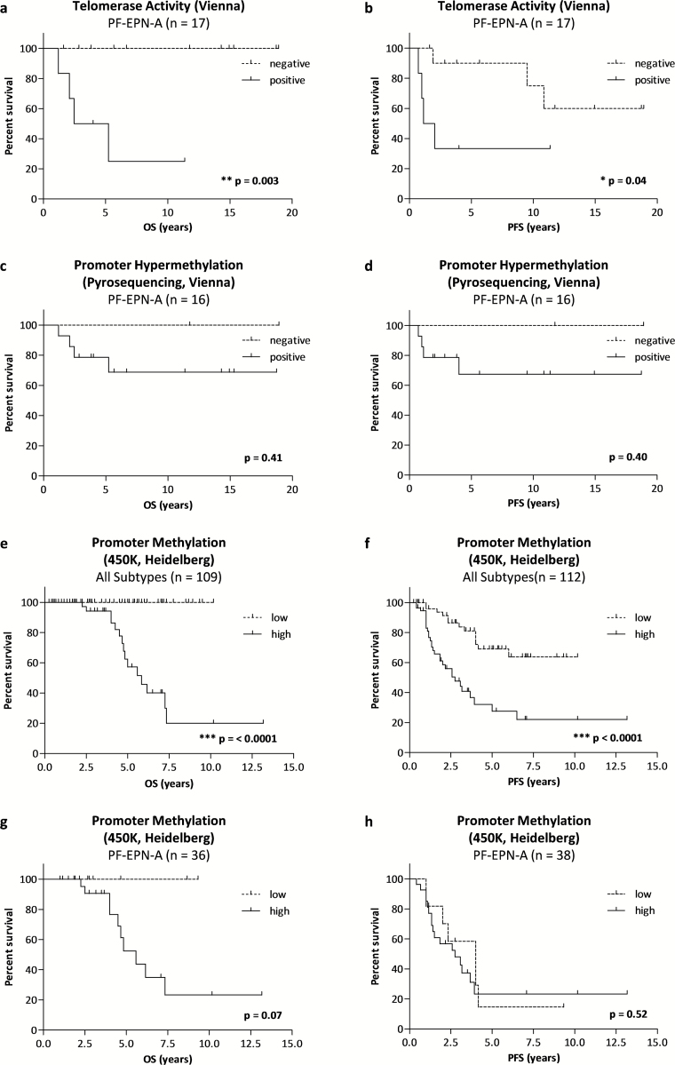
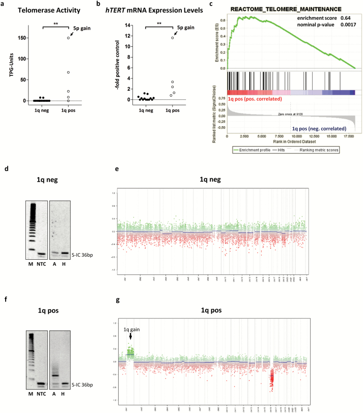
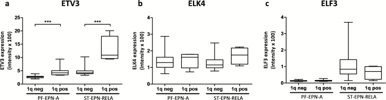
Results: In posterior fossa ependymoma group A (PF-EPN-A) tumors, telomerase activity varied and was significantly associated with dismal overall survival, whereas telomerase reactivation was present in all supratentorial RelA fusion-positive (ST-EPN-RELA) ependymomas. In silico analysis of methylation patterns showed that only these two subgroups harbor hypermethylated hTERT promoters suggesting telomerase reactivation via epigenetic mechanisms. Furthermore, chromosome 1q gain, a well-known negative prognostic factor, was strongly associated with telomerase reactivation in PF-EPN-A. Additional in silico analyses of gene expression data confirmed this finding and further showed enrichment of the E-twenty-six factor, Myc, and E2F target genes in 1q gained ependymomas. Additionally, 1q gained tumors showed elevated expression of ETV3, an E-twenty-six factor gene located on chromosome 1q.

Conclusion: Taken together we describe a subgroup-specific impact of telomerase reactivation on disease progression in pediatric ependymoma and provide preliminary evidence for the involved molecular mechanisms.

Keywords: RelA fusion; chromosome 1q; ependymoma; promoter methylation; telomerase.


Figures

Fig. 1
Fig. 1
Telomerase activation in ependymoma. (A) Telomerase activity and (B) hTERT mRNA expression levels of pilocytic astrocytoma (PA), myxopapillary ependymoma (SP-MPE), spinal ependymoma WHO grade II (SP-EPN), posterior fossa group A (PF-EPN-A) and B (PF-EPN-B), supratentorial subependymoma (ST-SE), RelA fusion-positive (ST-EPN-RELA), and Yap1 fusion-positive (ST-EPN-YAP1) ependymomas. The dotted line at zero highlights negative samples. (C) hTERT promoter methylation stratified for tumor localization. (D) Methylation analysis of the hTERT promoter derived from 450k methylation array data, blue indicating low and red high DNA methylation and stratified for molecular subgroups (GSE65362, n = 500). (E) Map of the hTERT promoter showing the investigated regions. Sites are annotated according to hg19 (University of California Santa Cruz Genome Browser). Statistical analyses were performed by Mann–Whitney U test (**P < .01). The mean of each group is depicted as a solid line. TSS, transcription start site.
Fig. 2
Fig. 2
Summary of telomerase-associated markers and clinical and molecular parameters in a cohort of 22 uniformly treated primary pediatric ependymoma patients. RTX, radiotherapy; CTX, chemotherapy.
Fig. 3
Fig. 3
Prognostic value of telomerase-associated markers. Kaplan–Meier survival curves for OS (A, C, E, G) and PFS (B, D, F, H) within the respective patient subgroups. Survival curves for PF-EPN-A within the uniformly treated cohort (Vienna, n = 17) stratified for (A, B) telomerase activity and (C, D) hTERT promoter hypermethylation detected by pyrosequencing. Survival curves for PF-EPN-A within the validation cohort (Heidelberg, n = 112) stratified for methylation levels at the cg11625005 CpG site in all ependymoma subtypes (E, F), and restricted to posterior fossa group A (PF-EPN-A) (G, H). Tables of “numbers at risk” are summarized in Supplementary Table S4. P-values (log-rank test) are indicated.
Fig. 4
Fig. 4
Association of chromosome 1q gain and telomerase reactivation in PF-EPN-A. (A) Telomerase activity and (B) hTERT mRNA expression levels segregated according to chromosome 1q gain. One single case with gain of chromosome 5p is indicated. The dotted line at zero highlights negative samples. (C) GSEA for the term “Reactome Telomere Maintenance.” TRAP gels and copy number profiles are shown for a 1q gain–negative (D, E) and a 1q gain–positive (F, G) tumor. Statistical analyses were performed by a Mann–Whitney U test (**P < .01); pos, positive; neg, negative; TPG, total product generated; M, marker; NTC, no template control; A, active sample; H, heat inactivated sample.
Fig. 5
Fig. 5
Overexpression of ETV3 in ependymoma with gain of chromosome 1q. Messenger RNA expression levels of the 3 Ets transcription factors located on chromosome 1q: (A) ETV3, (B) ELK4, and (C) ELF3 stratified for 1q status and molecular subtype.

Comment in

References

    1. Ostrom QT, Gittleman H, Liao P, et al. CBTRUS statistical report: primary brain and central nervous system tumors diagnosed in the United States in 2005–2009. Neuro Oncol. 2015;17 (supplOctober):iv1–iv62. - PMC - PubMed
    1. Ostrom QT, de Blank PM, Kruchko C, et al. Alex’s Lemonade Stand Foundation infant and childhood primary brain and central nervous system tumors diagnosed in the United States in 2007–2011. Neuro Oncol. 2015;16Suppl 10:x1–x36. - PMC - PubMed
    1. Ramaswamy V, Hielscher T, Mack SC, et al. Therapeutic impact of cytoreductive surgery and irradiation of posterior fossa ependymoma in the molecular era: a retrospective multicohort analysis. J Clin Oncol. 2016;34(21):2468–2477. - PMC - PubMed
    1. Massimino M, Miceli R, Giangaspero F, et al. Final results of the second prospective AIEOP protocol for pediatric intracranial ependymoma. Neuro Oncol. 2016;18(10):1451–1460. - PMC - PubMed
    1. Wu J, Armstrong TS, Gilbert MR. Biology and management of ependymomas. Neuro Oncol. 2016;18(7):902–913. - PMC - PubMed