

Metaplastic Carcinoma with Chondroid Differentiation Arising in Microglandular Adenosis
- PMID: 28372347
- PMCID: PMC5525030
- DOI: 10.4132/jptm.2016.10.06
Metaplastic Carcinoma with Chondroid Differentiation Arising in Microglandular Adenosis
Abstract
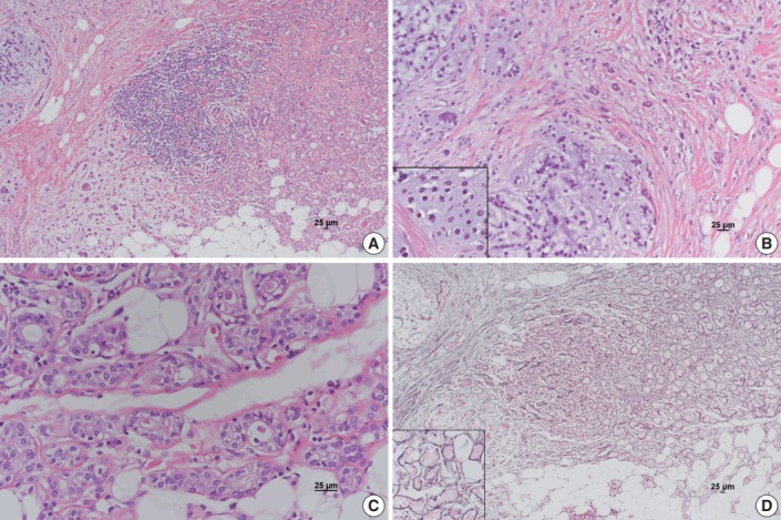
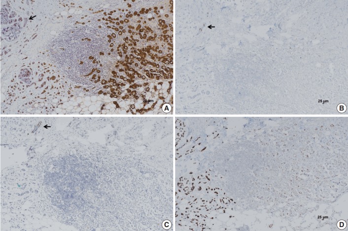
Microglandular adenosis (MGA) of the breast is a rare, benign proliferative lesion but with a significant rate of associated carcinoma. Herein, we report an unusual case of metaplastic carcinoma with chondroid differentiation associated with typical MGA. Histologically, MGA showed a direct transition to metaplastic carcinoma without an intervening atypical MGA or ductal carcinoma in situ component. The immunohistochemical profile of the metaplastic carcinoma was mostly similar to that of MGA. In both areas, all the epithelial cells were positive for S-100 protein, but negative for estrogen receptor, progesterone receptor, HER2/neu, and epidermal growth factor receptor. An increase in the Ki-67 and p53 labelling index was observed from MGA to invasive carcinoma. To the best of our knowledge, this is the first case of metaplastic carcinoma with chondroid differentiation arising in MGA in Korea. This case supports the hypothesis that a subset of MGA may be a non-obligate morphologic precursor of breast carcinoma, especially the triple-negative subtype.
Keywords: Breast; Fibrocystic breast disease; Metaplastic carcinoma.
Conflict of interest statement
No potential conflict of interest relevant to this article was reported.
Figures
References
-
- Shin SJ, Gobbi H. Microglandular adenosis, atypical microglandular adenosis and microglandular adenosis with carcinoma. In: Lakhani SR, Ellis IO, Schnitt SJ, Tan PH, van de Vijver MJ, editors. WHO classification of tumours of the breast. 4th ed. Lyon: IARC Press; 2012. pp. 113–4.
-
- Brogi E. Adenosis and microglandular adenosis. In: Hoda SA, Brogi E, Koerner FC, Rosen PP, editors. Rosen’s breast pathology. 4th ed. Philadelphia: Lippincott Williams & Wilkins; 2014. pp. 183–212.
-
- James BA, Cranor ML, Rosen PP. Carcinoma of the breast arising in microglandular adenosis. Am J Clin Pathol. 1993;100:507–13. - PubMed
-
- Koenig C, Dadmanesh F, Bratthauer GL, Tavassoli FA. Carcinoma arising in microglandular adenosis: an immunohistochemical analysis of 20 intraepithelial and invasive neoplasms. Int J Surg Pathol. 2000;8:303–15. - PubMed
-
- Khalifeh IM, Albarracin C, Diaz LK, et al. Clinical, histopathologic, and immunohistochemical features of microglandular adenosis and transition into in situ and invasive carcinoma. Am J Surg Pathol. 2008;32:544–52. - PubMed
LinkOut - more resources
Full Text Sources
Other Literature Sources
Research Materials
Miscellaneous
