

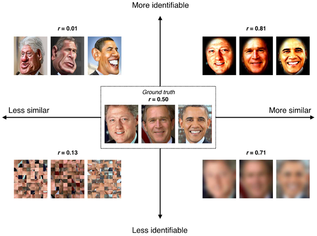
Can brain state be manipulated to emphasize individual differences in functional connectivity?
- PMID: 28373122
- PMCID: PMC8808247
- DOI: 10.1016/j.neuroimage.2017.03.064
Can brain state be manipulated to emphasize individual differences in functional connectivity?
Abstract
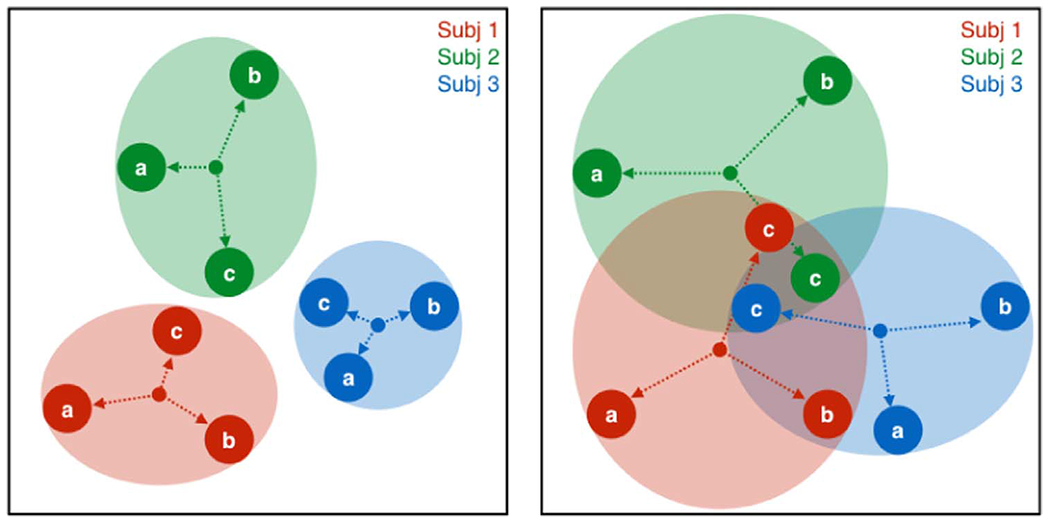
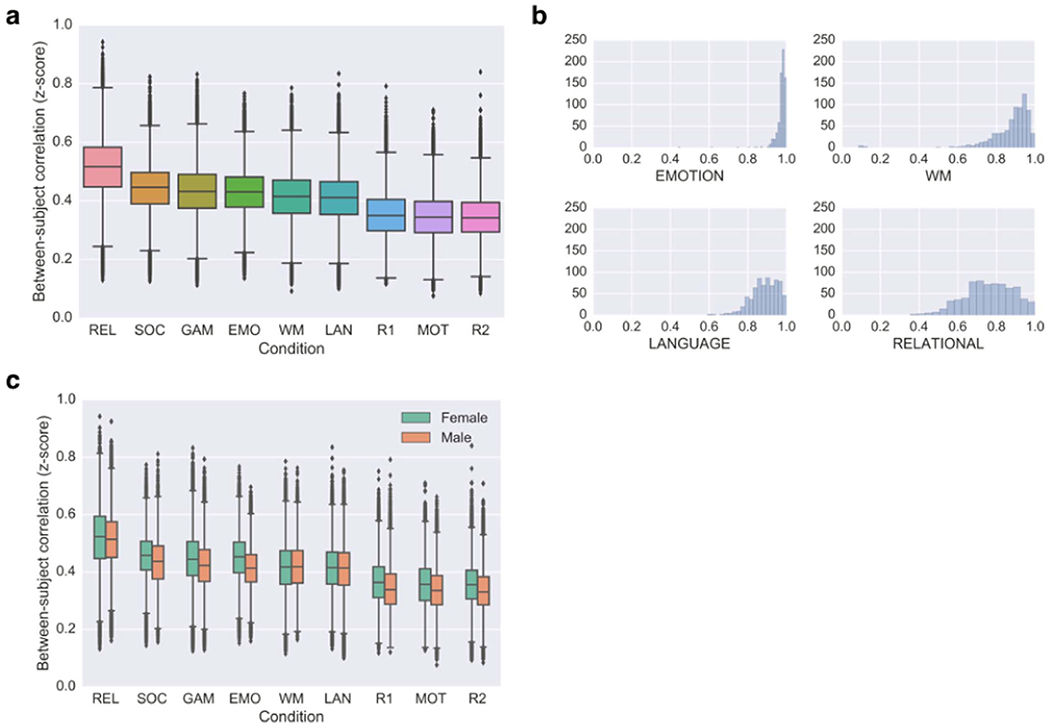
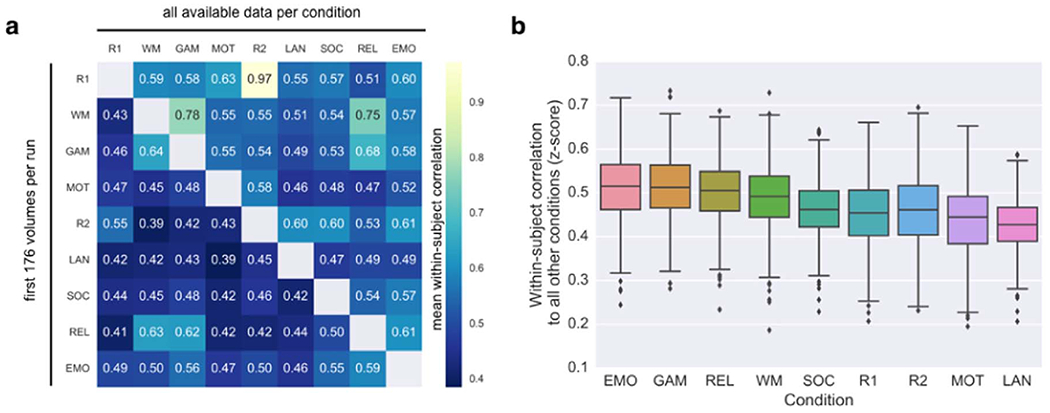
While neuroimaging studies typically collapse data from many subjects, brain functional organization varies between individuals, and characterizing this variability is crucial for relating brain activity to behavioral phenotypes. Rest has become the default state for probing individual differences, chiefly because it is easy to acquire and a supposed neutral backdrop. However, the assumption that rest is the optimal condition for individual differences research is largely untested. In fact, other brain states may afford a better ratio of within- to between-subject variability, facilitating biomarker discovery. Depending on the trait or behavior under study, certain tasks may bring out meaningful idiosyncrasies across subjects, essentially enhancing the individual signal in networks of interest beyond what can be measured at rest. Here, we review theoretical considerations and existing work on how brain state influences individual differences in functional connectivity, present some preliminary analyses of within- and between-subject variability across conditions using data from the Human Connectome Project, and outline questions for future study.
Keywords: Brain state; Functional connectivity; Human Connectome Project; Individual differences; Resting state; Scan condition; Task; fMRI.
Copyright © 2017 The Authors. Published by Elsevier Inc. All rights reserved.
Figures
References
-
- Allen JS, et al. 2003. Sexual dimorphism and asymmetries in the gray–white composition of the human cerebrum. Neuroimage 18, 880–894. - PubMed
-
- Bach S, et al. 2013. Print-specific multimodal brain activation in kindergarten improves prediction of reading skills in second grade. Neuroimage 82, 605–615. - PubMed
Publication types
MeSH terms
Grants and funding
LinkOut - more resources
Full Text Sources
Other Literature Sources
