. 2017 May 23;85(6):e00994-16.
doi: 10.1128/IAI.00994-16. Print 2017 Jun.

Clumping Factor B Promotes Adherence of Staphylococcus aureus to Corneocytes in Atopic Dermatitis

Affiliations

Clumping Factor B Promotes Adherence of Staphylococcus aureus to Corneocytes in Atopic Dermatitis

Orla M Fleury et al. Infect Immun. .

Abstract

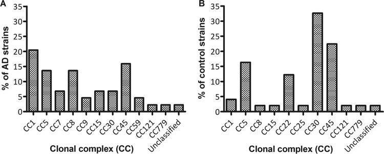
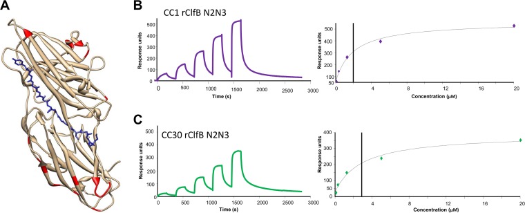
Staphylococcus aureus skin infection is a frequent and recurrent problem in children with the common inflammatory skin disease atopic dermatitis (AD). S. aureus colonizes the skin of the majority of children with AD and exacerbates the disease. The first step during colonization and infection is bacterial adhesion to the cornified envelope of corneocytes in the outer layer, the stratum corneum. Corneocytes from AD skin are structurally different from corneocytes from normal healthy skin. The objective of this study was to identify bacterial proteins that promote the adherence of S. aureus to AD corneocytes. S. aureus strains from clonal complexes 1 and 8 were more frequently isolated from infected AD skin than from the nasal cavity of healthy children. AD strains had increased ClfB ligand binding activity compared to normal nasal carriage strains. Adherence of single S. aureus bacteria to corneocytes from AD patients ex vivo was studied using atomic force microscopy. Bacteria expressing ClfB recognized ligands distributed over the entire corneocyte surface. The ability of an isogenic ClfB-deficient mutant to adhere to AD corneocytes compared to that of its parent clonal complex 1 clinical strain was greatly reduced. ClfB from clonal complex 1 strains had a slightly higher binding affinity for its ligand than ClfB from strains from other clonal complexes. Our results provide new insights into the first step in the establishment of S. aureus colonization in AD patients. ClfB is a key adhesion molecule for the interaction of S. aureus with AD corneocytes and represents a target for intervention.

Keywords: Staphylococcus aureus; atomic force microscopy; atopic dermatitis; corneocytes; filaggrin.


Figures

FIG 1
FIG 1
Clonal complex assignments of AD strains and control strains of S. aureus. Swab specimens were taken from a clinically infected site on the AD patient's skin (A) (AD strains) or from the anterior nares of asymptomatic carriers (B) (control strains). A single colony isolate from each patient was spa typed. One or more isolates of each unique spa type were subjected to multilocus sequence typing, and the clonal complex (CC) was assigned by eBURST analysis.
FIG 2
FIG 2
Adherence of S. aureus strains to L2v. Control strains (■) or AD strains (•) were grown to exponential phase, adjusted to an OD600 of 1.0, and incubated in wells coated with L2v at 37°C. Following incubation, the wells were washed, adherent bacteria were stained with crystal violet, and the absorbance was read at 570 nm. Each data point represents the mean adherence value for a single strain from three independent experiments, expressed as a percentage of the absorbance value measured for strain SH1000. Red, strains from CC1; blue, strains from CC8; green, strains from CC30; black, strains from other CCs. The horizontal lines represent the median adherence value for the population. Statistical analysis was performed using an unpaired t test. ***, P < 0.0001.
FIG 3
FIG 3
Characterization of a ClfB-deficient mutant of S. aureus AD08CC1. (A) S. aureus strain AD08CC1 (lane 1), its isogenic ClfB-deficient mutant AD08CC1 ΔclfB (lane 2), and complemented mutant AD08CC1 ΔclfB (pCU1::clfB) (lane 3) were grown to exponential phase. Cell wall extracts were separated on 7.5% acrylamide gels and blotted onto PVDF membranes, and ClfB was detected using polyclonal rabbit antibodies. Bound antibody was detected using horseradish peroxidase-conjugated protein A. Size markers (in kilodaltons) are indicated on the left. (B) S. aureus strain AD08CC1 (•) and AD08CC1 ΔclfB (■) were grown to exponential phase, washed, and incubated in microtiter plates coated with L2v. Adherent cells were stained with crystal violet, and the absorbance was read at 570 nm. The graph shown is representative of graphs from three independent experiments. (C) S. aureus strain AD08CC1 (red) or mutants AD08CC1 ΔclfB (blue), AD08CC1 ΔclfB(pCU1) (white), and AD08CC1 ΔclfB(pCU1::clfB) (green) were grown to exponential phase, washed, and incubated in microtiter plates coated with L2v (0.625 μg/ml). Adherent cells were stained with crystal violet, and the absorbance was read at 570 nm. Error bars represent the standard error of the mean values obtained from three independent experiments. Statistical significance was determined by Student's unpaired t test. **, P = 0.0059; ***, P < 0.0001.
FIG 4
FIG 4
Strength of the S. aureus-corneocyte interaction. (a) Box chart of the adhesion forces recorded between S. aureus AD08CC1 or S. aureus AD08CC1 ΔclfB and corneocytes from patient 1473; (b) representative force curves recorded under each condition for patient 1473; (c) box chart of the adhesion force between S. aureus AD08CC1 or S. aureus AD08CC1 ΔclfB and corneocytes from patient 1434; (d) representative force curves recorded under each condition for patient 1434. All curves were obtained using an applied force of 250 pN and an approach and retraction speed of 1.0 μm/s. In each case, values calculated from force curves recorded for 5 different S. aureus-corneocyte pairs were pooled and are represented as box charts, showing mean adhesion (full circle), the median, the first and third quartiles (box), the range of data without outliers (whiskers), the 99th percentile (open circle), and extreme outliers (triangles). Statistical analysis was performed using an unpaired t test. ***, P < 0.0001.
FIG 5
FIG 5
Nanoscale multiparametric imaging of corneocytes using single S. aureus probes. (a and b) Height image of a corneocyte coming from patient 1434 recorded in PBS using an S. aureus AD08CC1 cell probe (a) and the corresponding adhesion image (b). (c and d) Height image of a corneocyte coming from patient 1434 recorded in PBS using an S. aureus AD08CC1 ΔclfB cell probe (c) and the corresponding adhesion image (d). For both conditions, similar data were obtained in two other independent experiments.
FIG 6
FIG 6
Binding of rClfB N2N3 to L2v. (A) Ribbon representation of the crystal structure of the N2N3 subdomains of ClfB showing the residues that vary between the CC1 and CC30 sequences (red). The ClfB ligand bound in the trench between subdomains N2 and N3 is shown in stick format (blue). (B and C) Representative sensorgrams showing the binding of rClfB N2N3 to L2v in a single-cycle kinetics assay. GST-tagged L2v was captured on a CM5 chip coated with anti-GST IgG, and increasing concentrations of rClfB N2N3 with the CC1 (B) or CC30 (C) sequence were passed over the surface. (Left) Binding was plotted as response units against time. (Right) The affinities were calculated from curve fitting to a plot of the response unit values against the concentrations of rClfB N2N3. The data shown are representative of those from three individual experiments.

Similar articles

Cited by

References

    1. Park HY, Kim CR, Huh IS, Jung MY, Seo EY, Park JH, Lee DY, Yang JM. 2013. Staphylococcus aureus colonization in acute and chronic skin lesions of patients with atopic dermatitis. Ann Dermatol 25:410–416. doi:10.5021/ad.2013.25.4.410. - DOI - PMC - PubMed
    1. van Belkum A, Verkaik NJ, de Vogel CP, Boelens HA, Verveer J, Nouwen JL, Verbrugh HA, Wertheim HF. 2009. Reclassification of Staphylococcus aureus nasal carriage types. J Infect Dis 199:1820–1826. doi:10.1086/599119. - DOI - PubMed
    1. Kennedy EA, Connolly J, Hourihane JO, Fallon PG, McLean WH, Murray D, Jo JH, Segre JA, Kong HH, Irvine AD. 2017. Skin microbiome before development of atopic dermatitis: early colonization with commensal staphylococci at 2 months is associated with a lower risk of atopic dermatitis at 1 year. J Allergy Clin Immunol 139:166–172. doi:10.1016/j.jaci.2016.07.029. - DOI - PMC - PubMed
    1. Tauber M, Balica S, Hsu CY, Jean-Decoster C, Lauze C, Redoules D, Viode C, Schmitt AM, Serre G, Simon M, Paul CF. 2016. Staphylococcus aureus density on lesional and nonlesional skin is strongly associated with disease severity in atopic dermatitis. J Allergy Clin Immunol 137:1272–1274.e1–3. doi:10.1016/j.jaci.2015.07.052. - DOI - PubMed
    1. Palmer CN, Irvine AD, Terron-Kwiatkowski A, Zhao Y, Liao H, Lee SP, Goudie DR, Sandilands A, Campbell LE, Smith FJ, O'Regan GM, Watson RM, Cecil JE, Bale SJ, Compton JG, DiGiovanna JJ, Fleckman P, Lewis-Jones S, Arseculeratne G, Sergeant A, Munro CS, El Houate B, McElreavey K, Halkjaer LB, Bisgaard H, Mukhopadhyay S, McLean WH. 2006. Common loss-of-function variants of the epidermal barrier protein filaggrin are a major predisposing factor for atopic dermatitis. Nat Genet 38:441–446. doi:10.1038/ng1767. - DOI - PubMed