. 2017 Apr 18;114(16):4141-4146.
doi: 10.1073/pnas.1700530114. Epub 2017 Apr 3.

CMG-Pol epsilon dynamics suggests a mechanism for the establishment of leading-strand synthesis in the eukaryotic replisome

Affiliations

CMG-Pol epsilon dynamics suggests a mechanism for the establishment of leading-strand synthesis in the eukaryotic replisome

Jin Chuan Zhou et al. Proc Natl Acad Sci U S A. .

Abstract

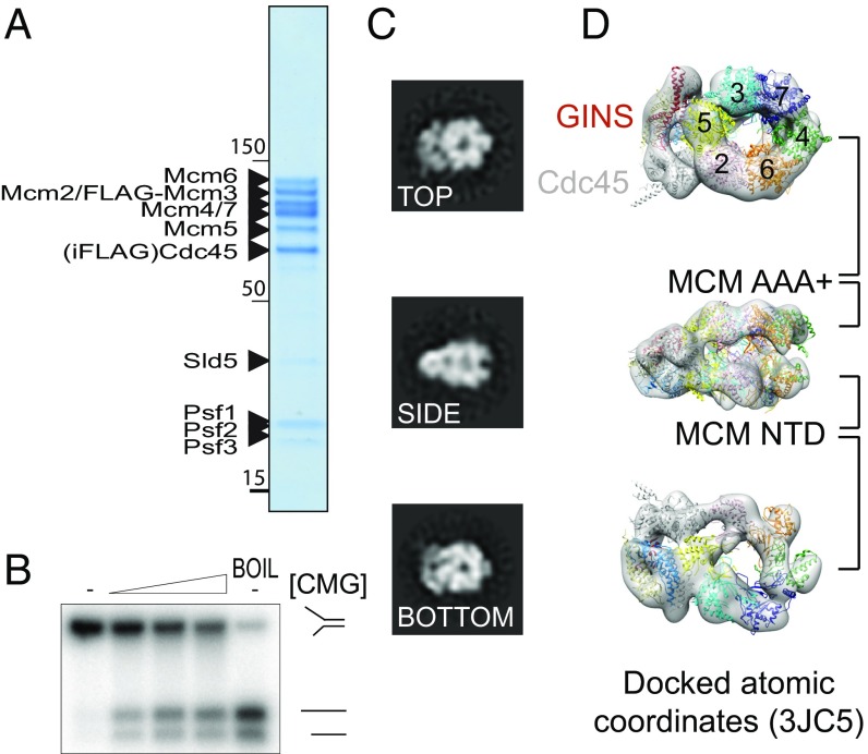
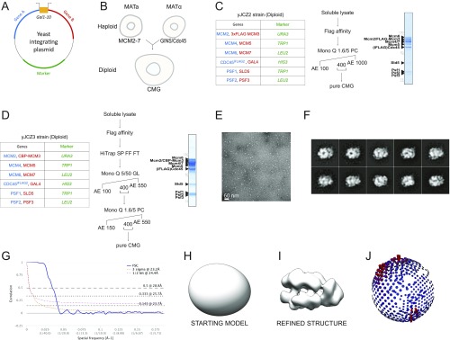
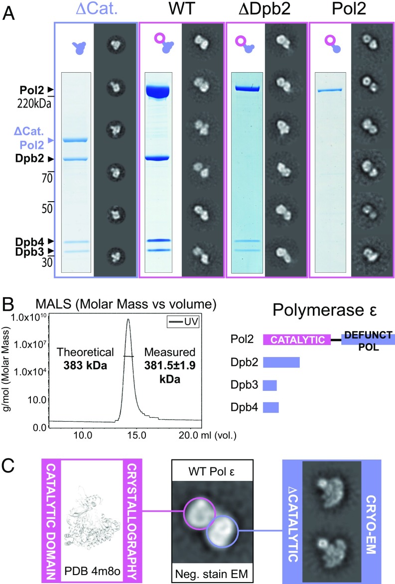
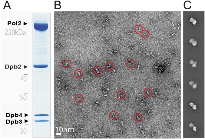
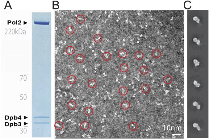
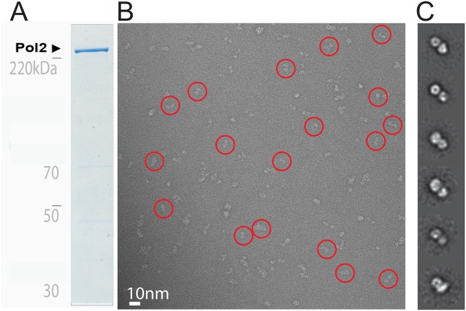
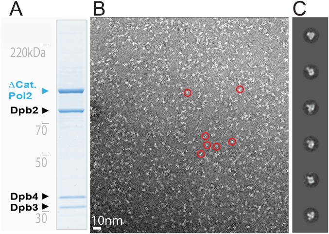
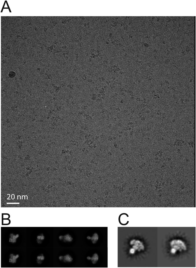
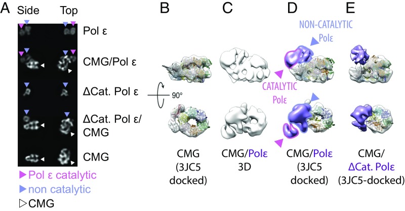
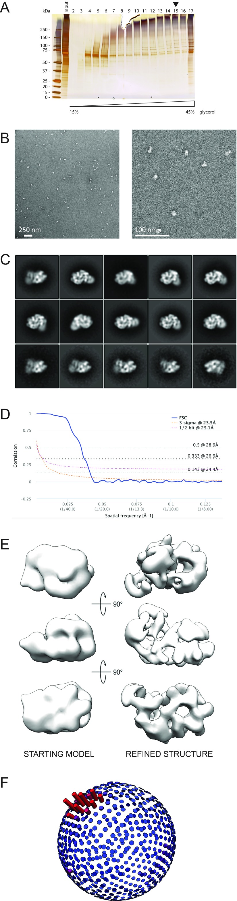
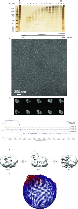
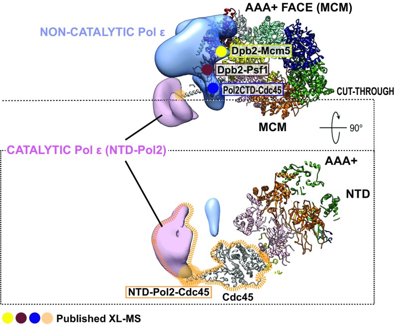
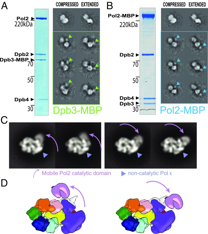
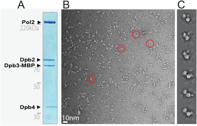
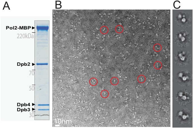
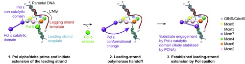
The replisome unwinds and synthesizes DNA for genome duplication. In eukaryotes, the Cdc45-MCM-GINS (CMG) helicase and the leading-strand polymerase, Pol epsilon, form a stable assembly. The mechanism for coupling DNA unwinding with synthesis is starting to be elucidated, however the architecture and dynamics of the replication fork remain only partially understood, preventing a molecular understanding of chromosome replication. To address this issue, we conducted a systematic single-particle EM study on multiple permutations of the reconstituted CMG-Pol epsilon assembly. Pol epsilon contains two flexibly tethered lobes. The noncatalytic lobe is anchored to the motor of the helicase, whereas the polymerization domain extends toward the side of the helicase. We observe two alternate configurations of the DNA synthesis domain in the CMG-bound Pol epsilon. We propose that this conformational switch might control DNA template engagement and release, modulating replisome progression.

Keywords: CMG helicase; DNA polymerase; DNA replication; single-particle electron microscopy.


Conflict of interest statement

The authors declare no conflict of interest.

Figures

Fig. 1.
Fig. 1.
Catalytically active yeast CMG. (A) Coomassie-stained gel of the yeast CMG obtained from strain yJCZ2. (B) A DNA unwinding assay shows that the overexpressed yeast CMG is catalytically active. CMG quantities used in the reactions are (from left to right) 0, 159, 397, and 793 fmol. (C) 2D image analysis showing that our CMG preparations contain stable CMG particles, suitable for 3D reconstruction. (D) 3D reconstruction with atomic docking of the 11 different CMG subunits (PDB ID code 3JC5) showing that the purified CMG is very similar to the published yeast and Drosophila CMG complexes.
Fig. S1.
Fig. S1.
Making homogeneous CMG from a diploid yeast strain and single-particle EM analysis. (A) Schematic representation of the integration plasmid containing two codon-optimized genes under the control of a bidirectional galactose-inducible promoter. (B) A diploid CMG overexpression strain was obtained by mating a Mat a Mcm2–7 overexpression strain and a Mat α GINS/Cdc45 strain. (C) Gene pairs and selection markers, purification protocol, and Coomassie-stained gel of the yeast CMG obtained from strain yJCZ2. (D) Gene pairs and selection markers, purification protocol, and Coomassie-stained gel of the yeast CMG obtained from strain yJCZ3. (E) Representative micrograph. (F) 2D class averages. (G) Fourier shell correlation indicates a resolution of 23.7 Å according to the 0.143 criterion. (H) Starting model for 3D refinement. (I) Refined 3D structure. (J) Angular distribution.
Fig. 2.
Fig. 2.
Architecture of the Pol epsilon complex. (A) From the left: A tetrameric complex lacking the catalytic domain of Pol2 (Δcat) forms a singly lobed assembly. The WT complex forms a bilobed assembly. A Dpb2 subunit dropout trimeric complex forms a bilobed assembly. Surprisingly, the isolated Pol2 is also bilobed. (B, Left) Size-exclusion chromatography with multiangle light scattering shows that the wild-type Pol epsilon complex is a single homotetramer in solution and not a dimer of tetramers. (Right) A stick diagram representing the Pol epsilon tetramer. (C) The Pol epsilon assembly is formed of two lobes. One lobe comprises the N-terminal catalytic domain of Pol2 (for which a crystal structure is available; PDB ID code 4m8o), and the second lobe comprises the noncatalytic portion of the complex (for which we determined a 2D cryo-EM structure).
Fig. S2.
Fig. S2.
Single-particle EM analysis of the wild-type yeast Pol epsilon. (A) Coomassie-stained gel of the purified Pol epsilon assembly. (B) Representative electron micrograph with a few circled particles, containing the characteristic bilobed structure. (C) 2D class averages.
Fig. S3.
Fig. S3.
Single-particle EM analysis of the ΔDpb2 yeast Pol epsilon. (A) Coomassie-stained gel of the purified Pol epsilon assembly. (B) Representative electron micrograph with a few circled particles, containing the characteristic bilobed structure. (C) 2D class averages.
Fig. S4.
Fig. S4.
Single-particle EM analysis of the Pol2 subunit of the yeast Pol epsilon. (A) Coomassie-stained gel of the purified Pol2 subunit. (B) Representative electron micrograph with a few circled particles, containing the characteristic bilobed structure. (C) 2D class averages.
Fig. S5.
Fig. S5.
Single-particle EM analysis of the Δcat yeast Pol epsilon. (A) Coomassie-stained gel of the purified Pol epsilon assembly. (B) Representative electron micrograph with a few circled particles, containing a singly lobed structure. (C) 2D class averages.
Fig. S6.
Fig. S6.
Single-particle cryo-EM analysis of the Δcat yeast Pol epsilon. (A) Representative sum of the aligned movie frames. (B) 2D class averages. (C) Notable anchor-shaped class averages visualized after imposing a 30–5 Å band-pass filter.
Fig. 3.
Fig. 3.
CMG–Pol epsilon reconstitution and 3D structure. (A) The isolated Pol epsilon has a bilobed structure (top row). In complex with Pol epsilon, the CMG is decorated with a bilobed feature (second row). The isolated Δcat Pol epsilon is a singly-lobed entity (third row). In complex with Δcat Pol epsilon, the CMG is decorated with a singly lobed feature (fourth row). Characteristic side and top views of the CMG (bottom row). (B) Yeast CMG reconstruction with docked atomic coordinates (PDB ID code 3JC5). (C) 3D structure of the CMG–Pol epsilon complex. (D) 3D structure of CMG–Pol epsilon complex with docked atomic coordinates of the CMG and assigned catalytic domain and noncatalytic portion of Pol epsilon. The catalytic domain departs radially from the core particle. Density corresponding to the polymerase is highlighted in purple. (E) 3D structure of the CMG–Δcat Pol epsilon complex color-coded as in C, with docked atomic coordinates.
Fig. S7.
Fig. S7.
Single-particle EM analysis of the CMG–Pol epsilon complex. (A) Silver-stained SDS/PAGE gel of the CMG–Pol epsilon preparation. The arrowhead indicates the lane analyzed by EM. (B) Representative micrograph (Left) and a magnified view thereof (Right). (C) 2D class averages. (D) Fourier shell correlation indicates a resolution of 24.4 Å according to the 0.143 criterion. (E) The yeast CMG was used as a starting model for 3D classification and refinement. The resulting structure is a CMG bound to a bilobed Pol epsilon assembly. (F) Angular distribution.
Fig. S8.
Fig. S8.
Single-particle EM analysis of the CMG–Δcat Pol epsilon complex. (A) Silver-stained SDS/PAGE gel of the CMG–Δcat Pol epsilon preparation. The arrowhead indicates the lane analyzed by EM. (B) Representative micrograph. (C) 2D class averages. (D) Fourier shell correlation indicates a resolution of 29.6 Å according to the 0.143 criterion. (E) The yeast CMG in complex with wild-type Pol epsilon was used as a starting model for 3D classification and refinement. The resulting structure is a CMG bound to a singly lobed Pol epsilon assembly. (F) Angular distribution.
Fig. 4.
Fig. 4.
Integration of the 3D EM structure of the CMG–Pol epsilon with published XL-MS data. The noncatalytic portion of Pol epsilon sits on top of the ATPase tier of the MCM. Dpb2 contacts Mcm5 (yellow) and Psf1 (brown), and Pol2-CTD contacts Cdc45 (blue). The catalytic domain of Pol2 contacts the tip of helix α6 in Cdc45 (orange).
Fig. 5.
Fig. 5.
CMG–Pol epsilon dynamics. (A) The isolated Pol epsilon complex with a fused MBP tag at the C terminus of Dpb3 contains two configurations: compressed (with the MBP tag mapping at the interface between lobes, equator) or extended (with the MBP tag mapping at the tip of one lobe, south pole). (B) The isolated Pol epsilon complex with a fused MBP tag at the C terminus of Pol2 also contains two configurations: compressed (MBP tag at the equator) or extended (MBP-tag at the south pole). (C) The CMG–Pol epsilon contains a flexible catalytic domain of Pol2. This can be found in proximity to the MCM ring (Left) or departing radially from the core particle (Right). (D) Cartoon representation depicting the flexibility of the CMG–Pol epsilon.
Fig. S9.
Fig. S9.
Single-particle EM analysis of the yeast CMG–Pol epsilon complex containing an MBP fused to the C terminus of Dpb3. (A) Coomassie-stained gel of the purified Pol epsilon assembly. (B) Representative electron micrograph with a few circled particles, containing the characteristic bilobed structure. (C) 2D class averages.
Fig. S10.
Fig. S10.
Single-particle EM analysis of the yeast CMG–Pol epsilon complex containing an MBP fused to the C terminus of Pol2. (A) Coomassie-stained gel of the purified Pol epsilon assembly. (B) Representative electron micrograph with a few circled particles, containing the characteristic bilobed structure. (C) 2D class averages.
Fig. 6.
Fig. 6.
A polymerase-switch mechanism for the establishment of leading-strand synthesis, facilitated by a reconfiguration of the Pol epsilon catalytic domain.

References

    1. Zhang D, O’Donnell M. The eukaryotic replication machine. Enzymes. 2016;39:191–229. - PubMed
    1. Yao NY, O’Donnell ME. Evolution of replication machines. Crit Rev Biochem Mol Biol. 2016;51:135–149. - PMC - PubMed
    1. Moyer SE, Lewis PW, Botchan MR. Isolation of the Cdc45/Mcm2-7/GINS (CMG) complex, a candidate for the eukaryotic DNA replication fork helicase. Proc Natl Acad Sci USA. 2006;103:10236–10241. - PMC - PubMed
    1. Ilves I, Petojevic T, Pesavento JJ, Botchan MR. Activation of the MCM2-7 helicase by association with Cdc45 and GINS proteins. Mol Cell. 2010;37:247–258. - PMC - PubMed
    1. Pellegrini L. The Pol α-primase complex. Subcell Biochem. 2012;62:157–169. - PubMed

Publication types

MeSH terms

LinkOut - more resources