

Recovery sleep after extended wakefulness restores elevated A1 adenosine receptor availability in the human brain
- PMID: 28373571
- PMCID: PMC5402442
- DOI: 10.1073/pnas.1614677114
Recovery sleep after extended wakefulness restores elevated A1 adenosine receptor availability in the human brain
Abstract
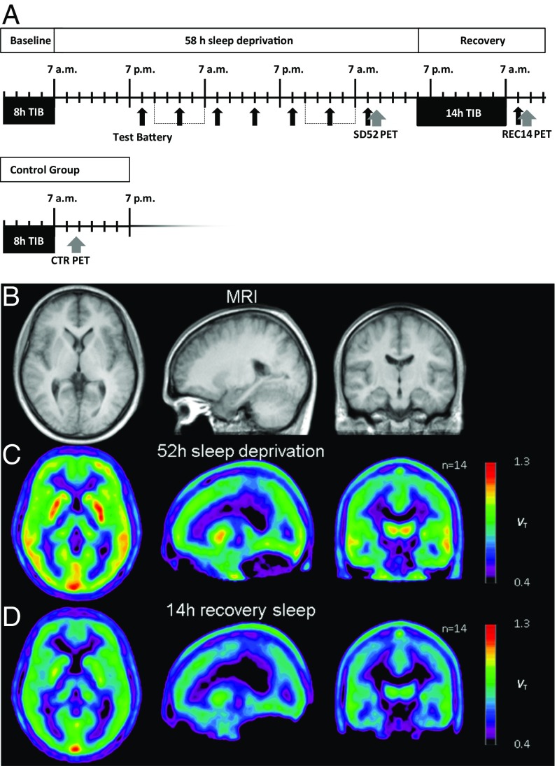
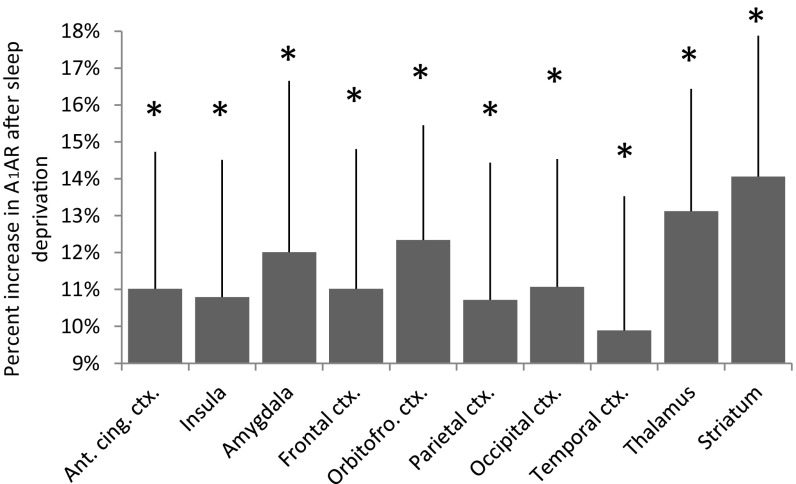
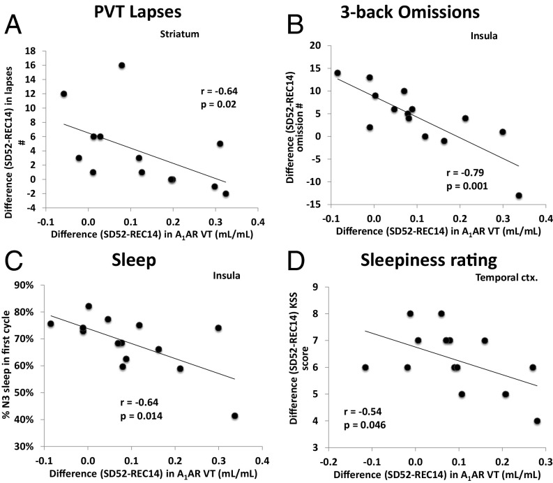
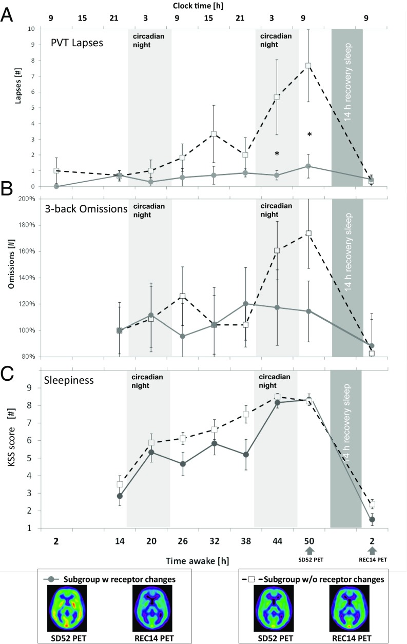
Adenosine and functional A1 adenosine receptor (A1AR) availability are supposed to mediate sleep-wake regulation and cognitive performance. We hypothesized that cerebral A1AR availability after an extended wake period decreases to a well-rested state after recovery sleep. [18F]CPFPX positron emission tomography was used to quantify A1AR availability in 15 healthy male adults after 52 h of sleep deprivation and following 14 h of recovery sleep. Data were additionally compared with A1AR values after 8 h of baseline sleep from an earlier dataset. Polysomnography, cognitive performance, and sleepiness were monitored. Recovery from sleep deprivation was associated with a decrease in A1AR availability in several brain regions, ranging from 11% (insula) to 14% (striatum). A1AR availabilities after recovery did not differ from baseline sleep in the control group. The degree of performance impairment, sleepiness, and homeostatic sleep-pressure response to sleep deprivation correlated negatively with the decrease in A1AR availability. Sleep deprivation resulted in a higher A1AR availability in the human brain. The increase that was observed after 52 h of wakefulness was restored to control levels during a 14-h recovery sleep episode. Individuals with a large increase in A1AR availability were more resilient to sleep-loss effects than those with a subtle increase. This pattern implies that differences in endogenous adenosine and A1AR availability might be causal for individual responses to sleep loss.
Keywords: cognitive performance; depression; interindividual differences; sleep deprivation; sleep homeostasis.
Conflict of interest statement
The authors declare no conflict of interest.
Figures
References
-
- Drummond SP, et al. Altered brain response to verbal learning following sleep deprivation. Nature. 2000;403:655–657. - PubMed
-
- Stickgold R. Sleep-dependent memory consolidation. Nature. 2005;437:1272–1278. - PubMed
-
- Van Dongen HP, Baynard MD, Maislin G, Dinges DF. Systematic interindividual differences in neurobehavioral impairment from sleep loss: Evidence of trait-like differential vulnerability. Sleep. 2004;27:423–433. - PubMed
-
- Nilsson JP, et al. Less effective executive functioning after one night’s sleep deprivation. J Sleep Res. 2005;14:1–6. - PubMed
-
- Killgore WD, Balkin TJ, Wesensten NJ. Impaired decision making following 49 h of sleep deprivation. J Sleep Res. 2006;15:7–13. - PubMed
MeSH terms
Substances
LinkOut - more resources
Full Text Sources
Other Literature Sources
Research Materials
