. 2018 Mar;23(3):713-722.
doi: 10.1038/mp.2017.54. Epub 2017 Apr 4.

A missense mutation in Katnal1 underlies behavioural, neurological and ciliary anomalies

Affiliations

A missense mutation in Katnal1 underlies behavioural, neurological and ciliary anomalies

G Banks et al. Mol Psychiatry. 2018 Mar.

Abstract

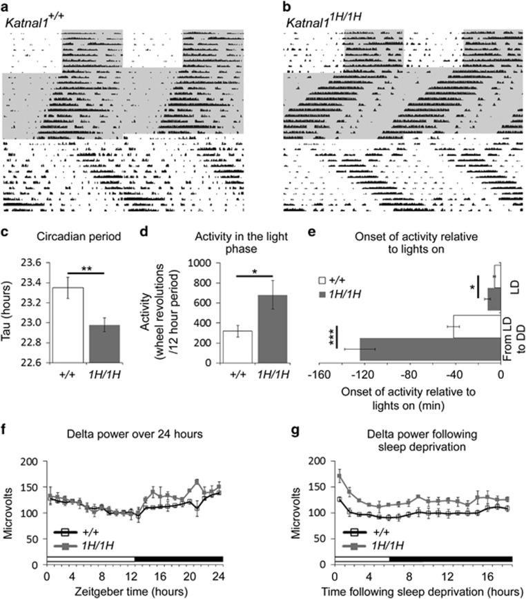
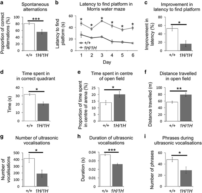
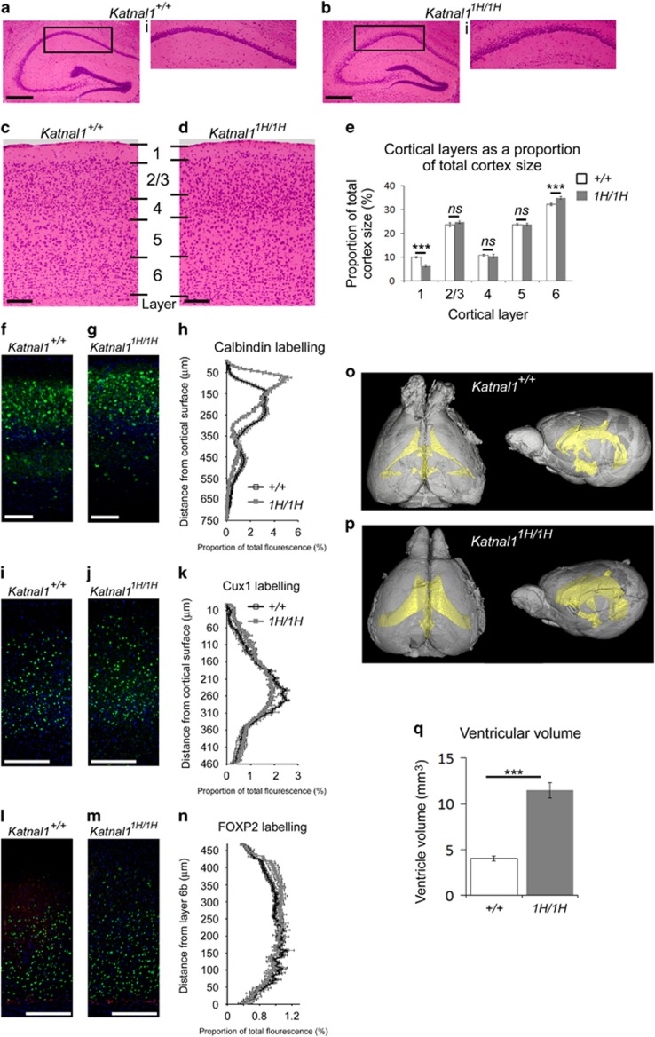
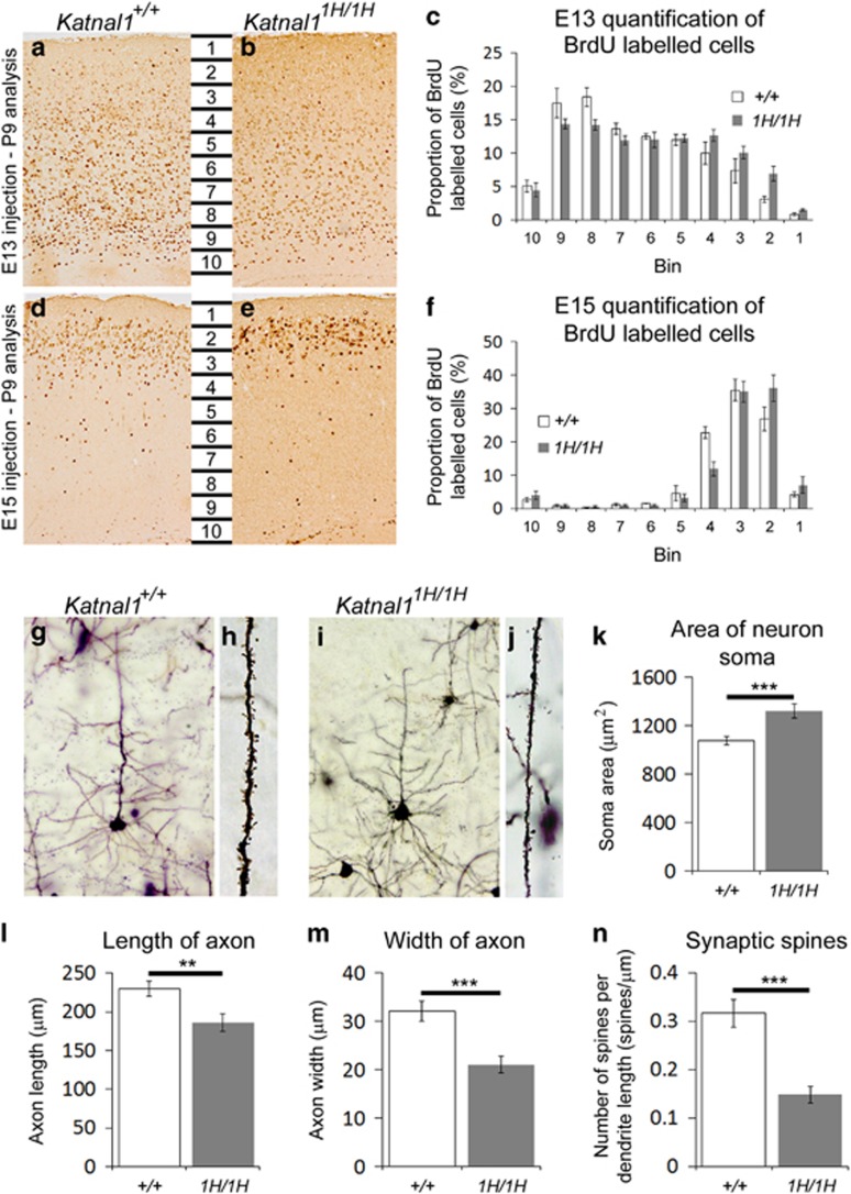
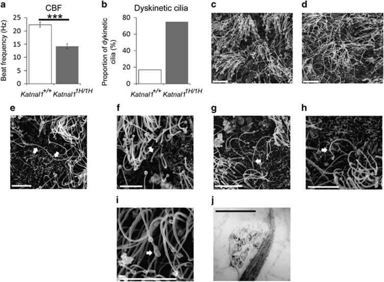
Microtubule severing enzymes implement a diverse range of tissue-specific molecular functions throughout development and into adulthood. Although microtubule severing is fundamental to many dynamic neural processes, little is known regarding the role of the family member Katanin p60 subunit A-like 1, KATNAL1, in central nervous system (CNS) function. Recent studies reporting that microdeletions incorporating the KATNAL1 locus in humans result in intellectual disability and microcephaly suggest that KATNAL1 may play a prominent role in the CNS; however, such associations lack the functional data required to highlight potential mechanisms which link the gene to disease symptoms. Here we identify and characterise a mouse line carrying a loss of function allele in Katnal1. We show that mutants express behavioural deficits including in circadian rhythms, sleep, anxiety and learning/memory. Furthermore, in the brains of Katnal1 mutant mice we reveal numerous morphological abnormalities and defects in neuronal migration and morphology. Furthermore we demonstrate defects in the motile cilia of the ventricular ependymal cells of mutants, suggesting a role for Katnal1 in the development of ciliary function. We believe the data we present here are the first to associate KATNAL1 with such phenotypes, demonstrating that the protein plays keys roles in a number of processes integral to the development of neuronal function and behaviour.


Conflict of interest statement

The authors declare no conflict of interest.

Figures

Figure 1
Figure 1
Circadian and sleep anomalies in Katnal11H/1H mice. (a and b): Double plotted actograms from wild-type (a) and Katnal11H/1H (b) animals. Wheel running activity is represented by vertical black bars, with each horizontal row representing two days of time; shaded regions show time spent in darkness, unshaded regions show time spent in light (see reference for descriptions of double plotted actograms). Compared to wild-type littermates, Katnal11H/1H animals have a shorter period (c), are more active in the light phase of the light/dark cycle (d) and show an earlier onset of activity in light/dark transitions and in the transition from light/dark cycles to constant darkness (e). In EEG recordings during sleep, Katnal11H/1H mice show increased non-REM delta power in the dark phase of the light/dark cycle (f) and following sleep deprivation (g). *P⩽0.05; **P⩽0.01; ***P⩽0.001. EEG, electroencephalography; DD, constant darkness; LD, light/dark cycle.
Figure 2
Figure 2
Katnal11H/1H mice display a spectrum of abnormal behaviours. Compared to wild-type littermates, Katnal11H/1H mice show: reduced spontaneous alternations in a T-maze (a); an increased latency to find the platform in Morris water maze trials (b); reduced improvement to find the platform in the Morris water maze (c); reduced time in the correct quadrant of the Morris water maze (d); increased time in the centre of an open field (e); greater movement in an open field (f); fewer USVs (g); shorter USVs (h); fewer phrases in their USVs (i). *P⩽0.05; **P⩽0.01; ***P⩽0.001. USV, ultrasonic vocalisation.
Figure 3
Figure 3
Aberrant brain histology in Katnal11H/1H mice. (a,b): Hippocampal histology in wild-type (a) and Katnal11H/1H (b) animals. Inserts (i) show CA1 layer. (c,d): Cortical layers in wild-type (c) and Katnal11H/1H (d) animals. (e: Katnal11H/1H animals have a narrower cortical layer 1 and a wider layer 6, compared to wild types. (f to n) Immunofluorescence of cortical layers: Calbindin immunofluorescence in cortical sections of wild-type (f) and Katnal11H/1H (g) animals. (h): Quantification of calbindin immunofluorescence demonstrates that Katnal11H/1H animals have a higher proportion of labelling towards the cortical surface than wild types. CUX1 immunofluorescence in cortical sections of wild-type (i) and Katnal11H/1H (j) animals. (k): Quantification of CUX1 immunofluorescence demonstrates that Katnal11H/1H animals have a higher proportion of labelling towards the cortical surface than wild types. FOXP2 (green) and CTGF (red) immunofluorescence in cortical sections of wild-type (l) and Katnal11H/1H (m) animals. (n): Quantification of FOXP2 immunofluorescence demonstrates that Katnal11H/1H animals have a higher proportion of labelling distant from layer 6b than wild-types. (o,p): μCT scans of the ventricular system (yellow) in wildwild-type (o) and Katnal11H/1H (p) brains. (q): Quantification of ventricular volume demonstrates that Katnal11H/1H mice have larger ventricles than wild types. Scale bars: 500 μm in a and b; 100 μm in dg. ***P⩽0.001.
Figure 4
Figure 4
Neuronal migration and morphology abnormalities in Katnal11H/1H mice. (a,b,d,e): BrdU labelling of cortical neurons following injection of BrdU at either E13 (a,b) or E15 (d,e) in wild-type (a,d) and Katnal11H/1H (b,e) animals. Quantification of BrdU immunohistochemistry shows that Katnal11H/1H animals have increased numbers of labelled neurons closer to the cortical surface following injection at both E13 (c) and E15 (f). (gj): Golgi labelling of neurons and dendrites in wild-type (g,h) and Katnal11H/1H (i,j) animals. (kn): Quantification of golgi labelling shows that Katnal11H/1H animals have larger soma (k), shorter axons (l), thinner axons (m) and fewer dendritic spines (n) than wild types. **P⩽0.01; ***P⩽0.001.
Figure 5
Figure 5
Ciliary dysfunction in Katnal11H/1H mice. (a,b): Ependymal motile cilia in the lateral ventricle of Katnal11H/1H animals have a significantly reduced CBF (a) and a higher proportion of ciliary dyskinesia (b) compared to wild-type littermates. (c,d): Scanning electron micrographs (SEM) of the ependymal motile cilia in the lateral ventricles of wiltype (c) and Katnal11H/1H (d) animals. (ei): Structural ciliary abnormalities found in Katnal11H/1H animals include abnormally long cilia (e) abnormally short cilia (f) bifurcated cilia (g) kinks in the cilia (h) and swellings along the length of the cilia (i). Transmission Electron Micrographs show vesicular aggregates within ciliary swellings (j). Arrows in (ei) indicate ciliary abnormalities. Scale bars: c–i=5 μm; j, 500 nm (***P⩽0.001). CBF, ciliary beat frequency.

References

    1. Wood JD, Landers JA, Binglet M, McDermott CJ, Thomas-McArthur V, Gleadall LJ et al. The microtubule-severing protein Spastin is essential for axon outgrowth in the zebrafish embryo. Hum Mol Genet 2006; 15: 2763–2771. - PubMed
    1. Trotta N, Orso G, Rossetto MG, Daga A, Broadie K. The hereditary spastic paraplegia gene Spastin regulates microtubule stability to modulate synaptic structure and function. Curr Biol 2004; 14: 1135–1147. - PubMed
    1. Hazen J, Fonknechten N, Mavel D, Paternotte C, Samson D, Artiguenave F et al. Spastin, a new AAA protein, is altered in the most frequent form of autosomal dominant spastic paraplegia. Nat Genet 1999; 23: 296–303. - PubMed
    1. Misha-Gorur K, Caglayan AO, Schaffer AE, Chabu C, Henegariu O, Vonhoff F et al. Mutations in KATNB1 cause complex cerebral malformations by disrupting asymmetrically dividing neural progenitors. Neuron 2014; 84: 1226–1239. - PMC - PubMed
    1. Hu WF, Pomp O, Ben-Omran T, Kodani A, Henke K, Mochida GH et al. Katanin p80 regulates human cortical development by limiting centriole and cilia number. Neuron 2014; 84: 1240–1257. - PMC - PubMed

Publication types

LinkOut - more resources