. 2017 Apr 3;7(1):539.
doi: 10.1038/s41598-017-00533-w.

The structures of a naturally empty cowpea mosaic virus particle and its genome-containing counterpart by cryo-electron microscopy

Affiliations

The structures of a naturally empty cowpea mosaic virus particle and its genome-containing counterpart by cryo-electron microscopy

Emma L Hesketh et al. Sci Rep. .

Abstract

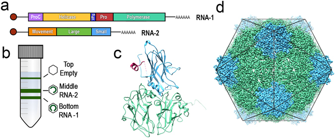
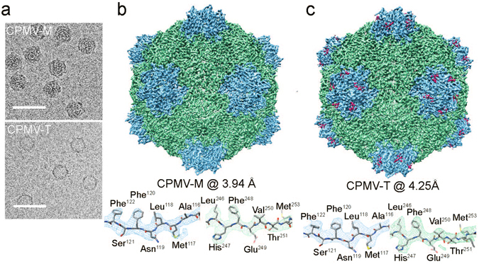
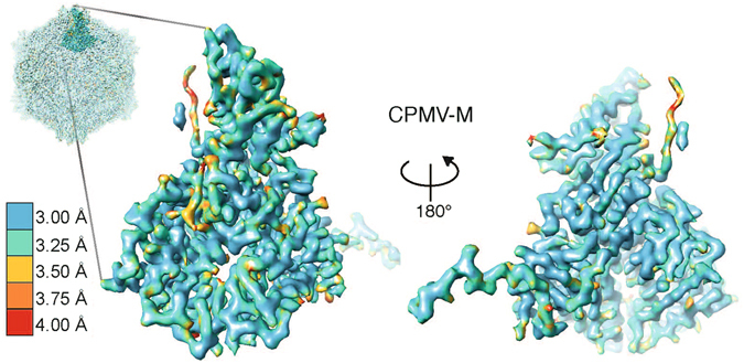
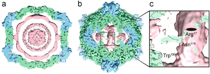
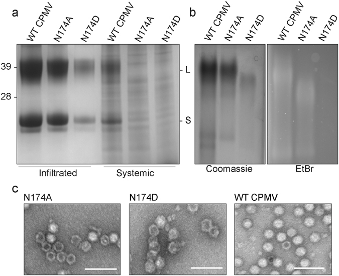
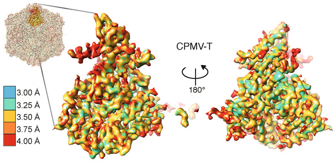
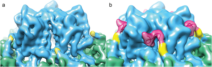
Cowpea mosaic virus (CPMV) is a picorna-like plant virus. As well as an intrinsic interest in CPMV as a plant pathogen, CPMV is of major interest in biotechnology applications such as nanotechnology. Here, we report high resolution cryo electron microscopy (cryo-EM) maps of wild type CPMV containing RNA-2, and of naturally-formed empty CPMV capsids. The resolution of these structures is sufficient to visualise large amino acids. We have refined an atomic model for each map and identified an essential amino acid involved in genome encapsidation. This work has furthered our knowledge of Picornavirales genome encapsidation and will assist further work in the development of CPMV as a biotechnological tool.


Conflict of interest statement

G.P.L. declares that he is a named inventor on granted patent WO 29087391 A1 which describes the transient expression system used in this manuscript.

Figures

Figure 1
Figure 1
An introduction to Cowpea mosaic virus (CPMV). (a) CPMVs single stranded bipartite RNA genome. RNA-1 is ~6 kb in length and encodes viral proteins required for replication. RNA-2 is ~3.5 kb in length and encodes the structural coat proteins and the movement protein required for moving CPMV virions from cell to cell. (b) Gradient centrifugation of wild-type CPMV permits separation into three components. Empty CPMV particles sediment at the top (CPMV-T), CPMV containing RNA-2 sediments in the middle (CPMV-M) and RNA-1 containing CPMV particles sediment at the bottom of a density gradient (CPMV-B). (c) An asymmetric unit of CPMV empty virus-like particle (eVLP), (PDB 5a33). The large coat protein subunit (L subunit, green) and the small coat protein subunit (S subunit, blue). The C terminal extension, only visualised in a eVLP is coloured pink. (d) The icosahedral organisation of CPMV using the EM derived map of eVLP (EMD-3014). Each of the 60 asymmetric units comprises one copy of the L subunit and the S subunit (coloured as in 1C). A view down the two-fold axis is shown.
Figure 2
Figure 2
Cryo-EM structure of CPMV-M and CPMV-T. (a) Electron micrographs of CPMV-M (top) and CPMV-T (bottom) to show particle distribution. Micrographs were imaged using a Titan Krios electron microscope and detected using a Falcon II direct electron detector (FEI). Scale bar in 50 nm. (b) EM density map of CPMV-M (CPMV particle containing RNA-2) determined by cryo-EM to a global resolution of 3.94 Å (EMD-3565). The Large (L) subunit is displayed in green and the Small (S) subunit shown in blue. The bottom panel shows an example of the EM density for an individual β strand from each the L and S subunits displayed as mesh representation. The atomic model within was refined against the EM density. (c) EM density map of the naturally occurring empty CPMV particles known as CPMV-T to 4.25 Å (EMD-3562). The view is identical to that shown in (b).
Figure 3
Figure 3
Local resolution of CPMV-M. A single asymmetric unit of CPMV-M (seen from outside the capsid) is shown coloured according to its local resolution. The interior of the asymmetric unit is also shown by rotating the asymmetric unit 180° around the y-axis. The highest resolution bin is 3.00 Å (blue) and the lowest resolution bin is 4.00 Å (red). A key is shown for reference.
Figure 4
Figure 4
Genome organisation of CPMV-M containing RNA-2. (a) A 40 Å thick central slab through the unsharpened CPMV-M EM map (at 4.25 Å resolution; also deposited with the EMD-3565 deposition. Suggested contour level is 0.012). A view down the two-fold axis is shown. The coat proteins are coloured as before with the extra EM density attributed to RNA coloured pink. (b) A view beneath the capsid at the two-fold axis showing the strongest density for RNA. (c) Zoomed in view to highlight RNA-protein interactions. The strongest density bridges between the capsid and RNA are at amino acids Asn174 and Arg17 from the L subunit. Trp190 binds strongly to RNA in CPMV-B virions (containing RNA-1), here we can see no density between Trp190 and the EM density attributed to RNA-2.
Figure 5
Figure 5
Asn174 in the Large subunit is essential for a systemic infection. (a) For each mutant and WT virion 15 grams of leaves were analysed. Coomassie blue stained SDS PAGE shows the mutation N174A results in similar levels of the Large (L) and small (S) coat proteins to those found with WT virus being produced in infiltrated leaves. By contrast the N174D mutation resulted in a substantial reduction in the amounts of the two coat proteins. Both mutations inhibit CPMV from causing a systemic infection, with the L and S coat proteins being undetectable in extracts. (b) Coomassie blue and ethidium bromide (EtBr) stained native agarose gels of particles isolated from infiltrated leaves. The N174A mutation allows genome encapsidation while N174D mutant does not appear to package RNA. (c) Negative stain EM shows N174A and N174D mutants appear to assemble as complete particles similar to WT CPMV (also shown for reference). Scale bar is 100 µm.
Figure 6
Figure 6
Local resolution of CPMV-T. A single asymmetric unit of CPMV-T is shown coloured according to its local resolution (the views and colour scheme are identical to those in Fig. 3). The interior of the asymmetric unit is also shown by rotating it 180° along the y-axis. The highest resolution bin is 3.00 Å (blue) and the lowest resolution bin is 4.00 Å (red). A key is shown for reference.
Figure 7
Figure 7
The C terminus extension of S subunit. (a) The unsharpened EM density map of CPMV-M. The S subunit is coloured blue and the L subunit is in green. The C terminal amino acid in the CPMV-M map is Ser183 and is coloured yellow. (b) The unsharpened EM density map of CPMV-T. Coloured as in (a). Amino acids Ser183 and Thr184 are coloured yellow. The C terminus extension, only visible in the CPMV-T map, amino acids Pro188 to Ile197 are coloured magenta.

References

    1. Lin T, et al. The refined crystal structure of cowpea mosaic virus at 2.8 A resolution. Virology. 1999;265:20–34. doi: 10.1006/viro.1999.0038. - DOI - PubMed
    1. Ochoa WF, Chatterji A, Lin T, Johnson JE. Generation and structural analysis of reactive empty particles derived from an icosahedral virus. Chem Biol. 2006;13:771–778. doi: 10.1016/j.chembiol.2006.05.014. - DOI - PubMed
    1. Lin T, Cavarelli J, Johnson JE. Evidence for assembly-dependent folding of protein and RNA in an icosahedral virus. Virology. 2003;314:26–33. doi: 10.1016/S0042-6822(03)00457-4. - DOI - PubMed
    1. Chen ZG, et al. Protein-RNA interactions in an icosahedral virus at 3.0 A resolution. Science. 1989;245:154–159. doi: 10.1126/science.2749253. - DOI - PubMed
    1. Lin T, et al. Structural fingerprinting: subgrouping of comoviruses by structural studies of red clover mottle virus to 2.4-A resolution and comparisons with other comoviruses. J Virol. 2000;74:493–504. doi: 10.1128/JVI.74.1.493-504.2000. - DOI - PMC - PubMed

Publication types