. 2017 Apr 3;7(1):545.
doi: 10.1038/s41598-017-00623-9.

Oral administration of Lactobacillus gasseri SBT2055 is effective in preventing Porphyromonas gingivalis-accelerated periodontal disease

Affiliations

Oral administration of Lactobacillus gasseri SBT2055 is effective in preventing Porphyromonas gingivalis-accelerated periodontal disease

R Kobayashi et al. Sci Rep. .

Abstract

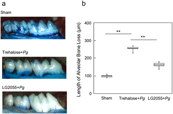
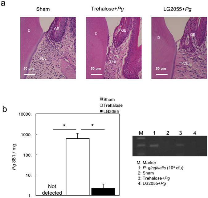
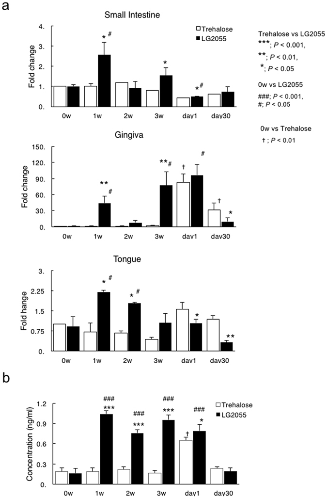
Probiotics have been used to treat gastrointestinal disorders. However, the effect of orally intubated probiotics on oral disease remains unclear. We assessed the potential of oral administration of Lactobacillus gasseri SBT2055 (LG2055) for Porphyromonas gingivalis infection. LG2055 treatment significantly reduced alveolar bone loss, detachment and disorganization of the periodontal ligament, and bacterial colonization by subsequent P. gingivalis challenge. Furthermore, the expression and secretion of TNF-α and IL-6 in gingival tissue was significantly decreased in LG2055-administered mice after bacterial infection. Conversely, mouse β-defensin-14 (mBD-14) mRNA and its peptide products were significantly increased in distant mucosal components as well as the intestinal tract to which LG2055 was introduced. Moreover, IL-1β and TNF-α production from THP-1 monocytes stimulated with P. gingivalis antigen was significantly reduced by the addition of human β-defensin-3. These results suggest that gastrically administered LG2055 can enhance immunoregulation followed by periodontitis prevention in oral mucosa via the gut immune system; i.e., the possibility of homing in innate immunity.


Conflict of interest statement

Toshiya Kobayashi, F.S., and T.H. are employees of Megmilk Snow Brand Co., Ltd. R.K., M.Y. and Tomoko Kurita-Ochiai declare no conflict of interest.

Figures

Figure 1
Figure 1
Reduction in P. gingivalis-induced alveolar bone loss by gastric intubation with LG2055. (a) Thirty days after P. gingivalis infection, the distance from the cementoenamel junction (CEJ) to the alveolar bone crest (ABC) at 14 predetermined sites in the defleshed maxilla were measured and totaled for each mouse. (b) Bone measurements were performed a total of three times by two evaluators using a random and blinded protocol. All values are presented as the means ± SEM of eight mice per group; **p < 0.01.
Figure 2
Figure 2
Suppression of P. gingivalis-induced detachment and disorganization of the periodontal ligament, and bacteria detection by gastric intubation with LG2055. (a) Histochemical analysis of gingival tissue. Thirty days after P. gingivalis infection, mouse lower jaws with gingival tissue were stained with hematoxylin and eosin. D: dentin, GE: gingival epithelium, PDL: periodontal ligament. (b) Detection of P. gingivalis-specific 16 S rRNA. Thirty days after P. gingivalis infection, DNA was extracted from gingival tissues of mice and amplified using real-time quantitative PCR with a pair of primers corresponding to P. gingivalis-specific 16 S rRNA. Different numbers of DNA from P. gingivalis 381 were used to generate a standard curve. All values are expressed as the means ± SEM per mg of tissue for eight mice per group; *p < 0.05.
Figure 3
Figure 3
Suppression of the P. gingivalis-induced inflammatory cytokine response in gingival tissues and GMCs by gastric intubation with LG2055. (a) Total RNA was extracted from gingival tissue 1 and 30 days after infection with P. gingivalis, and IL-6 and TNF-α mRNA levels were determined using real-time PCR. (b,c) GMCs (1 × 106/mL) were isolated 1 and 30 days after P. gingivalis infection, and then cultured for 3 days. (b) The culture supernatants were collected and subjected to IL-6- and TNF-α-specific ELISAs. (c) The cultured cells were harvested for RNA isolation and quantification of IL-6 and TNF-α mRNA using real-time PCR. All values are presented as the means ± SEM of eight mice per group at each time point; **p < 0.01, *p < 0.05.
Figure 4
Figure 4
β-Defensin-14 expression and protein production in mucosal components of mice administered LG2055 (a,b). At 0–3 weeks after LG2055 administration and 1 and 30 days after the final P. gingivalis infection, total RNA was extracted from gingival tissue, tongue, and small intestine, and mRNA levels were determined using quantitative real-time PCR (a). Similarly, saliva samples were collected and subjected to a β-defensin-3-specific ELISA (b). All values are presented as the means ± SEM of five mice per group at each time point; **p < 0.01, *p < 0.05.
Figure 5
Figure 5
Suppression of P. gingivalis antigen-induced inflammatory cytokine release in THP-1 by hBD3. PMA-primed THP-1 cells were pretreated with various doses of hBD3 for 30 min and stimulated with 1,000 ng/mL of P. gingivalis antigen for 23 h. The culture supernatants were collected and subjected to IL-1β- and TNF-α-specific ELISAs. The data are presented as the means SD (n = 3) of three independent experiments; **p < 0.01, *p < 0.05.
Figure 6
Figure 6
Experimental procedure. Eight-week-old female BALB/c mice were randomly divided into three groups (n = 36 per group); groups 1 and 2 were orally intubated with 25% trehalose or LG2055 (1 × 109 CFU/200 μL/mouse) throughout the experiment once per day for 5 weeks. After administration for 3 weeks, mice were orally infected with P. gingivalis (1 × 108 CFU/100 μL with 5% CMC/mouse) once per day for 2 weeks. The third group received 5% CMC without oral intubation. Mice were sacrificed 0, 1, 2, and 3 weeks (n = 5 at each time point) after LG2055 administration for a mouse β-defensin-3 assay, and were sacrificed 1 and 30 days (n = 8 at each time point) after P. gingivalis infection for mouse β-defensin-3 assay, GMC cultures, bacterial detection, histological analysis, and alveolar bone loss evaluation. The English in this document has been checked by at least two professional editors, both native speakers of English. For a certificate, please see: http://www.textcheck.com/certificate/bnmrY9.

References

    1. Socransky SS, Haffajee AD, Cugini MA, Smith C, Kent RL., Jr. Microbial complexes in subgingival plaque. J Clin Periodontol. 1998;25:134–144. doi: 10.1111/j.1600-051X.1998.tb02419.x. - DOI - PubMed
    1. Hajishengallis G, et al. Low-abundance biofilm species orchestrates inflammatory periodontal disease through the commensal microbiota and complement. Cell Host Microbe. 2011;10:497–506. doi: 10.1016/j.chom.2011.10.006. - DOI - PMC - PubMed
    1. Darveau RP, Hajishengallis G, Curtis MA. Porphyromonas gingivalis as a potential community activist for disease. J Dent Res. 2012;91:816–820. doi: 10.1177/0022034512453589. - DOI - PMC - PubMed
    1. Lamont RJ, Jenkinson HF. Life below the gum line: pathogenic mechanisms of Porphyromonas gingivalis. Microbiol Mol Biol Rev. 1998;62:1244–1263. - PMC - PubMed
    1. Holt SC, Kesavalu L, Walker S, Genco CA. Virulence factors of Porphyromonas gingivalis. Periodontol 2000. 1999;20:168–238. doi: 10.1111/j.1600-0757.1999.tb00162.x. - DOI - PubMed

Publication types

MeSH terms