. 2017 Apr 3;7(1):588.
doi: 10.1038/s41598-017-00665-z.

Delta-radiomics features for the prediction of patient outcomes in non-small cell lung cancer

Affiliations

Delta-radiomics features for the prediction of patient outcomes in non-small cell lung cancer

Xenia Fave et al. Sci Rep. .

Abstract

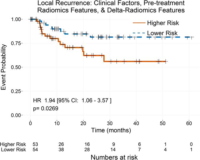
Radiomics is the use of quantitative imaging features extracted from medical images to characterize tumor pathology or heterogeneity. Features measured at pretreatment have successfully predicted patient outcomes in numerous cancer sites. This project was designed to determine whether radiomics features measured from non-small cell lung cancer (NSCLC) change during therapy and whether those features (delta-radiomics features) can improve prognostic models. Features were calculated from pretreatment and weekly intra-treatment computed tomography images for 107 patients with stage III NSCLC. Pretreatment images were used to determine feature-specific image preprocessing. Linear mixed-effects models were used to identify features that changed significantly with dose-fraction. Multivariate models were built for overall survival, distant metastases, and local recurrence using only clinical factors, clinical factors and pretreatment radiomics features, and clinical factors, pretreatment radiomics features, and delta-radiomics features. All of the radiomics features changed significantly during radiation therapy. For overall survival and distant metastases, pretreatment compactness improved the c-index. For local recurrence, pretreatment imaging features were not prognostic, while texture-strength measured at the end of treatment significantly stratified high- and low-risk patients. These results suggest radiomics features change due to radiation therapy and their values at the end of treatment may be indicators of tumor response.


Conflict of interest statement

The authors declare that they have no competing interests.

Figures

Figure 1
Figure 1
Workflow for the selection of feature specific image preprocessing. The images are all processed in four ways: no extra processing, smoothing with a Butterworth filter, resampling to an 8 bit depth, and both smoothing with a Butterworth filter and resampling with an 8 bit depth. Each feature is calculated from the four sets of processed images. Then the best processing is determined on a feature specific basis by evaluating the univariate significance, dependence on the CT model used to acquire the images, and volume dependence. Based on the results of these tests, one image preprocessing is selected for that feature. Then the process is repeated for the next feature. If no image preprocessing for a feature allows it to pass the tests, then the feature is removed entirely.
Figure 2
Figure 2
Workflow for building of multivariate models. A LOOCV loop is used to generate 3 models on each iteration: (1) Only clinical factors, (2) clinical factors and pre-treatment (TX) features, and (3) clinical factors, pre-TX features, and delta-radiomics features. After the three models have been built with each patient left out once, the number of times each covariate was selected is tabulated. Then those covariates that are selected in greater than 50% of the iterations are kept. These are then used to fit final versions of the 3 models.
Figure 3
Figure 3
Comparison of Kaplan-Meier curves for overall survival using the three nested models with patients stratified by the median prediction value of each model. The stratification was significant for all three models. The addition of a pretreatment radiomics feature, compactness2, to clinical factors alone had a small improving effect on the stratification, while the further addition of delta-radiomics features had almost no impact on the stratification.
Figure 4
Figure 4
Comparison of Kaplan-Meier curves for distant metastases using the two nested models with patients stratified by the median predicted value of each model. No delta-radiomics features were selected in the model-building process for distant metastases, so only two models were available. For distant metastases, the addition of a pretreatment radiomics feature, compactness2, significantly improved the stratification of the patients.
Figure 5
Figure 5
Kaplan-Meier curves for local-regional recurrence using the delta-radiomics model with patients stratified by the median predicted value from the model. The only covariate selected in the model building process for local recurrence, was the delta-radiomics feature texture strength measured at the end of treatment. When patients were stratified by the median of their predicted values, the resulting Kaplan-Meier curves were significantly stratified.

References

    1. Molina JR, Yang P, Cassivi SD, Schild SE, Adjei AA. Non-small cell lung cancer: Epidemiology, risk factors, treatment, and survivorship. Mayo Clin. Proc. 2008;83:584–594. doi: 10.1016/S0025-6196(11)60735-0. - DOI - PMC - PubMed
    1. SEER stat fact sheets: Lung and bronchus cancer. Available at: http://seer.cancer.gov/statfacts/html/lungb.html (Accessed: 23rd September 2016) (2014).
    1. Kumar V, et al. Radiomics: the process and the challenges. Magn. Reson. Imaging. 2012;30:1234–1248. doi: 10.1016/j.mri.2012.06.010. - DOI - PMC - PubMed
    1. Lambin P, et al. Radiomics: Extracting more information from medical images using advanced feature analysis. Eur. J. Cancer. 2012;48:441–446. doi: 10.1016/j.ejca.2011.11.036. - DOI - PMC - PubMed
    1. Wang H, et al. Multilevel binomial logistic prediction model for malignant pulmonary nodules based on texture features of CT image. Eur. J. Radiol. 2010;74:124–129. doi: 10.1016/j.ejrad.2009.01.024. - DOI - PubMed

Publication types

MeSH terms