. 2017 Jan 2;6(1):e7-e13.
doi: 10.1016/j.eats.2016.08.029. eCollection 2017 Feb.

Arthroscopic Curettage and Bone Grafting of Bone Cysts of the Talar Body

Affiliations

Arthroscopic Curettage and Bone Grafting of Bone Cysts of the Talar Body

Tun Hing Lui. Arthrosc Tech. .

Abstract

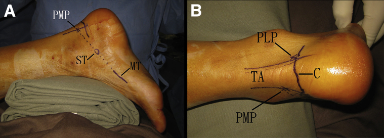
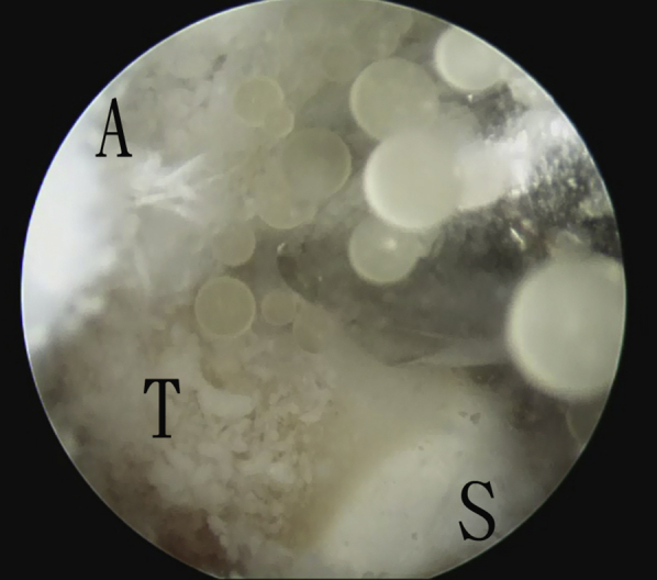
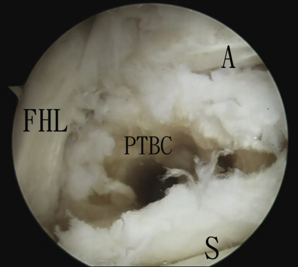
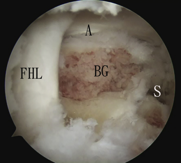
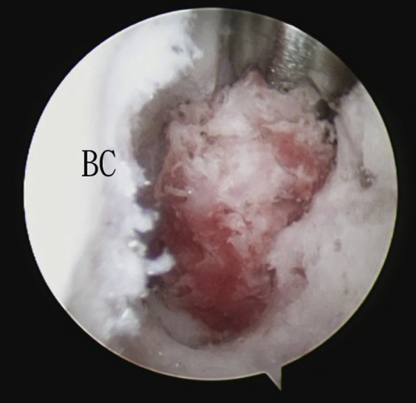
Talar bone cysts can develop as a result of osteochondral lesions of the talus. This can be a source of deep ankle pain. Open debridement and bone grafting of the bone cysts requires extensive soft tissue dissection and malleolar osteotomy. Removal of normal cartilage of the talus is frequently required to approach the bone cysts. Alternatively, the cysts can be grafted arthroscopically with minimal disruption of the normal cartilage surface. The key to success is careful preoperative planning with a computed tomogram of the ankle. Bone cyst of the posterior half of the talar body can be grafted via posterior ankle endoscopy. Bone cyst of the anterior half of the talar body can be debrided and grafted via anterior talar osseous portals. The purpose of this technical note is to describe a minimally invasive approach of curettage and bone grafting of the talar bone cysts with preservation of the articular surfaces.


Figures

Fig 1
Fig 1
Preoperative computed tomogram of the illustrated case. Transverse (A) and sagittal (B) views of the posterior talar cyst confirmed that the cyst is best approached by the posterior ankle endoscopy (arrows). Transverse (C) and sagittal (D) views of the anterior talar cyst showed that the cyst can be approached via the anterolateral bone portal (arrowheads).
Fig 2
Fig 2
Posterior ankle endoscopy, with the patient in a prone position. (A) The posteromedial portal (PMP) is located at the intersection of the medial border of the Achilles tendon and the line joining the undersurfaces of the sustentaculum tali (ST) and first metatarsal (MT). (B) The posterolateral portal (PLP) is located at the lateral side of the Achilles tendon (TA) just above the posterior calcaneal tubercle (C).
Fig 3
Fig 3
Posterior ankle endoscopy, with the patient in a prone position. The posteromedial portal is the viewing portal, and the posterolateral portal is the working portal. Extrusion of viscid gelatinous material during drilling of the posterior talus (T) implies that the posterior bone cyst is entered. (A, ankle joint; S, posterior subtalar joint.)
Fig 4
Fig 4
Posterior ankle endoscopy, with the patient in a prone position. Using the posterolateral portal as the viewing portal, the posterior cyst wall is resected and the posterior talar bone cyst is exposed. (A, ankle joint; FHL, flexor hallucis longus tendon; PTBC, posterior talar bone cyst; S, subtalar joint.)
Fig 5
Fig 5
Posterior ankle endoscopy, with the patient in a prone position. The posteromedial portal is the viewing portal. The posterior talar bone cyst is packed with cancellous bone graft. (A, ankle joint; BG, bone graft; FHL, flexor hallucis longus tendon; S, subtalar joint.)
Fig 6
Fig 6
The patient is in a supine position. (A) Ankle arthroscopy. With the anteromedial ankle portal as the viewing portal, a Wissinger rod is inserted into the trans--osteochondral lesion (OCL) portal tract via the anterolateral ankle portal. (B) The arthroscope cannula is inserted into the trans-OCL portal tract along the Wissinger rod. (C) The arthroscope is in the trans-OCL portal (ToP). The anterolateral bone portal (ALBP) is created with a cannulated drill. (D) With the trans-OCL portal as the viewing portal, a guidewire is inserted from lateral (Lat) to medial (med). (C, anterior ankle capsule; OCL, osteochondral lesion; T, talar body.)
Fig 7
Fig 7
The patient is in a supine position, and the trans--osteochondral lesion portal is the viewing portal. The membranous lining (M) of the anterior talar bone cyst (BC) is debrided with an arthroscopic shaver via the anterolateral bone portal.
Fig 8
Fig 8
The patient is in a supine position, and the trans--osteochondral lesion portal is the viewing portal. Microfracture of the cyst wall (BC) is performed with an arthroscopic awl via the anterolateral bone portal.
Fig 9
Fig 9
The patient is in a supine position, and the anterolateral bone portal is the viewing portal. The bone cyst (BC) is packed with cancellous bone graft by means of a drill sleeve via the trans--osteochondral lesion portal.
Fig 10
Fig 10
The patient is in a supine position. Bone grafting is completed. (A) With the anteromedial ankle portal as the viewing portal, the cyst is grafted up to the surface of the osteochondral lesion (OCL). (B) With the anterolateral ankle portal as the viewing portal, the cyst is grafted up to the surface of the anterolateral bone portal (ALBP). The transverse (C) and sagittal (D) views of the postoperative computed tomogram show that the talar bone cysts have been filled up with bone graft. (LT, lateral facet of talar body; T, talar dome.)

References

    1. van Dijk C.N., Reilingh M.L., Zengerink M., van Bergen C.J.A. Osteochondral defects in the ankle: Why painful? Knee Surg Sports Traumatol Arthrosc. 2010;18:570–580. - PMC - PubMed
    1. Koulalis D., Schultz W. Massive intraosseous ganglion of the talus: Reconstruction of the articular surface of the ankle joint. Arthroscopy. 2000;16:E14. - PubMed
    1. Ogilvie-Harris D.J., Sarrosa E.A. Arthroscopic treatment of post-traumatic cysts of the talus. Arthroscopy. 2000;16:197–201. - PubMed
    1. Uysal M., Akpinar S., Ozalay M. Arthroscopic debridement and grafting of an intraosseous talar ganglion. Arthroscopy. 2005;21:1269e1–1269e4. - PubMed
    1. Lui T.H. Arthroscopic bone grafting of talar bone cyst using posterior ankle arthroscopy. J Foot Ankle Surg. 2013;52:529–532. - PubMed

LinkOut - more resources