

The Basolateral Amygdalae and Frontotemporal Network Functions for Threat Perception
- PMID: 28374005
- PMCID: PMC5368167
- DOI: 10.1523/ENEURO.0314-16.2016
The Basolateral Amygdalae and Frontotemporal Network Functions for Threat Perception
Abstract
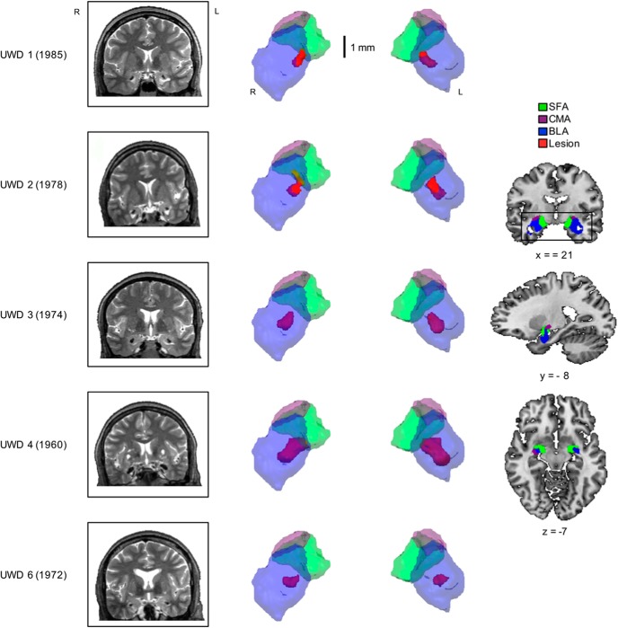
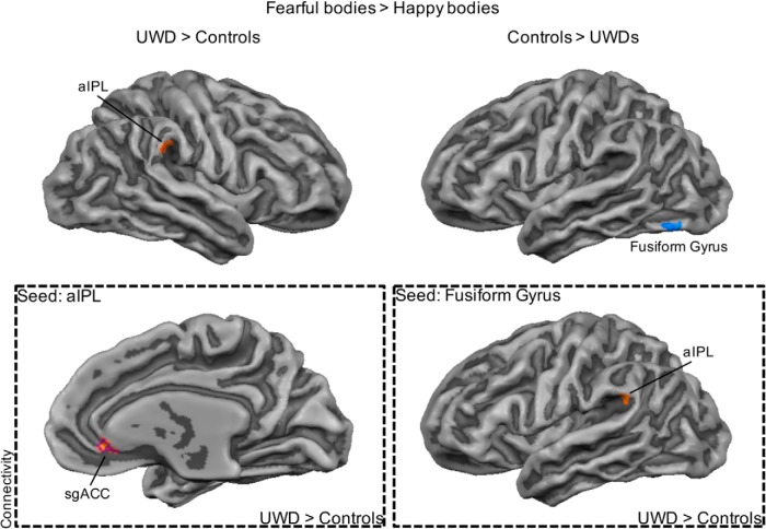
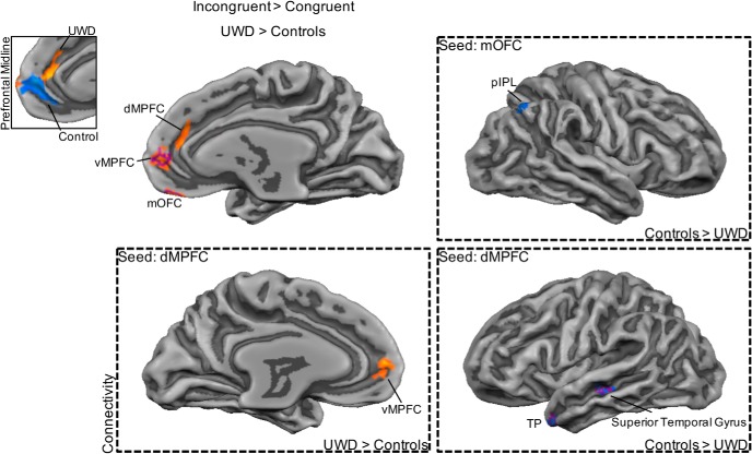
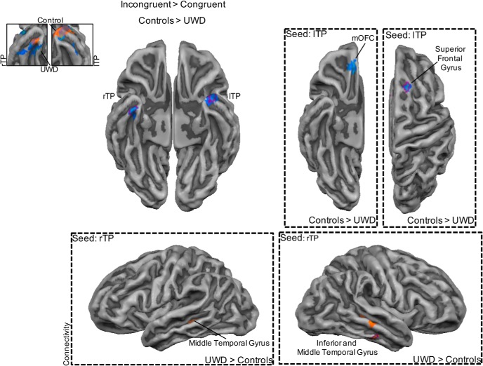
Although the amygdalae play a central role in threat perception and reactions, the direct contributions of the amygdalae to specific aspects of threat perception, from ambiguity resolution to reflexive or deliberate action, remain ill understood in humans. Animal studies show that a detailed understanding requires a focus on the different subnuclei, which is not yet achieved in human research. Given the limits of human imaging methods, the crucial contribution needs to come from individuals with exclusive and selective amygdalae lesions. The current study investigated the role of the basolateral amygdalae and their connection with associated frontal and temporal networks in the automatic perception of threat. Functional activation and connectivity of five individuals with Urbach-Wiethe disease with focal basolateral amygdalae damage and 12 matched controls were measured with functional MRI while they attended to the facial expression of a threatening face-body compound stimuli. Basolateral amygdalae damage was associated with decreased activation in the temporal pole but increased activity in the ventral and dorsal medial prefrontal and medial orbitofrontal cortex. This dissociation between the prefrontal and temporal networks was also present in the connectivity maps. Our results contribute to a dynamic, multirole, subnuclei-based perspective on the involvement of the amygdalae in fear perception. Damage to the basolateral amygdalae decreases activity in the temporal network while increasing activity in the frontal network, thereby potentially triggering a switch from resolving ambiguity to dysfunctional threat signaling and regulation, resulting in hypersensitivity to threat.
Keywords: Urbach–Wiethe disease; amygdalae; basolateral amygdalae; emotion; threat.
Conflict of interest statement
Authors report no conflict of interest.
Figures
Similar articles
-
Temporal lobe epilepsy and emotion recognition without amygdala: a case study of Urbach-Wiethe disease and review of the literature.Epileptic Disord. 2014 Dec;16(4):518-27. doi: 10.1684/epd.2014.0696. Epileptic Disord. 2014. PMID: 25465029 Review.
-
The role of the basolateral amygdala in the perception of faces in natural contexts.Philos Trans R Soc Lond B Biol Sci. 2016 May 5;371(1693):20150376. doi: 10.1098/rstb.2015.0376. Philos Trans R Soc Lond B Biol Sci. 2016. PMID: 27069053 Free PMC article.
-
Hypervigilance for fear after basolateral amygdala damage in humans.Transl Psychiatry. 2012 May 15;2(5):e115. doi: 10.1038/tp.2012.46. Transl Psychiatry. 2012. PMID: 22832959 Free PMC article.
-
Impaired threat prioritisation after selective bilateral amygdala lesions.Cortex. 2015 Feb;63:206-13. doi: 10.1016/j.cortex.2014.08.017. Epub 2014 Sep 10. Cortex. 2015. PMID: 25282058 Free PMC article.
-
Translational neuroscience of basolateral amygdala lesions: Studies of Urbach-Wiethe disease.J Neurosci Res. 2016 Jun;94(6):504-12. doi: 10.1002/jnr.23731. J Neurosci Res. 2016. PMID: 27091312 Review.
Cited by
-
Ultrahigh Field fMRI Reveals Different Roles of the Temporal and Frontoparietal Cortices in Subjective Awareness.J Neurosci. 2024 May 15;44(20):e0425232023. doi: 10.1523/JNEUROSCI.0425-23.2023. J Neurosci. 2024. PMID: 38531633 Free PMC article.
-
The Implementation of Infant Anoesis and Adult Autonoesis in the Retrogenesis and Staging System of the Neurocognitive Disorders: A Proposal for a Multidimensional Person-Centered Model.Geriatrics (Basel). 2025 Feb 2;10(1):20. doi: 10.3390/geriatrics10010020. Geriatrics (Basel). 2025. PMID: 39997519 Free PMC article. Review.
-
Dynamic Interactions between Emotion Perception and Action Preparation for Reacting to Social Threat: A Combined cTBS-fMRI Study.eNeuro. 2018 Jul 2;5(3):ENEURO.0408-17.2018. doi: 10.1523/ENEURO.0408-17.2018. eCollection 2018 May-Jun. eNeuro. 2018. PMID: 29971249 Free PMC article.
-
Resting-state functional connectivity of the amygdala subregions in unmedicated patients with obsessive-compulsive disorder before and after cognitive behavioural therapy.J Psychiatry Neurosci. 2021 Nov 16;46(6):E628-E638. doi: 10.1503/jpn.210084. Print 2021 Nov-Dec. J Psychiatry Neurosci. 2021. PMID: 34785511 Free PMC article.
-
Looking at the face and seeing the whole body. Neural basis of combined face and body expressions.Soc Cogn Affect Neurosci. 2018 Jan 1;13(1):135-144. doi: 10.1093/scan/nsx130. Soc Cogn Affect Neurosci. 2018. PMID: 29092076 Free PMC article.
References
-
- Aggleton JP, Burton MJ, Passingham RE (1980) Cortical and subcortical afferents to the amygdala of the rhesus monkey (Macaca mulatta). Brain Res 190:347–368. - PubMed
-
- Amunts K, Kedo O, Kindler M, Pieperhoff P, Mohlberg H, Shah NJ, Habel U, Schneider F, Zilles K (2005) Cytoarchitectonic mapping of the human amygdala, hippocampal region and entorhinal cortex: intersubject variability and probability maps. Anat Embryol 210:343–352. 10.1007/s00429-005-0025-5 - DOI - PubMed
Publication types
MeSH terms
LinkOut - more resources
Full Text Sources
Other Literature Sources