. 2017 Mar 29;4(2):ENEURO.0129-16.2017.
doi: 10.1523/ENEURO.0129-16.2017. eCollection 2017 Mar-Apr.

Divergent Modulation of Nociception by Glutamatergic and GABAergic Neuronal Subpopulations in the Periaqueductal Gray

Affiliations

Divergent Modulation of Nociception by Glutamatergic and GABAergic Neuronal Subpopulations in the Periaqueductal Gray

Vijay K Samineni et al. eNeuro. .

Abstract

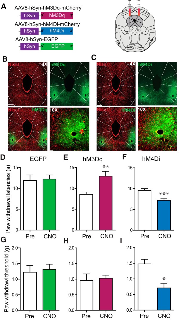
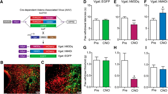
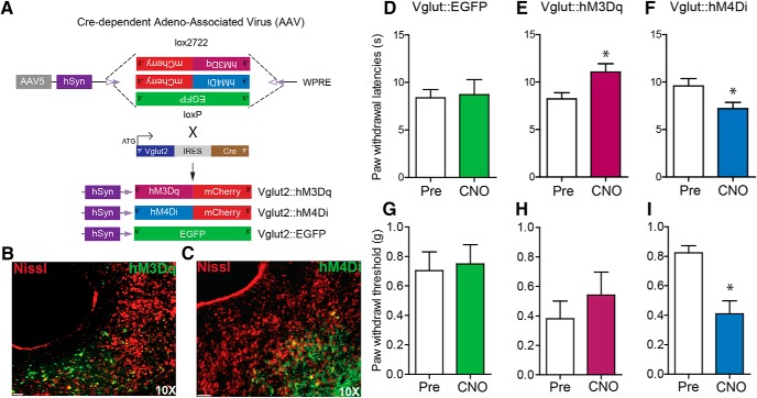
The ventrolateral periaqueductal gray (vlPAG) constitutes a major descending pain modulatory system and is a crucial site for opioid-induced analgesia. A number of previous studies have demonstrated that glutamate and GABA play critical opposing roles in nociceptive processing in the vlPAG. It has been suggested that glutamatergic neurotransmission exerts antinociceptive effects, whereas GABAergic neurotransmission exert pronociceptive effects on pain transmission, through descending pathways. The inability to exclusively manipulate subpopulations of neurons in the PAG has prevented direct testing of this hypothesis. Here, we demonstrate the different contributions of genetically defined glutamatergic and GABAergic vlPAG neurons in nociceptive processing by employing cell type-specific chemogenetic approaches in mice. Global chemogenetic manipulation of vlPAG neuronal activity suggests that vlPAG neural circuits exert tonic suppression of nociception, consistent with previous pharmacological and electrophysiological studies. However, selective modulation of GABAergic or glutamatergic neurons demonstrates an inverse regulation of nociceptive behaviors by these cell populations. Selective chemogenetic activation of glutamatergic neurons, or inhibition of GABAergic neurons, in vlPAG suppresses nociception. In contrast, inhibition of glutamatergic neurons, or activation of GABAergic neurons, in vlPAG facilitates nociception. Our findings provide direct experimental support for a model in which excitatory and inhibitory neurons in the PAG bidirectionally modulate nociception.

Keywords: DREADDs; Descending modulation; PAG; RVM; chemogenetics; pain.


Figures

Figure 1.
Figure 1.
Global chemogenetic manipulation of vlPAG activity suggests parallel bidirectional modulation of nociceptive behaviors. A, Constructs used in viral targeting of AAV8 hM3Dq–mCherry, AAV8 hM4Di–mCherry and AAV8–EGFP via bilateral injections into the vlPAG. B, C, Representative images of coronal sections containing vlPAG demonstrating restricted viral expression following microinjection of the AAV8 hM3Dq (B) and hM4Di (C) into the vlPAG. D, G, Relative to pretreatment baseline values, CNO (1 mg/kg, i.p.) did not have any significant effects on PWLs in mice expressing the control EGFP construct. E, H, CNO (1 mg/kg, i.p.) administration in hM3Dq-injected mice resulted in a significant increase in PWLs but not in PWTs. F, I, CNO (1 mg/kg, i.p.) administration in hM4Di-injected mice resulted in a significant decrease in PWLs and PWTs. ∗p < 0.05, ∗∗p < 0.005, ∗∗∗p < 0.0001. Scale bars, 300 and 35 μm, 4× and 10×, respectively.
Figure 2.
Figure 2.
RNA-FISH demonstrates segregation of vlPAG GABAergic and glutamatergic neurons and specificity of Cre in targeting Vgat+ neurons in the Vgat-IRES-Cre mice or Vglut2+ neurons in the Vglut2-IRES-Cre mice. A, Double RNA-FISH for Vgat (green) and Vglut2 (red) shows that GABAergic and glutamatergic neurons in the PAG are nonoverlapping populations. Scale bar, 200 μm. Counterstaining (blue) is DAPI. B, High-magnification image showing no colocalization of GABAergic and glutamatergic neurons in the PAG. Scale bar, 60 μm. C, 79 ± 4.1% of cells positive for Vglut2 transcripts in the vlPAG colabel with Vglut2 Cre-expressing neurons, and 97.5 ± 2.5% of Vglut2 Cre-expressing neurons in the vlPAG colabel with Vglut2+ transcripts (N = 2 mice). D, 92 ± 4.5% of cells positive for Vgat transcripts in the vlPAG colabel with Vgat Cre-expressing neurons, and 95.5 ± 4.2% of Vgat Cre-expressing neurons in the vlPAG colabel with Vgat+ transcripts (N = 2 mice). E–G, Double RNA-FISH for Vglut2 (red) and Cre (green) shows extensive colocalization of Vglut2 + transcripts with Cre-expressing neurons in the vlPAG obtained from Vglut2 Cre mice. Scale bar, 60 μm. H–J, High-magnification image shows extensive colocalization of Vglut2+ transcripts with Cre-expressing neurons in the vlPAG. Scale bar, 15 μm. K, M, Double RNA-FISH for Vgat (red) and Cre (green) shows extensive colocalization of Vgat+ transcripts with Cre-expressing neurons in the vlPAG obtained from Vgat Cre mice. Scale bar, 60 μm. N–P, High-magnification image shows extensive colocalization of Vgat+ transcripts with Cre-expressing neurons in the vlPAG. Scale bar, 15 μm.
Figure 3.
Figure 3.
Functional characterization of Gq- and Gi-DREADDs in vlPAG neurons of Vgat-Cre and Vglut2-Cre mice. A, Infrared DIC image of vlPAG Vgat+ neuron expressing hM4Di-mCherry. Images were acquired following CNO stimulation. B, Whole-cell current-clamp recording from an hM3Dq-expressing PAG neuron. Brief bath application of 10 µM CNO caused a transient depolarization and robust action potential firing in Vgat+ and Vglut+ neurons. C, Voltage trace showing that bath perfusion with 10 µM CNO caused prolonged membrane hyperpolarization and silencing of both Vgat+ and Vglut+ vlPAG neurons. Dashed lines in B and C represent the membrane potential of the cells before application of CNO. D, G, Quantification of the CNO effects on membrane potential and input resistance in grouped Vgat+ and Vglut2+ neurons (N = 8 for Vgat+ and Vglut2+ neurons). E–I, Voltage traces showing responses to a hyperpolarizing current of -20 pA and a depolarizing current injection of either 1× rheobase (purple traces) or 2× rheobase (blue traces) in both Vgat+ (E, H) and Vglut2+ (F, I) neurons. In hM3Dq-expressing neurons, bath application of CNO elicited increased action potential firing in response to the same stimulus (E, H, green traces). In hM4Di+ neurons, CNO perfusion decreased neuronal excitability to supratheshold stimuli. B, C, Scale bars, 20 mv and 10 s; E–I, Scale bars, 10 mv and 100 ms. All values are mean ± SEM.
Figure 4.
Figure 4.
Chemogenetic manipulation of vlPAG GABAergic neurons bidirectionally modulates nociceptive behaviors. A, Illustration showing viral targeting strategy of AAV5-hSyn-DIO-hM3Dq–mCherry, AAV5-hSyn-DIO-hM4Di–mCherry, and AAV5-hSyn-DIO-EGFP bilaterally injected into the vlPAG of Vgat Cre mice. B, C, Representative images of coronal sections containing vlPAG showing restricted viral expression following microinjection of AAV5-hSyn-DIO-hM3Dq (B) or AAV5-hSyn-DIO-hM4Di (C) into the vlPAG of Vgat Cre mice. E, H, CNO (3 mg/kg, i.p.) administration resulted in a significant decrease in PWLs and PWTs in Vgat::hM3Dq mice. F, I, CNO administration resulted in a significant increase in PWLs but not in PWTs in Vgat::hM4Di mice. D, G, CNO had no significant effect on PWLs or PWTs in Vgat Cre mice expressing the control EGFP construct compared with baseline PWLs and PWTs before CNO administration. All values are mean ± SEM. Student’s t test; ∗p < 0.05, ∗∗p < 0.005. Scale bars, 25 μm.
Figure 5.
Figure 5.
Chemogenetic modulation of vlPAG glutamatergic neurons bidirectionally modulates nociceptive behaviors. A, Illustration showing the strategy for viral targeting of AAV5-hSyn-DIO-hM3Dq–mCherry, AAV5-hSyn-DIO-hM4Di–mCherry, and AAV5-hSyn-DIO-EGFP bilaterally injected into the vlPAG of Vglut2 Cre mice. B, C, Representative images of a coronal sections containing vlPAG showing restricted viral expression following microinjection of the AAV5-hSyn-DIO-hM3Dq–mCherry and AAV5-hSyn-DIO-hM4Di–mCherry into the vlPAG of Vglut2 Cre mice. E, H, CNO (2 mg/kg, i.p.) administration resulted in a significant increase in PWLs but not on PWTs in Vglut2::hM3Dq mice. F, I, CNO (2 mg/kg, i.p.) administration resulted in a significant decrease in PWLs and PWTs in Vglut2::hM4Di mice. D, G, CNO had no significant effect on PWLs or PWTs in Vglut2 Cre mice expressing the control EGFP construct compared with baseline PWLs and PWTs before CNO administration. All values are mean ± SEM, Student’s t test; ∗p < 0.05. Scale bars, 25 μm.

Similar articles

Cited by

References

    1. Anseloni VC, Gold MS (2008) Inflammation-induced shift in the valence of spinal GABA-A receptor-mediated modulation of nociception in the adult rat. J Pain 9:732–738. 10.1016/j.jpain.2008.03.004 - DOI - PMC - PubMed
    1. Antal M, Odeh F (1998) The projections of the midbrain periaqueductal gray to serotonergic and noradrenergic nuclei of the pons and medulla oblongata in the rat. Eur J Neurosci 10:218–218. - PubMed
    1. Bandler R, Keay KA (1996) Columnar organization in the midbrain periaqueductal gray and the integration of emotional expression. Prog Brain Res 107:285–300. - PubMed
    1. Basbaum AI, Fields HL (1978) Endogenous pain control mechanisms: review and hypothesis. Ann Neurol 4:451–462. 10.1002/ana.410040511 - DOI - PubMed
    1. Basbaum AI, Fields HL (1979) The origin of descending pathways in the dorsolateral funiculus of the spinal cord of the cat and rat: further studies on the anatomy of pain modulation. J Comp Neur 187:513–531. 10.1002/cne.901870304 - DOI - PubMed

Publication types

MeSH terms

LinkOut - more resources