

Accuracy of bone mineral density quantification using dual-layer spectral detector CT: a phantom study
- PMID: 28374079
- PMCID: PMC5579207
- DOI: 10.1007/s00330-017-4801-4
Accuracy of bone mineral density quantification using dual-layer spectral detector CT: a phantom study
Abstract
Objectives: To investigate the accuracy of bone mineral density (BMD) quantification using dual-layer spectral detector CT (SDCT) at various scan protocols.
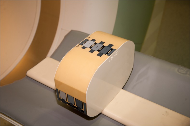
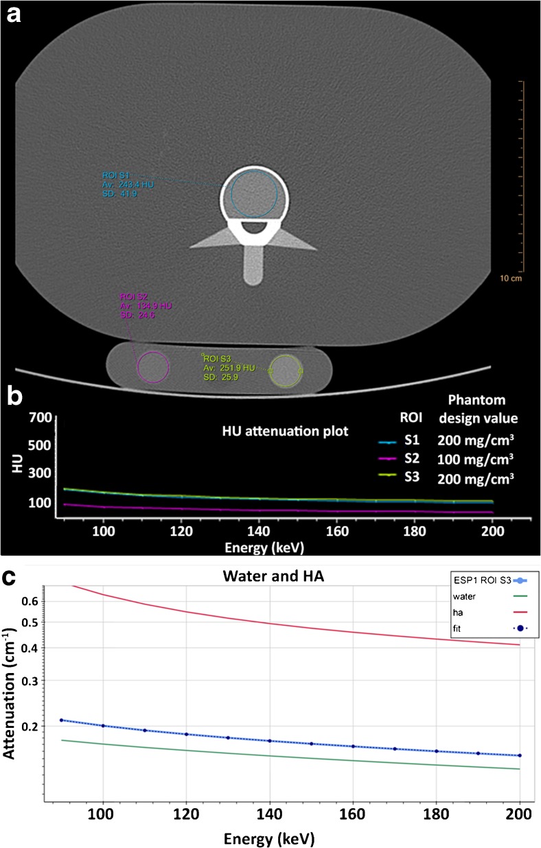
Methods: Two validated anthropomorphic phantoms containing inserts of 50-200 mg/cm3 calcium hydroxyapatite (HA) were scanned using a 64-slice SDCT scanner at various acquisition protocols (120 and 140 kVp, and 50, 100 and 200 mAs). Regions of interest (ROIs) were placed in each insert and mean attenuation profiles at monochromatic energy levels (90-200 keV) were constructed. These profiles were fitted to attenuation profiles of pure HA and water to calculate HA concentrations. For comparison, one phantom was scanned using dual energy X-ray absorptiometry (DXA).
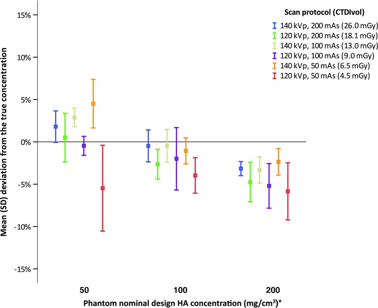
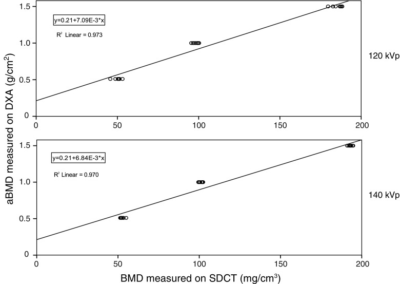
Results: At both 120 and 140 kVp, excellent correlations (R = 0.97, P < 0.001) were found between true and measured HA concentrations. Mean error for all measurements at 120 kVp was -5.6 ± 5.7 mg/cm3 (-3.6 ± 3.2%) and at 140 kVp -2.4 ± 3.7 mg/cm3 (-0.8 ± 2.8%). Mean measurement errors were smaller than 6% for all acquisition protocols. Strong linear correlations (R2 ≥ 0.970, P < 0.001) with DXA were found.
Conclusions: SDCT allows for accurate BMD quantification and potentially opens up the possibility for osteoporosis evaluation and opportunistic screening in patients undergoing SDCT for other clinical indications. However, patient studies are needed to extend and translate our findings.
Key points: • Dual-layer spectral detector CT allows for accurate bone mineral density quantification. • BMD measurements on SDCT are strongly linearly correlated to DXA. • SDCT, acquired for several indications, may allow for evaluation of osteoporosis. • This potentially opens up the possibility for opportunistic osteoporosis screening.
Keywords: Bone mineral density; Dual energy X-Ray absorptiometry; Dual-energy CT; Dual-layer spectral detector CT; Material decomposition.
Conflict of interest statement
Guarantor
The scientific guarantor of this publication is prof. dr. Tim Leiner.
Conflict of interest
The authors of this manuscript declare no relationships with any companies whose products or services may be related to the subject matter of the article.
Funding
The authors state that this work has not received any funding.
Statistics and biometry
One of the authors has significant statistical expertise.
Ethical approval
Institutional Review Board approval was not required because this concerns a phantom study.
Methodology
• experimental
• performed at one institution
Figures
References
MeSH terms
LinkOut - more resources
Full Text Sources
Other Literature Sources
Medical
