Review
. 2017 Jul;235(7):1953-1997.
doi: 10.1007/s00221-017-4950-3. Epub 2017 Apr 3.

Experimental investigations of control principles of involuntary movement: a comprehensive review of the Kohnstamm phenomenon

Affiliations
Review

Experimental investigations of control principles of involuntary movement: a comprehensive review of the Kohnstamm phenomenon

Jack De Havas et al. Exp Brain Res. 2017 Jul.

Abstract

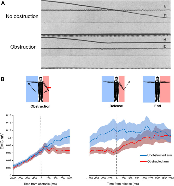
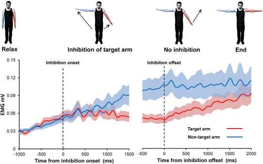
The Kohnstamm phenomenon refers to the observation that if one pushes the arm hard outwards against a fixed surface for about 30 s, and then moves away from the surface and relaxes, an involuntary movement of the arm occurs, accompanied by a feeling of lightness. Central, peripheral and hybrid theories of the Kohnstamm phenomenon have been advanced. Afferent signals may be irrelevant if purely central theories hold. Alternatively, according to peripheral accounts, altered afferent signalling actually drives the involuntary movement. Hybrid theories suggest afferent signals control a centrally-programmed aftercontraction via negative position feedback control or positive force feedback control. The Kohnstamm phenomenon has provided an important scientific method for comparing voluntary with involuntary movement, both with respect to subjective experience, and for investigating whether involuntary movements can be brought under voluntary control. A full review of the literature reveals that a hybrid model best explains the Kohnstamm phenomenon. On this model, a central adaptation interacts with afferent signals at multiple levels of the motor hierarchy. The model assumes that a Kohnstamm generator sends output via the same pathways as voluntary movement, yet the resulting movement feels involuntary due to a lack of an efference copy to cancel against sensory inflow. This organisation suggests the Kohnstamm phenomenon could represent an amplification of neuromotor processes normally involved in automatic postural maintenance. Future work should determine which afferent signals contribute to the Kohnstamm phenomenon, the location of the Kohnstamm generator, and the principle of feedback control operating during the aftercontraction.

Keywords: Action awareness; Action inhibition; Aftercontraction; Involuntary movement; Muscle afferents; Posture.


Conflict of interest statement

The authors declare no competing financial interests.

Figures

Fig. 1
Fig. 1
Kohnstamm phenomenon. The first documented image of the Kohnstamm phenomenon (a). Dr. Alberto Salmon has one of his patients push outwards against his arms. Upon relaxation, the patient’s arms rise involuntarily due to an aftercontraction of the lateral deltoid muscles (Adapted from Salmon 1916). b Modern recording of the Kohnstamm phenomenon showing the basic kinematics, average duration, and a typical EMG trace from the right lateral deltoid muscle
Fig. 2
Fig. 2
Evidence for muscle thixotropy underlying the Kohnstamm phenomenon. The first panel a shows arm movement during the conditioning procedure. Normally, the full conditioning procedure was performed on one arm (control) and a reduced version, with some steps omitted was performed on the other arm (test). However, the upper panel here shows single trials when the full procedure was performed for both arms. This consisted of: (1) voluntary arm abduction up against solid surface; (2) forceful, voluntary abductor contraction against solid surface (5–10 s; filled bar on graph); (3) relaxation with experimenter holding the arms in place (4–8 s); and (4) experimenter assisted lowering of arms. After step 4, the aftercontraction occurred. The lower panel (a) shows a single trial, where performing the induction contraction with the arm partially abducted for the test arm (longer muscle length) leads to an absence of aftercontraction, while an aftercontraction was clearly present for the control arm (short muscle length). The second panel b shows the size of aftercontractions after omitting steps from the induction (C control arm, T test arm). For Trial A, the same conditioning procedure was used on both arms. For trial B, the initial arm abduction was omitted for the test arm, for trial C, the voluntary isometric contraction was omitted for the test arm, for trial D, the experimenter-assisted relaxation period was omitted for the test arm, while for trial E, the test arm was returned rapidly instead of slowly. The third panel c shows that warming the test arm significantly reduced the size of the aftercontraction, while cooling produced a trend in the other direction, relative to the control arm (Figure Adapted from Hagbarth and Nordin 1998)
Fig. 3
Fig. 3
Results of physically obstructing of the aftercontraction. The first panel a shows an early experiment to determine whether physical obstruction of the aftercontraction resulted in a cessation of muscle activity. Arm position (lines labelled M) and electromyography (lines labelled E) are shown when no obstacle was present (upper graph) and when the arm was obstructed at around 20° of abduction (lower graph). Only single traces could be recorded at that time, but the experiment confirmed that electrical activity could be detected by a string galvanometer following obstruction, disproving an earlier claim that electrical activity detected during the aftercontraction was due to the movement itself, rather than a reflection of involuntary muscle activity (Adapted from Forbes et al. 1926) The second panel b shows the results of a more recent experiment involving unpredictably obstructing one arm for 2 s during a bilateral aftercontraction. Group average EMG is shown (error bars show SEM). It was found that physical obstruction caused a significant reduction in the slope of the aftercontraction EMG, relative to no obstruction, indicating that the output of the Kohnstamm generator is modified by afferent signals. Upon removal of the obstacle the previously obstructed arm immediately resumed its previous involuntary abduction and accompanying pattern of increasing EMG. Final arm angle and EMG level was the same as for the never obstructed arm, indicating that afferent information did not alter the state of the Kohnstamm generator itself, but rather only attenuated its output (Adapted from De Havas et al. 2015). (Color figure online)
Fig. 4
Fig. 4
Brain regions active during Aftercontraction and TVR. Brain regions showing a significant increase in BOLD-signal in 11 subjects during a voluntary induction contraction of wrist extensor muscle, b vibration of wrist extensor tendon, c involuntary aftercontraction of wrist extensor muscle (here referred to as a post-contraction), and d post-vibration response (more commonly known as TVR) in contrast with a rest period (no movement; false discovery rate, P < 0.005). Note the large regions of sensorimotor cortex active during the Kohnstamm aftercontraction (Adapted from Duclos et al. 2007). (Color figure online)
Fig. 5
Fig. 5
Applying TMS to M1 during aftercontraction shows cortical involvement in Kohnstamm phenomenon. A Kohnstamm aftercontraction was induced by having the participants push against a wall and then step away and relax the deltoid muscle (a). Kinematic and EMG traces of the Kohnstamm induction and aftercontraction are shown from a single representative participant (b). TMS of the motor cortex during aftercontraction (d) and matched voluntary movements (c) results in a prolonged silent period, suggesting a cortical origin (representative participant’s data). Mean muscle silent period duration following application of TMS did not differ across aftercontraction and voluntary movement conditions (e). Muscular contractions made a full recovery after the silent period for both Kohnstamm aftercontractions and voluntary movements (f). Adapted from Ghosh et al. (2014)
Fig. 6
Fig. 6
Mean firing rate of motor units significantly lower during aftercontraction compared to voluntary movements. The first panel a shows a raw EMG recorded in human triceps muscle showing recruitment of a motor unit during the first 2 s of an aftercontraction. Solid line shows elbow joint angle change. Motor unit firing rate progressively increases after the latent period, followed by a relatively steady state of firing. Aftercontractions were compared to voluntary movements of matched velocity (b). It was found that across participants motor units showed lower firing rates (c) during aftercontraction compared to voluntary movements (Adapted from Kozhina et al. 1996)
Fig. 7
Fig. 7
Voluntary inhibition of Kohnstamm aftercontraction. The effect of inhibiting, and releasing inhibition, of a single ‘target’ arm during bilateral Kohnstamm aftercontraction on rectified, smoothed deltoid EMG. Dashed lines show time of inhibition onset and offset. Error bars show SEM. Note the significant increase in EMG for the non-target arm relative to the plateauing of EMG in the target arm, beginning approximately 500 ms after the instruction to inhibit. After participants were instructed to stop inhibiting, target arm EMG increased and the arm began to involuntarily rise once more. Final arm angle and EMG level was the same for both arms across participants, indicating that the Kohnstamm generator itself was not modified by voluntary inhibition (Adapted from De Havas et al. 2016). (Color figure online)
Fig. 8
Fig. 8
Reduced aftercontraction EMG in response to decreased muscle loading. Participants pushed upwards against the force transducer (60% MVC, 60 s) to induce an aftercontraction of the anterior deltoid muscle (a). A movable counter-weight attached to the arm via a lever allowed the loading on the muscle to be systematically reduced across conditions. EMG and arm angle results of a single participant are shown (b), including the last 10 s of the induction and the entire aftercontraction. Group average results of reducing the muscle load on EMG across joint angles are shown (c). A load of 1 means that the arm was of normal weight, while a load of 0 meant that the counterweight perfectly balanced the arm weight, meaning that there should have been negligible loading on anterior deltoid. Reducing the load from 1 down to 0 produced a reliable decrease in aftercontraction EMG across joint angles (Adapted from Parkinson and McDonagh 2006)
Fig. 9
Fig. 9
Subjective experience of inhibiting the Kohnstamm aftercontraction. The first panel a shows the results of an experiment in which the subjective experience of voluntarily bringing the arm down (adduction) during an aftercontraction was rated (1 strong disagreement, 5 strong agreement). Participants clearly perceived an upward resistance, most closely resembling an air balloon (Adapted from Ghosh et al. 2014). The second panel b shows the results of an experiment when the subjective upward drive from the Kohnstamm generator was compared to the actual muscle contraction strength during voluntary inhibition of an aftercontraction (b), compared to a range of isometric voluntary contractions (a). Participants rated how much force their arm could support during inhibition of aftercontraction (arm held stationary, partially abducted). This rating was plotted (c left graph; red squares; single illustrative participant) together with the relation between perceived and actual force from voluntary trials (c left graph; green diamonds). Interpolating this relation allowed an estimation of the equivalent Kohnstamm forces that would be required to generate percepts similar to those on voluntary trials. The level of voluntary EMG required to generate the equivalent Kohnstamm force was calculated, using the relation between EMG and actual force for voluntary trials (c right graph). This perceived aftercontraction was compared to the actual level of aftercontraction EMG during the period of inhibition across participants (d). Subjective aftercontraction strength was significantly overestimated, suggesting the Kohnstamm generator does not produce efference copies to cancel against the sensory inflow (Adapted from De Havas et al. 2016). (Color figure online)
Fig. 10
Fig. 10
A model of the Kohnstamm phenomenon. The left panel shows a model of how an aftercontraction is induced from a strong, sustained voluntary contraction (V). Efferent output produces a contraction in the muscle, which will, upon relaxation (cessation of voluntary signal), display an aftercontraction. The Kohnstamm generator (K) is centrally located and must receive input during the induction. However, it is not known whether the necessary signal to the Kohnstamm generator originates from the muscle, and/or directly from central regions (V). The right panel shows how the aftercontraction is controlled once it has begun. The Kohnstamm generator (K) does not output directly to the muscle. Rather a positive signal is sent to an efferent output stage (E likely M1), which in turn produces the involuntary muscle contraction. The strength of the signal sent from the Kohnstamm generator can be reduced via both voluntary inhibition and via afferent signals resulting from the limb being arrested by a physical obstacle. While the limb is moving, it is not known if the Kohnstamm generator receives modulatory positive force feedback or negative position feedback from the muscle. Alternatively, this putative feedback might not modify the Kohnstamm generator directly, and instead operate at a lower level (E)

Similar articles

Cited by

References

    1. Adamson G, McDonagh M (2004) Human involuntary postural aftercontractions are strongly modulated by limb position. Eur J Appl Physiol, 92(3), 343–351. 10.1007/s00421-004-1091-8 - DOI - PubMed
    1. Allen F. The post-contraction of the muscles of the arm. Q J Exp Physiol. 1937;26(4):305–317.
    1. Allen F, O’Donoghue C. The post-contraction proprioceptive reflex, its augmentation and inhibition. Q J Exp Physiol. 1927;18(3):199–242.
    1. Archambault PS, Mihaltchev P, Levin MF, Feldman AG. Basic elements of arm postural control analyzed by unloading. Exp Brain Res. 2005;164(2):225–241. doi: 10.1007/s00221-005-2245-6. - DOI - PubMed
    1. Ball T, Schreiber A, Feige B, Wagner M, Lücking CH, Kristeva-Feige R. The role of higher-order motor areas in voluntary movement as revealed by high-resolution EEG and fMRI. NeuroImage. 1999;10(6):682694. doi: 10.1006/nimg.1999.0507. - DOI - PubMed

LinkOut - more resources