Review
. 2017 Sep;136(9):1015-1042.
doi: 10.1007/s00439-017-1790-y. Epub 2017 Apr 3.

Alternative splicing: the pledge, the turn, and the prestige : The key role of alternative splicing in human biological systems

Affiliations
Review

Alternative splicing: the pledge, the turn, and the prestige : The key role of alternative splicing in human biological systems

L M Gallego-Paez et al. Hum Genet. 2017 Sep.

Abstract

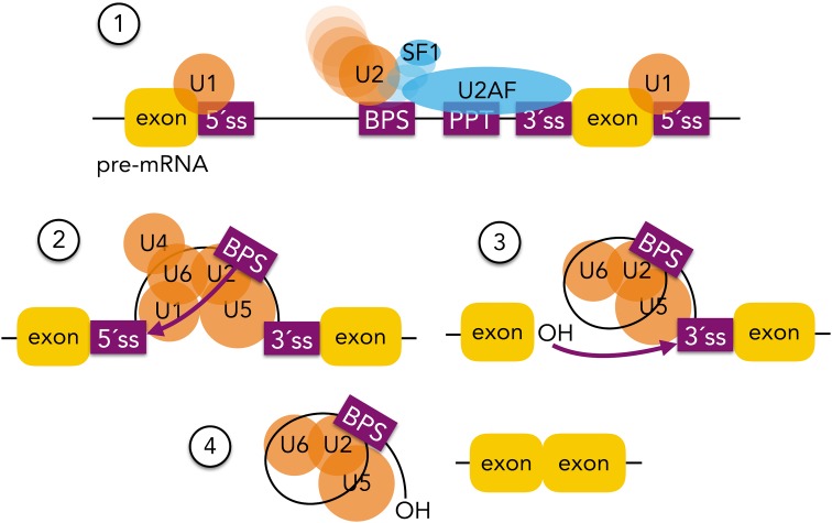
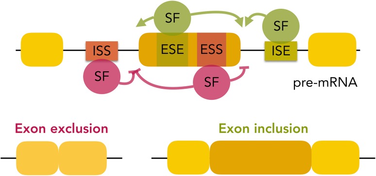
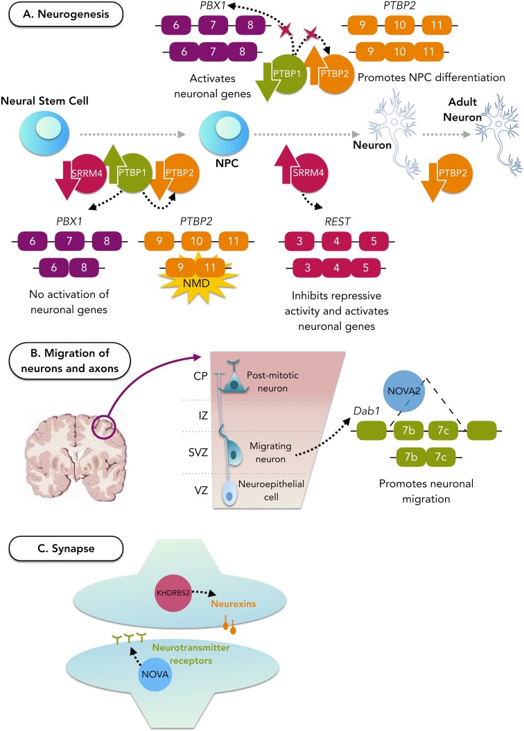
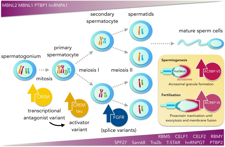
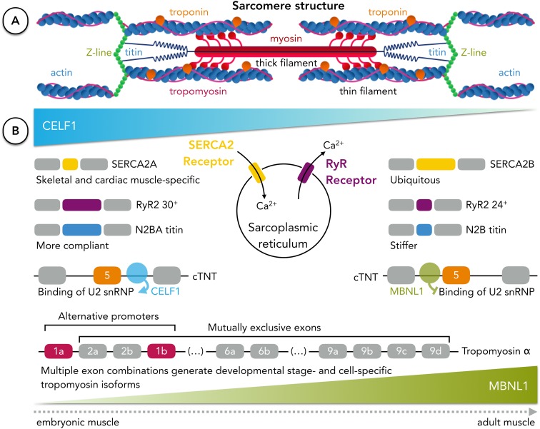
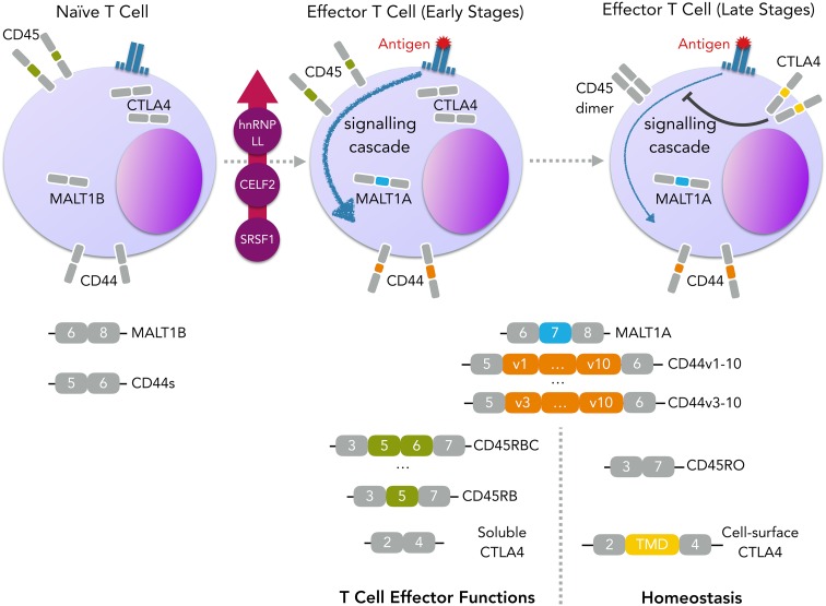
Alternative pre-mRNA splicing is a tightly controlled process conducted by the spliceosome, with the assistance of several regulators, resulting in the expression of different transcript isoforms from the same gene and increasing both transcriptome and proteome complexity. The differences between alternative isoforms may be subtle but enough to change the function or localization of the translated proteins. A fine control of the isoform balance is, therefore, needed throughout developmental stages and adult tissues or physiological conditions and it does not come as a surprise that several diseases are caused by its deregulation. In this review, we aim to bring the splicing machinery on stage and raise the curtain on its mechanisms and regulation throughout several systems and tissues of the human body, from neurodevelopment to the interactions with the human microbiome. We discuss, on one hand, the essential role of alternative splicing in assuring tissue function, diversity, and swiftness of response in these systems or tissues, and on the other hand, what goes wrong when its regulatory mechanisms fail. We also focus on the possibilities that splicing modulation therapies open for the future of personalized medicine, along with the leading techniques in this field. The final act of the spliceosome, however, is yet to be fully revealed, as more knowledge is needed regarding the complex regulatory network that coordinates alternative splicing and how its dysfunction leads to disease.

Keywords: Alternative Splice; Duchenne Muscular Dystrophy; Round Spermatid; Spinal Muscular Atrophy; Splice Factor.


Conflict of interest statement

On behalf of all authors, the corresponding author states that there is no conflict of interest.

Figures

Fig. 1
Fig. 1
Spliceosome assembly and splicing reactions. (1) U1 snRNP binds to the 5′ splice site (5′ss), whereas the splicing factor 1 (SF1) and U2AF proteins bind to the branch point site (BPS), the polypyrimidine tract (PPT), and 3′ splice site (3′ss). The interaction between U1 and U2 snRNPs results in the formation of the pre-spliceosome. (2) The first splicing reaction is performed after the recruitment of the U4/5/6 snRNPs through a nucleophilic attack from the adenosine in the BPS to the 5′ss of the upstream exon. (3) The intron lariat is then formed. The free 3′ hydroxyl group performs a nucleophilic attack to the phosphate of the 3′ splice site of the downstream exon. (4) Finally, the intron lariat is released and both exons are ligated
Fig. 2
Fig. 2
AS regulation by RNA-binding splicing factors. Binding of specific splicing factors (SF) to intronic or exonic splicing enhancers (ISE and ESE, respectively) promotes the inclusion of the alternative exon, whereas binding of given splicing factors to intronic or exonic splicing silencers (ISS and ESS, respectively) inhibits the splicing of the alternative exon
Fig. 3
Fig. 3
Role of splicing factors during neurogenesis and neuron maturation. A PTBP1 is responsible for repressing the activation of neuronal genes and is highly expressed in neuronal stem cells and neuronal progenitor cells. Upon differentiation, PTBP1 becomes downregulated, allowing the induction of PTBP2 and PBX1 that will activate neuronal genes. SRRM4 also becomes expressed during neuronal differentiation and contributes to it by inactivating REST, a repressor of activation of neuronal genes. After the neurons become mature, the levels of PTBP2 decrease, giving rise to an adult neuronal splicing programme. NMD nonsense-mediated decay, NPC neural progenitor cell. B Once the neuronal cell fate commitment is achieved, neurons can migrate to generate the laminar structure of the brain. NOVA2 is a splicing factor particularly important for the cortical lamination since it regulates AS of Dab1 to promote neuronal migration. VZ ventricular zone, SVZ subventricular zone, IZ intermediate zone, CP cortical plate. C For the maturation process, neurons form synapses. This process is equally controlled by splicing factors, namely, KHDRBS2 that regulates neurexins (presynaptic cell-adhesion proteins), which are essential for synapse formation and transmission, and the NOVA family that regulates AS of neurotransmitter receptors
Fig. 4
Fig. 4
Graphical representation of spermatogenesis and its associated AS program. Temporal expression of key splicing factors and splice variants during meiotic division and spermatid maturation is represented by violet gradients. Bottom gradient panel shows the upregulation of the non-germ cell-specific splicing factors SPF27, RBM5, PTBP2, Tra2b, CELF1, and CELF2 and the germ cell-specific splicing factors (Sam68, T-STAR, hnRNPGT, and RBMY). Top gradient panel shows downregulation of the splicing factors PTBP1, MBNL1, MBNL2, and hnRNPA1. AS of the mRNA of the transcription factor CREM induces a functional switch from a transcriptional repressor in premeiotic cells to a transcriptional activator in the pachytene spermatocyte stage. Studies in mouse suggest that two splice variants of the proacrosin-binding protein ACRBP, ACRBP-V5, and ACRBP-W, participate in transport/packaging of proacrosin into acrosomal granules during spermiogenesis and in the promotion of acrosin release from the acrosome during acrosomal exocytosis, respectively. Similarly, splice variants of the fibroblast growth factor receptors (FGFRs) are expressed in spermatocytes and round spermatids and localise to the acrosomal region and the flagellum of mature sperm cells in humans
Fig. 5
Fig. 5
Alternative splicing of sarcomeric and membrane receptor proteins tunes muscular function. a Muscle contraction is achieved through the sliding between thin (rich in actin) and thick (rich in myosin) myofilaments of the sarcomere, shortening its length. Diversity of isoforms of sarcomeric proteins (such as titin, tropomyosin or troponin) required for tissue- or developmental stage-specific functions in muscular tissues arises by alternative splicing (sarcomere structure based on (Seeley et al. 2006)). b RNA-binding proteins MBNL1 and CELF1 are two major regulators of muscle-specific AS whose levels shift during the transition from embryonic to mature tissue. The calcium equilibrium needed for contraction of muscle cells is achieved by the coordinated activities of Ca2+ receptors at the membrane of the sarcoplasmic reticulum. Developmentally regulated AS of the sarcoplasmic/endoplasmic reticulum ATPase Ca2+ transporting (SERCA2) and ryanodine receptors (RyR) shapes calcium handling, controlling sarcomere contraction. Titin isoforms with different levels of stiffness change their relative abundance ratio in muscle cells during the transition from embryonic to adult tissue, altering myocardial compliance. The levels of the larger and more compliant titin isoform N2BA decrease with development, while the smaller and stiffer isoform N2B levels increase in mature and healthy muscle tissue. Troponin, one of the thin filament proteins, tunes the interactions between actin and myosin. MBNL1 and CELF1 regulate the inclusion of exon 5 of the cardiac troponin (cTNT) pre-mRNA by binding in the upstream or downstream intron, respectively. Tissue and developmental stage specificity of tropomyosin is achieved through the usage of alternative promoters and mutually exclusive exons of three of the four tropomyosin mammalian genes. In the case of the tropomyosin α gene, two alternative first exons and three sets of mutually exclusive exons contribute to the variability of tropomyosin isoforms
Fig. 6
Fig. 6
Isoform shifts following T-cell activation. T-cell activation upon antigen recognition leads to global changes in AS, from which the inclusion of MALT1 exon 7 and CD44 variable exons are highlighted. In the case of MALT1, inclusion of TRAF-binding domains contained in its exon 7 leads to a higher recruitment of TRAF6 to the CARMA1-BCL10-MALT1 (CBM) signalling complex, which facilitates IKK activation (Meininger et al. 2016). This results in an enhancement of signalling pathways downstream of TCR signalling and promotion of T-cell activation. As for the transmembrane glycoprotein CD44, ten variable exons are located in the extracellular domain of the protein, which can be excluded or included in different combinations, leading to differences in binding affinity to extracellular matrix components, namely, hyaluronic acid (Naor et al. 1997). While in resting T cells the CD44 variable exons are skipped (isoform CD44s), these are included upon activation (Arch et al. 1992). Even though the importance of this event is not yet clear, CD44 is known to be involved in T-cell homing (DeGrendele et al. 1997) and survival (Baaten et al. 2010). Later stage changes in alternative pre-mRNA splicing often impact genes involved in homeostasis and immunologic memory, from which we take CD45 and CTLA4 as examples. Skipping of alternative exons 4–6 of CD45, results in the production of an isoform more prone to dimerisation, which inhibits the role of CD45 in TCR-signalling transduction. CTLA4, on the other hand, competes with CD28 for ligand binding (van der Merwe et al. 1997), and delivers inhibitory signals that counteract the co-stimulatory signal conferred by CD28 (Krummel and Allison 1995). Upon activation, CTLA4 expression is increased and exon 3, encoding a transmembrane domain, is included (Oaks et al. 2000), drastically increasing the expression of CTLA on the cell surface and empowering the T-cell inhibitory signal. TCR T-cell receptor, TMD transmembrane domain
Fig. 7
Fig. 7
Dual RNA-seq workflow. Due to the relative difference in total RNA abundance between host cells and pathogen in most infection models, deep sequencing is required to obtain a more precise profiling of transcriptomic changes associated with the infection process. This strategy allows the transcriptomic analysis of both host and pathogen at different time points during infection, with the discrimination between the two taking place only at the bioinformatics stage
Fig. 8
Fig. 8
Alterations in the expression of splicing factors in cancer. A pan-cancer analysis using TCGA data revealed 132 splicing factors differentially expressed between tumour and normal samples (x-axis). Patterns of upregulation and downregulation across different tumor types (y-axis) are shown in green and violet gradients, respectively. The color intensity indicates the log2-fold change (log2 FC). Splicing factors are clustered into three groups according to the incidence of each expression pattern in the analysed tumours: frequently downregulated (left), frequently upregulated (right), and tendency to show an opposite pattern between the three kidney and the rest of tumor types (Opposing). The bar plot in the top indicates the frequency of tumor types with up-(green) or down-(violet) regulation for each factor. Kidney Chr, kidney chromophobe; Kidney RC, kidney renal clear cell carcinoma; Kidney RP, kidney renal papillary cell carcinoma; Lung Ad, lung adenocarcinoma; Lung Sq, lung squamous cell carcinoma. Image adapted with permission, from Sebestyén et al. (2016)
Fig. 9
Fig. 9
Splicing therapy methods. a Antisense oligonucleotides (ASOs) are being used for spinal muscular atrophy to correct the aberrant splicing of exon 7 of SMN2. The ASO binds to the unique GC-rich sequence located within the downstream intron to promote the exon 7 inclusion. b Spliceosome-mediated RNA trans-splicing (SMaRT) method relies on the correction of alterations at the post-transcriptional level by modifying the mRNA sequence. An exogenous RNA is introduced in targeted cells to induce a splicing event in trans with the target endogenous sequence, generating a chimeric RNA with exons from the exogenous and the endogenous RNA free of mutations. 5′ss, 5′ splice site; 3′ ss, 3′ splice site; BPS, branching point site; PPT, polypyrimidine tract; pA, polyadenylation signal

References

    1. Aartsma-Rus A, Krieg AM. FDA approves eteplirsen for duchenne muscular dystrophy: the next chapter in the eteplirsen saga. Nucleic Acid Ther. 2016 - PMC - PubMed
    1. Abbas AK, Lichtman AHH, Pillai S (2014) Cellular and molecular immunology. Elsevier Health Sciences
    1. Ahmadi Rastegar D, Sharifi Tabar M, Alikhani M, et al. Isoform-level gene expression profiles of human Y chromosome azoospermia factor genes and their X chromosome paralogs in the testicular tissue of non-obstructive azoospermia patients. J Proteome Res. 2015;14:3595–3605. doi: 10.1021/acs.jproteome.5b00520. - DOI - PubMed
    1. Ajiro M, Zheng Z-M. E6E7, a novel splice isoform protein of human papillomavirus 16, stabilizes viral E6 and E7 oncoproteins via HSP90 and GRP78. MBio. 2015;6:e02068. doi: 10.1128/mBio.02068-14. - DOI - PMC - PubMed
    1. Albert BJ, McPherson PA, O’Brien K, et al. Meayamycin inhibits pre-messenger RNA splicing and exhibits picomolar activity against multidrug-resistant cells. Mol Cancer Ther. 2009;8:2308–2318. doi: 10.1158/1535-7163.MCT-09-0051. - DOI - PMC - PubMed

Publication types