. 2017 Oct;21(10):2298-2307.
doi: 10.1111/jcmm.13150. Epub 2017 Apr 4.

A Novel CXCR4 antagonist enhances angiogenesis via modifying the ischaemic tissue environment

Affiliations

A Novel CXCR4 antagonist enhances angiogenesis via modifying the ischaemic tissue environment

Xiaoqing Yan et al. J Cell Mol Med. 2017 Oct.

Abstract

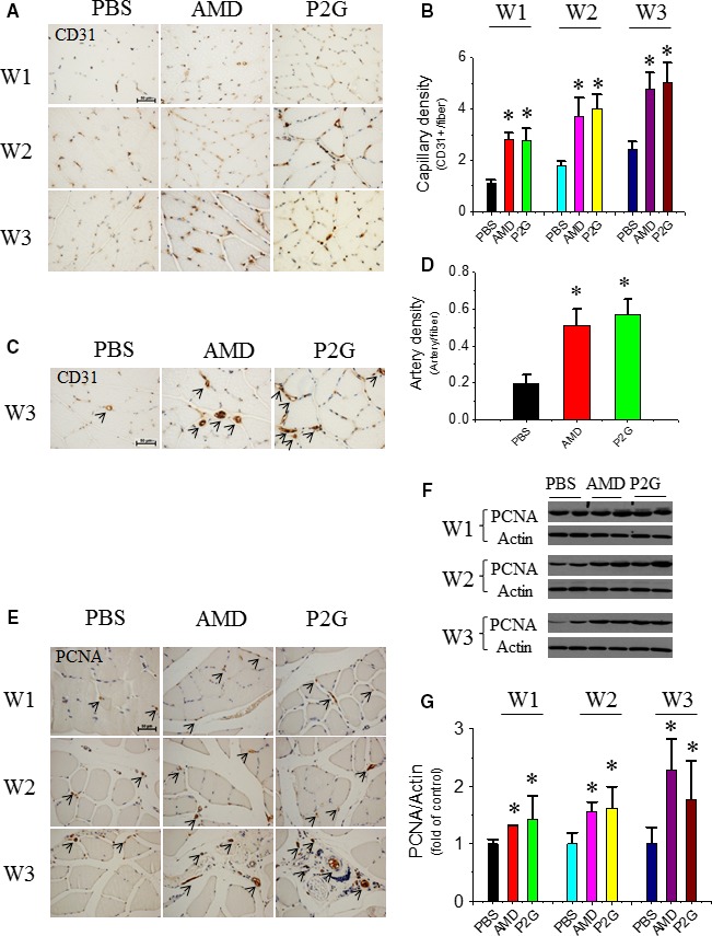
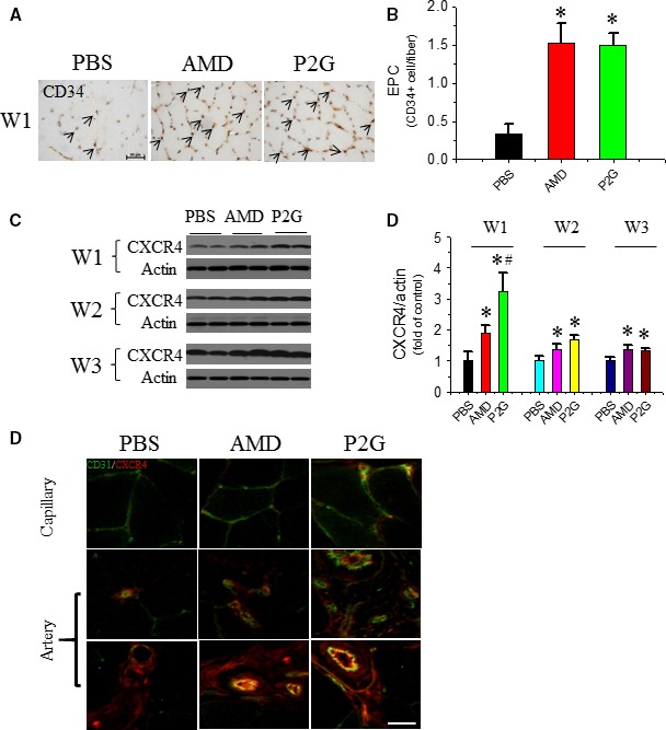
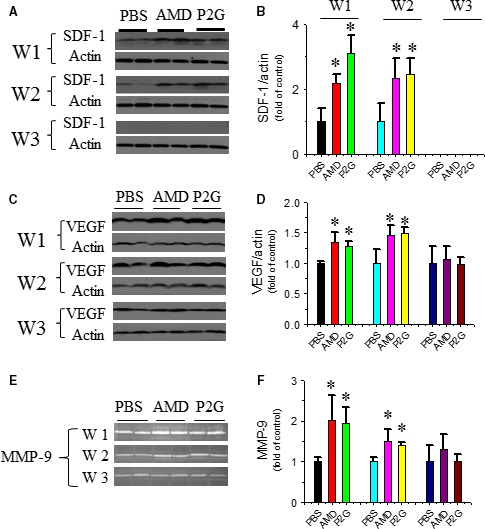
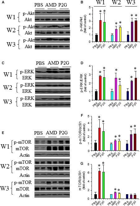
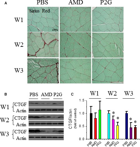
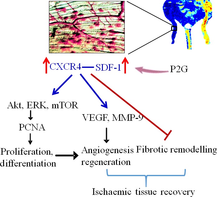
Endothelial progenitor cells (EPCs) play a capital role in angiogenesis via directly participating in neo-vessel formation and secreting pro-angiogenic factors. Stromal cell-derived factor 1 (SDF-1) and its receptor CXCR4 play a critical role in the retention and quiescence of EPCs within its niche in the bone marrow. Disturbing the interaction between SDF-1 and CXCR4 is an effective strategy for EPC mobilization. We developed a novel CXCR4 antagonist P2G, a mutant protein of SDF-1β with high antagonistic activity against CXCR4 and high potency in enhancing ischaemic angiogenesis and blood perfusion. However, its direct effects on ischaemic tissue remain largely unknown. In this study, P2G was found to possess a robust capability to promote EPC infiltration and incorporation in neo-vessels, enhance the expression and function of pro-angiogenic factors, such as SDF-1, vascular endothelial growth factor and matrix metalloprotein-9, and activate cell signals involved in angiogenesis, such as proliferating cell nuclear antigen, protein kinase B (Akt), extracellular regulated protein kinases and mammalian target of rapamycin, in ischaemic tissue. Moreover, P2G can attenuate fibrotic remodelling to facilitate the recovery of ischaemic tissue. The capability of P2G in direct augmenting ischaemic environment for angiogenesis suggests that it is a potential candidate for the therapy of ischaemia diseases.

Keywords: AMD3100; P2G; angiogenesis; endothelial progenitor cells.


Figures

Figure 1
Figure 1
P2G enhances neo‐vessel formation in ischaemic gastrocnemius muscles. Representative microscopic photographs of capillaries (brown dots) identified by CD31 staining were provided (A), and the quantitative summaries of capillary density were expressed as capillary number per muscle fibre at the 1st (W1), 2nd (W2) and 3rd (W3) week after surgery (B); representative microscopic photographs of small blood vessels identified by CD31 staining were provided (C), and the quantitative summaries of small blood vessel density were expressed as vessel number per muscle fibre at W3 (D). Cell proliferation indicator PCNA was evaluated by immunohistochemical staining (E) and further confirmed by Western blot (F, G). = 5 for PBS, 4 for AMD and 8 for P2G group, respectively. *< 0.05 versus PBS, Bar = 50 μm. PBS: Phosphate‐buffered saline; AMD: AMD3100.
Figure 2
Figure 2
P2G enhances endothelial progenitor cell (EPC) infiltrating in ischaemic gastrocnemius muscles. At the indicated time‐points after surgery, EPC localization in the ischaemic gastrocnemius muscle tissue was identified by a EPC marker CD34 by immunohistochemical staining (A), and the quantitative analysis of EPC density was expressed as capillary number per muscle fibre at W1 (B); The expression of another EPC marker CXCR4 (C) in the ischaemic gastrocnemius muscle tissue was detected by Western blot assay, followed by quantitative analyses of the optical density relative to the loading control actin (D). The localization of CXCR4 +  EPC in capillary and small blood vessels was determined by co‐immunofluorescent staining of both CD31 and CXCR4 (E). = 5 for PBS, 4 for AMD and 8 for P2G group respectively. *< 0.05 versus PBS, # < 0.05 versus AMD3100, Bar = 50 μm.
Figure 3
Figure 3
P2G up‐regulates pro‐angiogenic factors in ischaemic gastrocnemius muscles. The expression of SDF‐1 (A, B) and VEGF (C, D) was detected by Western blot, and the activity of MMP‐9 was detected by gelatin zymography (E, F). = 5 for PBS, 4 for AMD and 8 for P2G group, respectively. *< 0.05 versus PBS.
Figure 4
Figure 4
P2G enhances pro‐angiogenic signal activation in ischaemic gastrocnemius muscles. The expression and phosphorylation of Akt (A, B), ERK (C, D) and mTOR (E, F, G) were assayed by Western blot and quantified by optical density relative to the loading control actin and/or the total target protein, respectively. = 5 for PBS, 4 for AMD and 8 for P2G group, respectively. *< 0.05 versus PBS.
Figure 5
Figure 5
P2G attenuates fibrotic remodelling in ischaemic gastrocnemius muscles. The accumulation of collagen in ischaemic gastrocnemius muscles was assayed by Sirius‐red staining (A). The expression of fibrotic marker connective tissue growth factor (CTGF) in ischaemic gastrocnemius muscle was assayed by Western blot (B) and quantified by optical density relative to the loading control actin (C). = 5 for PBS, 4 for AMD and 8 for P2G group, respectively. *< 0.05 versus PBS.
Figure 6
Figure 6
Schematic diagram of P2G promoting ischaemic angiogenesis. P2G treatment enhances SDF‐1 production and CXCR4 expression in the ischaemic tissue. The interaction of SDF‐1 and CXCR4 up‐regulates VEGF expression and MMP‐9 activity, which promotes angiogenesis and tissue regeneration. Moreover, the interaction between SDF‐1 and CXCR4 also activates Akt, ERK and mTOR pathway, then up‐regulates PCNA expression and further enhances cell proliferation and differentiation, which contribute to angiogenesis and tissue regeneration, resulting in ordered remodelling. [Colour figure can be viewed at wileyonlinelibrary.com]

References

    1. Asahara T, Murohara T, Sullivan A, et al Isolation of putative progenitor endothelial cells for angiogenesis. Science. 1997; 275: 964–7. - PubMed
    1. Yoder MC. Human endothelial progenitor cells. Cold Spring Harb Perspect Med. 2012; 2: 14. - PMC - PubMed
    1. Zhao YD, Courtman DW, Deng Y, et al Rescue of monocrotaline‐induced pulmonary arterial hypertension using bone marrow‐derived endothelial‐like progenitor cells: efficacy of combined cell and eNOS gene therapy in established disease. Circ Res. 2005; 96: 442. - PubMed
    1. Sainz J, Sata M. CXCR4, a key modulator of vascular progenitor cells. Arterioscl Throm Vas. 2007; 27: 263. - PubMed
    1. Shantsila E, Watson T, Lip GYH. Endothelial progenitor cells in cardiovascular disorders. J Am Coll Cardiol. 2007; 49: 741–52. - PubMed

Publication types

MeSH terms

LinkOut - more resources