

The role of digital breast tomosynthesis in the breast assessment clinic: a review
- PMID: 28374502
- PMCID: PMC5587657
- DOI: 10.1002/jmrs.230
The role of digital breast tomosynthesis in the breast assessment clinic: a review
Abstract
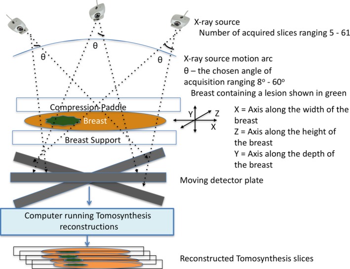
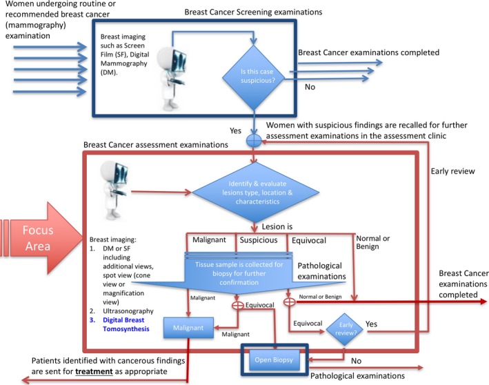
Mammography has long been considered as the primary technique in breast cancer detection and assessment. Despite low specificity, mammography has been preferred over other contemporary techniques such as magnetic resonance imaging (MRI), computed tomography (CT) and ultrasonography (US) due to superior sensitivity and significant health economic benefits. The development of a new technique, a limited angle cone beam pseudo-three-dimensional tomosynthesis, digital breast tomosynthesis (DBT), has gained momentum. Several preliminary studies and ongoing trials are showing evidence of the benefits of DBT in improving lesion visibility, accuracy of cancer detection and observer performance. This raises the possibility of adoption of DBT in the breast cancer assessment clinic, wherein confirming or dismissing the presence of malignancy (at the potential site identified during screening) is of utmost importance. Identification of suspected malignancy in terms of lesion characteristics and location is also essential in assessment. In this literature review, we evaluate the role of DBT for use in breast cancer assessment and its future in biopsy.
Keywords: DBT; Assessment; biopsy; breast cancer; diagnosis; digital breast tomosynthesis.
© 2017 The Authors. Journal of Medical Radiation Sciences published by John Wiley & Sons Australia, Ltd on behalf of Australian Society of Medical Imaging and Radiation Therapy and New Zealand Institute of Medical Radiation Technology.
Figures
References
-
- Kopans DB. Breast Imaging, 3rd edn. Lippincott Williams & Wilkins, Philadelphia, PA, 2007.
-
- Hologic Inc . The use of breast tomosynthesis in clinical practice. Applied Radiology, 2012. Available from: http://www.hologic.com/sites/default/files/white-papers/2012 Nov ‐ Applied Radiology Tomosynthesis Supplement ‐ FINAL.pdf.)
-
- Alakhras M, Bourne R, Rickard M, Ng KH, Pietrzyk M, Brennan PC. Digital tomosynthesis: A new future for breast imaging? Clin Radiol 2013; 68: e225–36. - PubMed
-
- Poplack SP, Tosteson TD, Kogel CA, Nagy HM. Digital breast tomosynthesis: Initial experience in 98 women with abnormal digital screening mammography. Am J Roentgenol 2007; 189: 616–23. - PubMed
-
- Michell MJ, Iqbal A, Wasan RK, et al. A comparison of the accuracy of film‐screen mammography, full‐field digital mammography, and digital breast tomosynthesis. Clin Radiol 2012; 67: 976–81. - PubMed
Publication types
MeSH terms
LinkOut - more resources
Full Text Sources
Other Literature Sources
Medical
