. 2017 Apr 4:8:14831.
doi: 10.1038/ncomms14831.

Mechano-regulated surface for manipulating liquid droplets

Affiliations

Mechano-regulated surface for manipulating liquid droplets

Xin Tang et al. Nat Commun. .

Abstract

The effective transfer of tiny liquid droplets is vital for a number of processes such as chemical and biological microassays. Inspired by the tarsi of meniscus-climbing insects, which can climb menisci by deforming the water/air interface, we developed a mechano-regulated surface consisting of a background mesh and a movable microfibre array with contrastive wettability. The adhesion of this mechano-regulated surface to liquid droplets can be reversibly switched through mechanical reconfiguration of the microfibre array. The adhesive force can be tuned by varying the number and surface chemistry of the microfibres. The in situ adhesion of the mechano-regulated surface can be used to manoeuvre micro-/nanolitre liquid droplets in a nearly loss-free manner. The mechano-regulated surface can be scaled up to handle multiple droplets in parallel. Our approach offers a miniaturized mechano-device with switchable adhesion for handling micro-/nanolitre droplets, either in air or in a fluid that is immiscible with the droplets.


Conflict of interest statement

The authors declare no competing financial interests.

Figures

Figure 1
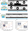
Figure 1. The design and principle of the MRS.
(a) Schematic illustration of the assembly of an MRS, which consists of a superhydrophobic mesh and an array of hydrophilic microfibres. (b) Optical micrographs showing the structure of a four-fibre MRS; a water droplet (stained blue) is locked onto the MRS. Scale bar, 300 μm. Scanning electron microscopy images of (c) a hydrophilic microfibre wrapped in a protective jacket, scale bar, 300 μm; (d) a superhydrophobic mesh coated with graphene nanoplatelets, scale bar, 30 μm; and (e) a typical magnified region in (d), scale bar, 500 nm. (f) A side-view schematic illustration of the mechanism of the MRS: the hydrophilic array (green) advances towards the superhydrophobic mesh (orange) (Capturing) until the array protrudes from the pores of the mesh, at which time the water droplet is pinned (Pinned); when the array is retracted through the mesh (Releasing), the surface returns to its non-wettable state, allowing the water droplet to roll (Released). (g) Schematic illustrations and optical micrographs showing the rolling and pinned states, respectively, of a water droplet on an MRS. Scale bar, 1 mm.
Figure 2
Figure 2. The adhesive performance of the MRS.
(a) The liquid capacity of an MRS increases as the number of regulating fibres increases. (b) The measured adhesive force of an MRS increases as the number of fibres increases. The red and orange lines represent the measured adhesion of MRSs with hydrophobic and hydrophilic fibres, respectively, based on ref. . The purple line represents the estimated adhesion of an MRS with hydrophilic fibres based on equation (2). The error bars indicate the standard deviations over five independent measurements. A series of optical microscopy images showing (c) the capillary bridges with receding contact lines on MRSs with numbers of hydrophobic fibres varying from 1 to 6 and (d) the thinning capillary bridges between the droplets and MRSs with numbers of hydrophilic fibres varying from 1 to 6. Scale bars, 200 μm.
Figure 3
Figure 3. The manipulation of aqueous droplets in air using an MRS.
Schematic illustrations and time-sequence images showing the capture and complete release of water droplets of (a) 10 μl and (b) 55 nl onto a superhydrophobic substrate in air. Scale bar, 1 mm. (c) The volume of the water droplet prepared using a hydrophilic-fibre MRS increases as the protrusion length of the fibres increases. The inset presents an optical micrograph showing a stained water column trapped by four hydrophilic fibres. Scale bar, 300 μm. The error bars indicate the standard deviations over five independent measurements. (d) The drop volume remains the same before and after transfer using a 4-hydrophilic-fibre MRS. The insets present optical microscopy images showing almost no water residue on the regulating fibres after the transfer process. Scale bar, 200 μm. The error bars indicate the standard deviations over five independent measurements.
Figure 4
Figure 4. The manipulation of oil droplets underwater using an MRS.
Schematic illustrations and time-sequence images showing (a) the underwater capture of a 5-μl droplet of DCE using an MRS with hydrophobic fibres and (b) the subsequent release of the DCE droplet underwater. Scale bar, 1 mm.
Figure 5
Figure 5. The durability of an MRS.
(a) A nine-cycle measurement of reversible switching between the adhesive and slippery states for a 4-hydrophilic-fibre MRS. For the sliding angle measurements, 5-μl water droplets were used. (b) The contact angle of a water droplet on an MRS remains constant for pH values varying from 1 to 14, demonstrating the excellent chemical resistance of the MRS. The error bars indicate the standard deviations over five independent measurements. The insets present the silhouettes of droplets with pH values of 1 and 14 on the superhydrophobic mesh.
Figure 6
Figure 6. MRS-assisted micro-reactors.
(a) Time-sequence images showing an MRS-based process using droplet-based micro-reactors for the generation of Fe(OH)3 precipitates. Scale bar, 3 mm. (b) Time-sequence images showing an MRS-based process using droplet-based micro-reactors for the synthesis of silver nanoparticles. Scale bars, 2 mm. Also presented in a transmission electron microscopy image showing the synthesized silver nanoparticles. Scale bar, 50 nm.
Figure 7
Figure 7. Manipulation of multiple droplets using a scaled-up MRS.
Schematic illustration and time-sequence images showing the in-parallel manipulation of multiple droplets using an MRS with a larger-scale fibre array. The scaled-up MRS successively captures three aqueous droplets (dyed red, magenta and blue) of different volumes and then releases them simultaneously. Scale bar, 3 mm.

References

    1. Dorvee J. R., Derfus A. M., Bhatia S. N. & Sailor M. J. Manipulation of liquid droplets using amphiphilic, magnetic one-dimensional photonic crystal chaperones. Nat. Mater. 3, 896–899 (2004). - PubMed
    1. Velev O. D., Prevo B. G. & Bhatt K. H. On-chip manipulation of free droplets. Nature 426, 515–516 (2003). - PubMed
    1. Aussillous P. & Quéré D. Liquid marbles. Nature 411, 924–927 (2001). - PubMed
    1. Chaudhury M. K. & Whitesides G. M. How to make water run uphill. Science 256, 1539–1541 (1992). - PubMed
    1. Cho S. K., Moon H. & Kim C.-J. Creating, transporting, cutting, and merging liquid droplets by electrowetting-based actuation for digital microfluidic circuits. J. Microelectromech. Syst. 12, 70–80 (2003).

Publication types

MeSH terms