. 2017 Jul 4;18(13):1305-1316.
doi: 10.1002/cbic.201700083. Epub 2017 May 11.

Sialylation Is Dispensable for Early Murine Embryonic Development in Vitro

Affiliations

Sialylation Is Dispensable for Early Murine Embryonic Development in Vitro

Markus Abeln et al. Chembiochem. .

Abstract

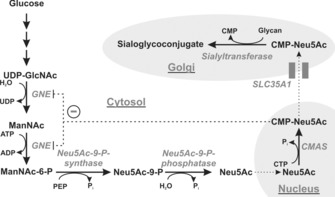
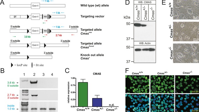
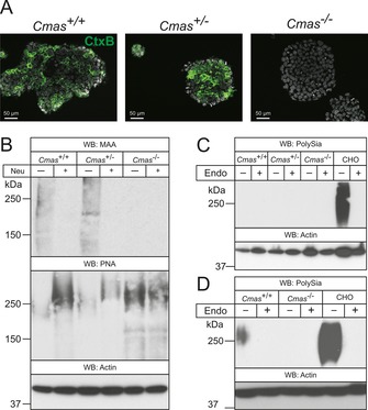
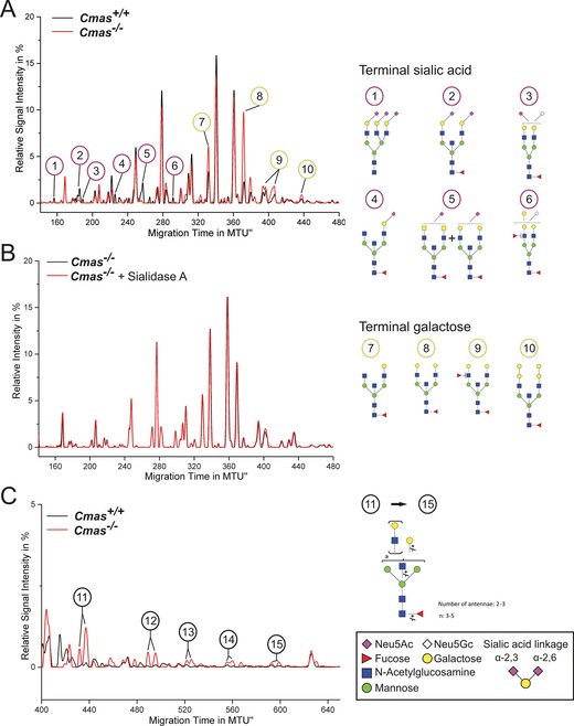
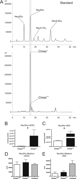
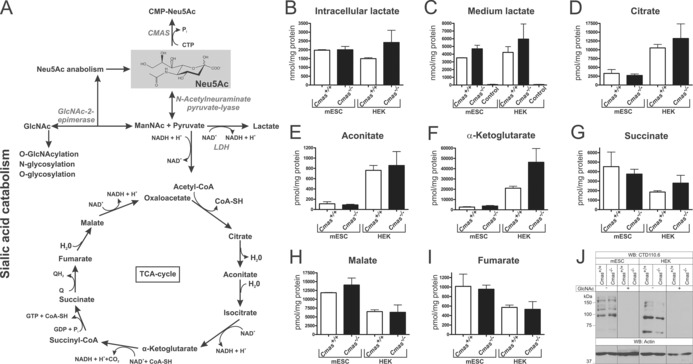
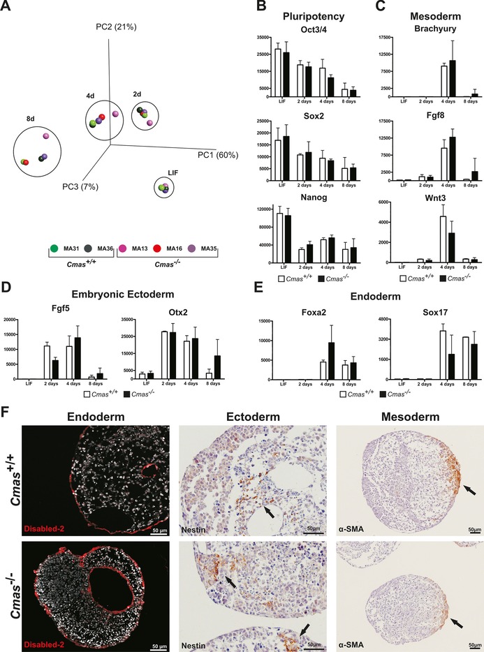
The negatively charged nonulose sialic acid (Sia) is essential for murine development in vivo. In order to elucidate the impact of sialylation on differentiation processes in the absence of maternal influences, we generated mouse embryonic stem cell (mESC) lines that lack CMP-Sia synthetase (CMAS) and thereby the ability to activate Sia to CMP-Sia. Loss of CMAS activity resulted in an asialo cell surface accompanied by an increase in glycoconjugates with terminal galactosyl and oligo-LacNAc residues, as well as intracellular accumulation of free Sia. Remarkably, these changes did not impact intracellular metabolites or the morphology and transcriptome of pluripotent mESC lines. Moreover, the capacity of Cmas-/- mESCs for undirected differentiation into embryoid bodies, germ layer formation and even the generation of beating cardiomyocytes provides first and conclusive evidence that pluripotency and differentiation of mESC in vitro can proceed in the absence of (poly)sialoglycans.

Keywords: CMP-sialic acid synthase; differentiation; glycosylation; metabolism; sialic acids.


Figures

Scheme 1
Scheme 1
Biosynthesis of sialoglycoconjugates in vertebrates. Enzymes are italicised: UDP‐GlcNAc 2‐epimerase/ManNAc kinase (GNE), CMP‐sialic acid synthetase (CMAS), CMP‐sialic acid transporter (SLC35A1). Cell organelles are shaded in grey. CMP‐Neu5Ac is a feedback inhibitor of GNE (−).
Figure 1
Figure 1
Targeting strategy and characterisation of CMAS‐deficient mESC. A) Cmas targeting strategy. Targeting vector with diphtheria toxin cassette (DT) to increase homologous recombination, frt‐flanked neomycin resistance cassette (neo), exon 4 and neo flanked by LoxP sites. Correct homologous integration of the targeting vector into ES cells was confirmed by the neo and a 5′ outside primer pair (green arrows; amplifying a 3.8 kb fragment) and by neo and a 3′ outside primer pair (red arrows; amplifying a 2.7 kb fragment). Inside primers were used as a control (light blue arrows; 1 kb fragment). Correctly targeted mESCs were used to generate mutant mice. The neo cassette was deleted by inter‐crosses with ACTFLPe mice, and exon 4 and the remaining frt site were deleted by crosses with Zp3‐cre mice, thereby resulting in the Cmas knock‐out allele. B) PCR analysis of homologous integration. A PCR product of 3.8 kb was amplified with neo‐ and 5′ outside primers (green in A), and a 2.7 kb product was amplified with neo‐ and 3′ outside primers (red in A). Both fragments occurred correctly only in the targeted Cmas neo mESC (lane 2), not in wild‐type mESC (lane 3) or in wild‐type mouse tail tissue (lane 4). A 1 kb PCR fragment from inside primers served as control for the PCR reaction (blue in A). 1 kb marker in lane 1. C) Quantitative PCR of Cmas expression from feeder‐free cultures of Cmas +/+, Cmas +/− and Cmas −/− mESCs (n=3 of one representative cell line with the respective genotype). n.d.=not detectable. D) Cell lysates of Cmas +/+, Cmas +/− and Cmas −/− mESC were separated by SDS‐PAGE, blotted and immunostained with anti‐CMAS antibody. The 48 kDa CMAS protein was detected in Cmas +/+ and Cmas +/− lysates but not in Cmas −/−. Anti‐actin staining was used as loading control. E) Morphology of Cmas +/+, Cmas +/− and Cmas −/− mESC cultured on MEFs supplemented with LIF. F) Indirect immunofluorescence staining of Oct3/4 in Cmas +/+, Cmas +/− and Cmas −/− mESC cultured feeder‐free with LIF supplementation. Representative results from one cell line per genotype are shown in D)–F).
Figure 2
Figure 2
Cmas −/− mESC lack Sia on gangliosides and proteins. A) Direct immunofluorescence staining of the ganglioside GM1 with FITC‐conjugated CTXB in undifferentiated mESC lines. B) Whole‐cell lysates of Cmas +/+, Cmas +/− and Cmas −/− mESC were analysed before and after neuraminidase (Neu) treatment by SDS‐PAGE and western blotting. Detection of α2,3‐linked sialic acids was performed with Maackia amurensis agglutinin (MAA); galactose‐capped glycans were stained with peanut agglutinin (PNA). Cell lysates of C) undifferentiated Cmas +/+, Cmas +/− and Cmas −/− mESCs, and D) differentiated WT and CMAS‐depleted mESC were analysed with or without prior endoneuraminidase NF treatment (Endo) by SDS‐PAGE and western blotting. Detection of polysialic acid was performed with mAb 735. Anti‐actin staining was used as loading control. In order to maintain the pluripotent state, mESC lines were cultured feeder‐free with LIF supplementation. For undirected monolayer differentiation, mESC lines were cultivated for eleven days without LIF supplementation. Representative results from one cell‐line per genotype are shown.
Figure 3
Figure 3
Loss of CMAS activity in mESCs entails increased exposure of galactose and oligo‐LacNAc residues at the cell surface. The y‐axis of normalised electropherogram is divided by the summed peak height of all quantifiable peaks (S/N≥9); relative signal intensity [%] of total peak height is plotted. A) and C) Differential xCGE‐LIF analysis of N‐glycans from undifferentiated Cmas +/+ and Cmas −/− mESCs: electropherogram regions from A) 140 to 480 normalised migration time units (MTU) and C) 400 to 650 MTU. B) Electropherogram of undifferentiated Cmas −/− mESCs before (black) and after sialidase treatment (red). N‐Glycan structures in xCGE‐LIF analyses are annotated: sialylated N‐glycans (1–6), galactose capped N‐ glycans (7–10) and oligo‐LAcNAc capped N‐glycans (11–15). Example N‐glycan structures 1 to 15 are depicted (right; detailed N‐glycan annotation in Figure S4). Activity and specificity of A. urefaciens sialidase was confirmed by treatment of bovine fetuin and subsequent xCGE‐LIF analysis of the well‐defined N‐glycans (Figure S3 A). Structures are presented following the Consortium for Functional Glycomics notation (www.functionalglycomics.org/glycomics/molecule/jsp/carbohydrate/carbMoleculeHome.jsp). Linkage positions of sialic acids are indicated by differing angles. All mESC lines were cultured feeder‐free with LIF supplementation to maintain the pluripotent state (Cmas +/+ n=3, Cmas +/− n=5, Cmas −/− n=4). Representative results from one cell line per genotype are shown.
Figure 4
Figure 4
Cmas −/− mESC accumulate intracellular Neu5Ac. A) Reversed‐phase HPLC elution profile of the DMB‐labelled Sia derivatives. Sia standards (top) and samples obtained from intracellular metabolite extracts (bottom) of Cmas +/+ (‐ ‐ ‐ ‐) and Cmas −/− (—) mESCs. B) and C) Quantification of intracellular Neu5Ac by integration of peak areas from RP‐HPLC analysis of B) Cmas +/+ and Cmas −/− mESCs and C) CMAS +/+ and CMAS −/− HEK293 cells (peak area per mg protein; Student's t‐test * p<0.05, n=3 with three technical replicates per biological replicate). Generation and biochemical characterisation of CMAS −/− HEK293 cells in Figure S4. Quantification of free Neu5Ac in the cell culture supernatant of WT and CMAS‐deficient D) mESCs and E) HEK cells by integration of peak areas of DMB‐derivatised Sia from RP‐HPLC analyses (Student's t‐test * p<0.05, n=2). Cell‐free medium was used as control. All mESC lines were cultured feeder‐free with LIF supplementation to maintain the pluripotent state.
Figure 5
Figure 5
Increased levels of Neu5Ac affect neither related metabolite levels nor global O‐GlcNAcylation. A) Sialic acid anabolism and catabolism in mammals. Enzymes are italicised: CMP‐Sia synthetase (CMAS), lactate dehydrogenase (LDH). Quantification of B) intracellular and C) cell‐culture lactate levels from WT and CMAS‐deficient mESC and HEK metabolite extracts by HPLC‐MS/MS (nmol per mg protein). D–I) Quantification of intracellular TCA cycle metabolite levels from WT and CMAS‐depleted mESC and HEK metabolite extracts by HPLC‐MS/MS (pmol per mg protein). J) Analysis of global O‐GlcNAcylation. Whole‐cell lysates of WT and CMAS‐deficient mESC and HEK cells were analysed by SDS‐PAGE and western blotting with mAb CTD110.6. Specificity of antibody staining was controlled by preincubation of the primary antibody with N‐Acetylglucosamine (GlcNAc). Anti‐actin staining was used as loading control. All mESC lines were cultured feeder‐free with LIF supplementation to maintain the pluripotent state (Cmas +/+ mESC n=2, Cmas −/− mESC n=3, for all HEK cell lines n=3).
Figure 6
Figure 6
Loss of sialylation does not affect primary germ layer formation of mESCs in vitro. A) Principal component analysis of transcriptomic data from undifferentiated (LIF) mESC and after two, four and eight days of EB differentiation of Cmas +/+ (green, n=2) and Cmas −/− (red and purple, n=3) mESC lines. B)–E) mRNA expression quantified by normalised fluorescence intensity of B) pluripotency factors Oct3/4, Sox2 and Nanog; C) mesodermal markers Brachyury, Fgf8 and Wnt3; D) embryonic ectodermal markers Fgf5 and Otx2; and E) endodermal markers Foxa2 and Sox17 obtained from transcriptome array data. F) Indirect immunofluorescence and immuno‐histochemical analysis of sections from paraffin‐embedded Cmas +/+ and Cmas −/− EBs after eight days of differentiation. The capacity to form primary germ layers was detected by staining for the marker proteins Disabled‐2, Nestin and α‐smooth muscle actin (α‐SMA).

References

    1. Ioffe E., Stanley P., Proc. Natl. Acad. Sci. USA 1994, 91, 728–732; - PMC - PubMed
    2. Xia L., Ju T., Westmuckett A., An G., Ivanciu L., McDaniel J. M., Lupu F., Cummings R. D., McEver R. P., J. Cell Biol. 2004, 164, 451–459. - PMC - PubMed
    1. Kornfeld S., Kornfeld R., Neufeld E. F., O'Brien P. J., Proc. Natl. Acad. Sci. USA 1964, 52, 371–379. - PMC - PubMed
    1. Zhuo Y., Bellis S. L., J. Biol. Chem. 2011, 286, 5935–5941. - PMC - PubMed
    1. Weinhold B., Sellmeier M., Schaper W., Blume L., Philippens B., Kats E., Bernard U., Galuska S. P., Geyer H., Geyer R., Worthmann K., Schiffer M., Groos S., Gerardy-Schahn R., Münster-Kühnel A. K., J. Am. Soc. Nephrol. 2012, 23, 1319–1328. - PMC - PubMed
    1. Celeste F. V., Vilboux T., Ciccone C., de Dios J. K., Malicdan M. C., Leoyklang P., McKew J. C., Gahl W. A., Carrillo-Carrasco N., Huizing M., Hum. Mutat. 2014, 35, 915–926. - PMC - PubMed

Publication types

MeSH terms