. 2017 Apr 3;12(4):e1311437.
doi: 10.1080/15592324.2017.1311437.

Take a look at plant DNA replication: Recent insights and new questions

Affiliations

Take a look at plant DNA replication: Recent insights and new questions

Savannah D Savadel et al. Plant Signal Behav. .

Abstract

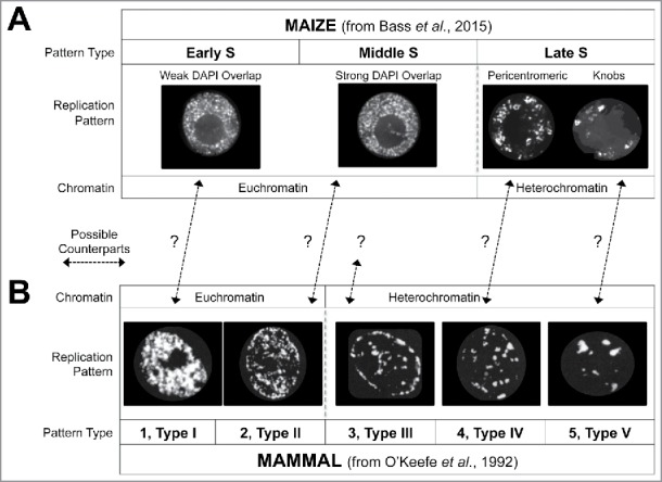
Recent advances in replicative DNA labeling technology have allowed new ways to study DNA replication in living plants. Temporal and spatial aspects of DNA replication programs are believed to derive from genomic structure and function. Bass et al. (2015) recently visualized DNA synthesis using 3D microscopy of nuclei at three sub-stages of S phase: early, middle and late. This addendum expands on that study by comparing plant and animal DNA replication patterns, by considering implications of the two-compartment model of euchromatin, and by exploring the meaning of the DNA labeling signals inside the nucleolus. Finally, we invite the public to explore and utilize 300 image data sets through OMERO, a teaching and research web resource for visualization, management, or analysis of microscopic data.

Keywords: 3D microscopy; Chromatin; DNA synthesis; maize; omero; rDNA.


Figures

Figure 1.
Figure 1.
Cross-kingdom comparison of temporal patterns of DNA replication. (A) Maize nuclei from early, middle, or late S-phase, showing representative examples from maize EdU-labeled root-tip nuclei (from Bass et al.6). (B) Chinese Hamster Ovary nuclei from 5 sequential stages of S-phase, showing BrdU-labeled cell culture nuclei (from Fig. 4 of O'Keefe et al., 199210). The replication pattern types are labeled as “1–5” (O'Keefe et al.10) and also “type I-V” (From Zink 20061). The question marks reflect untested counterpart assignments.
Figure 2.
Figure 2.
Predicted chromatin features based on the mini-domain chromatin fiber replication timing model. The maize interphase nucleoplasm was observed to be partitioned into 2 types of euchromatin, early-S or middle-S. (A) DAPI image of a maize nucleus at interphase showing a large nucleolus (n) and thick, ∼300 nm chromatin fibers (dashed box). (B) Diagram of the model showing the 2 types of euchromatin, MID-S (thick/black) and EARLY-S (thin/gray). (C) Table of chromatin features that might show differential assortment into the 2 types of chromatin on the basis of known or reported genomic features and epigenomic marks (as reviewed for example by Fuchs and Schubert17).
Figure 3.
Figure 3.
Replicative labeling signals can be seen inside the nucleoli as discrete punctate foci observed in early and middle, but not late S-phase nuclei. (A) Examples of EdU-labeled DNA replication from Early-S or Middle-S nuclei showing intra-nucleolar labeling (white dashed boxes). (B) Diagram of maize chromosome 6 showing the NOR with copy number estimate from Rivin et al. (C) Enlarged diagram of 3 rDNA repeats. (D) Enlarged diagram of one ∼10kb rDNA repeat showing the 200 bp sub-repeats (thin hatch lines, as per McMullen et al.16) and the major transcription start site (TSS). (E) Table relating real-space dimensions of DNA as a function of packaging, including Watson-Crick B-form DNA (Linear DNA), “beads-on-a-string” chromatin (10 nm fiber), and one of the basic higher order chromatin fibers (30 nm fiber).
Figure 4.
Figure 4.
OMERO Image Database Example. (A) Screenshot of the file browser showing 3 partitions: DATA FOLDERS (Left) listing projects, data sets, and image files; IMAGE THUMBNAILS (Middle) displaying individual data sets; and SINGLE FILE METADATA (Right) displaying metadata and file specs auto-extracted during upload or user-added annotations such as tagging, value inputs, comments, or attachments. (B) Screenshot of the split channel view for a single nucleus. The image data display window and display settings control panel is shown. Basic controls allow for viewing as normal (single Z-sections), max intensity (through-focus projection), or split view (shown here). Display settings give user control over wavelength colors, brightness and contrast, scale bars, and image link for URL sharing.

Erratum for

  • Addendum to: Bass HW, Hoffman GG, Lee TJ, Wear EE, Joseph SR, Allen GC, Hanley-Bowdoin L, Thompson WF. Defining multiple, distinct, and shared spatiotemporal patterns of DNA replication and endoreduplication from 3D image analysis of developing maize (Zea mays L.) root tip nuclei. Plant Mol Biol 2015; 89:339-51; http://dx.doi.org/10.1007/s11103-015-0364-4.

References

    1. Zink D. The temporal program of DNA replication: new insights into old questions. Chromosoma 2006; 115:273-87; PMID:16552593; http://dx.doi.org/10.1007/s00412-006-0062-8 - DOI - PubMed
    1. Gilbert DM, Takebayashi SI, Ryba T, Lu J, Pope BD, Wilson KA, Hiratani I. Space and time in the nucleus: developmental control of replication timing and chromosome architecture. Cold Spring Harb Symp Quant Biol 2010; 75:143-53; PMID:21139067; http://dx.doi.org/10.1101/sqb.2010.75.011 - DOI - PubMed
    1. Jackson D, Wang X, Rudner DZ. Spatio-temporal organization of replication in bacteria and eukaryotes (nucleoids and nuclei). Cold Spring Harb Perspect Biol 2012; 4:a010389; PMID:22855726 - PMC - PubMed
    1. Dileep V, Rivera-Mulia JC, Sima J, Gilbert DM. Large-scale chromatin structure-function relationships during the cell cycle and development: insights from replication timing. Cold Spring Harb Symp Quant Biol 2015; 80:53-63; PMID:26590169; http://dx.doi.org/10.1101/sqb.2015.80.027284 - DOI - PubMed
    1. Bass HW, Wear EE, Lee TJ, Hoffman GG, Gumber HK, Allen GC, Thompson WF, Hanley-Bowdoin L. A maize root tip system to study DNA replication programmes in somatic and endocycling nuclei during plant development. J Exp Bot 2014; 65:2747-56; PMID:24449386; http://dx.doi.org/10.1093/jxb/ert470 - DOI - PubMed

Publication types

LinkOut - more resources