. 2017 May 1;127(5):1714-1724.
doi: 10.1172/JCI84598. Epub 2017 Apr 4.

Megakaryocytes compensate for Kit insufficiency in murine arthritis

Megakaryocytes compensate for Kit insufficiency in murine arthritis

Pierre Cunin et al. J Clin Invest. .

Abstract

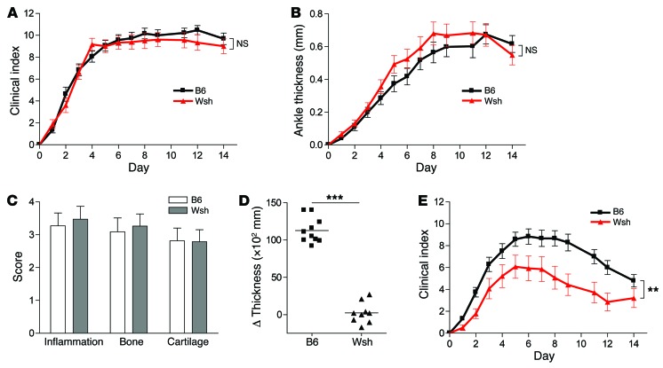
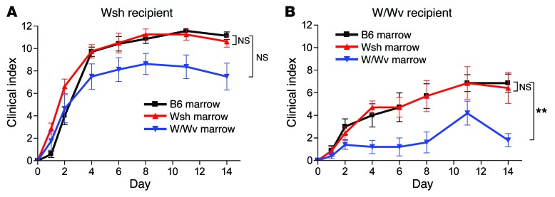
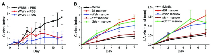
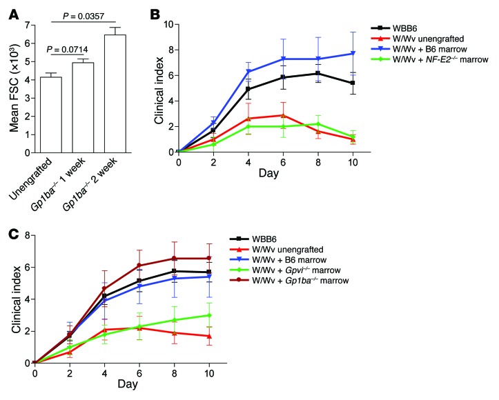
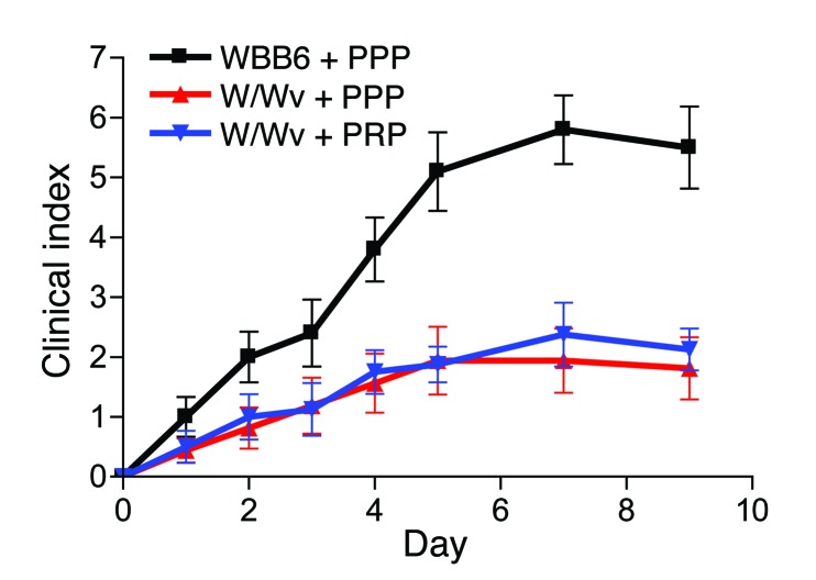
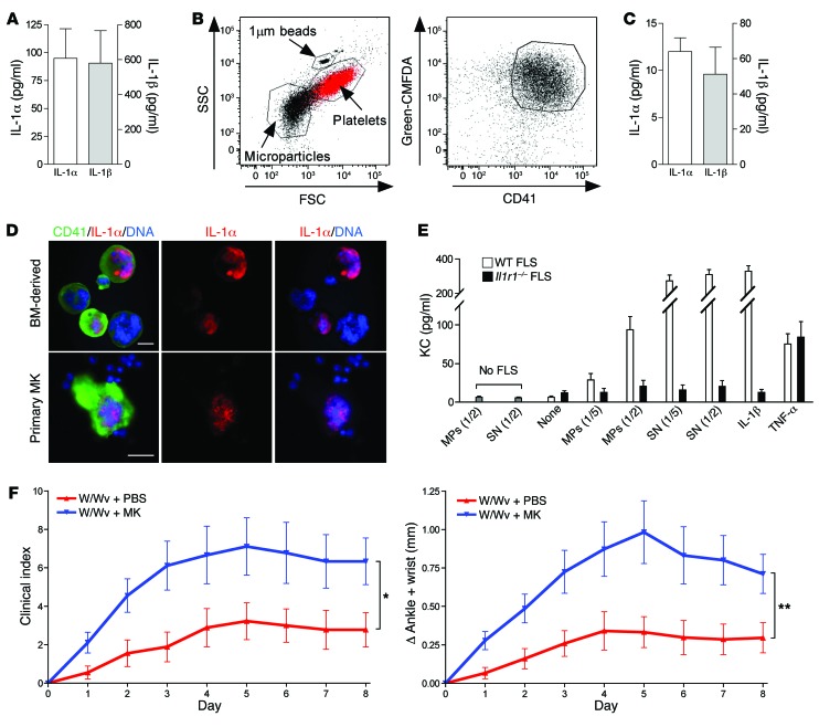
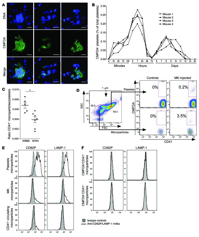
The growth factor receptor Kit is involved in hematopoietic and nonhematopoietic development. Mice bearing Kit defects lack mast cells; however, strains bearing different Kit alleles exhibit diverse phenotypes. Herein, we investigated factors underlying differential sensitivity to IgG-mediated arthritis in 2 mast cell-deficient murine lines: KitWsh/Wsh, which develops robust arthritis, and KitW/Wv, which does not. Reciprocal bone marrow transplantation between KitW/Wv and KitWsh/Wsh mice revealed that arthritis resistance reflects a hematopoietic defect in addition to mast cell deficiency. In KitW/Wv mice, restoration of susceptibility to IgG-mediated arthritis was neutrophil independent but required IL-1 and the platelet/megakaryocyte markers NF-E2 and glycoprotein VI. In KitW/Wv mice, platelets were present in numbers similar to those in WT animals and functionally intact, and transfer of WT platelets did not restore arthritis susceptibility. These data implicated a platelet-independent role for the megakaryocyte, a Kit-dependent lineage that is selectively deficient in KitW/Wv mice. Megakaryocytes secreted IL-1 directly and as a component of circulating microparticles, which activated synovial fibroblasts in an IL-1-dependent manner. Transfer of WT but not IL-1-deficient megakaryocytes restored arthritis susceptibility to KitW/Wv mice. These findings identify functional redundancy among Kit-dependent hematopoietic lineages and establish an unanticipated capacity of megakaryocytes to mediate IL-1-driven systemic inflammatory disease.


Conflict of interest statement

Conflict of interest: J.N. Thon and J.E. Italiano are founders of Platelet BioGenesis. M.M. Chen is an employee of Takeda Pharmaceuticals. M.F. Gurish has served as a consultant for Karos Pharmaceuticals. P.A. Nigrovic receives salary support as a member of the steering committee of the Childhood Arthritis and Rheumatology Research Alliance, authorship royalties from UpToDate Inc. and the American Academy of Pediatrics, and holds investigator-initiated grants from Novartis and Sobi.

Figures

Figure 1
Figure 1. Dose-dependent susceptibility of Wsh mice to K/BxN serum transfer arthritis.
(AD) Male B6 (n = 19) and Wsh (n = 18) mice were treated with 150 μl i.p. K/BxN serum on days 0 and 2 and assessed for arthritis over 2 weeks. Results were pooled from 4 identical experiments using a single pooled batch of K/BxN serum. (A) Clinical scoring on a 0 to 12 scale (P = NS). (B) Ankle thickness change (P = NS). (C) Histological scoring on ankle sections (19 to 22 ankles/group, all comparisons P = NS). (D) Acute increase in ankle and wrist thickness (flare) assessed 30 minutes after injection of K/BxN serum (assessed in n = 10/group, P < 0.0001). (E) 50 μl i.p. K/BxN serum on days 0 and 2 (B6, n = 24; Wsh, n = 14 pooled from 4 experiments; P = 0.0088). **P < 0.01, ***P < 0.001.
Figure 2
Figure 2. Mixed hematopoietic and radioresistant contributions to differential arthritis susceptibility in Wsh and W/Wv mice.
(A and B) Wsh and W/Wv mice were lethally irradiated and engrafted for 10 weeks with whole donor marrow from W/Wv, Wsh, or B6 donors, followed by initiation of arthritis with 150 μl i.p. K/BxN serum on days 0 and 2 (n = 5–8 mice/group pooled from 2 experiments; W/Wv arms, P = 0.0096). B6 recipient data are in Supplemental Figure 4C. **P < 0.01. (A) B6 vs. Wsh: P = NS; B6 vs. W/Wv: P =NS; Wsh vs W/Wv: P =0.0410. (B) B6 vs. Wsh: P =NS; B6 vs. W/Wv: P =0.0096; Wsh vs W/Wv: P =0.0155.
Figure 3
Figure 3. A nonneutrophil IL-1–expressing marrow lineage bypasses the requirement for MC in W/Wv mice.
(A) W/Wv mice were injected i.v. with purified B6 bone marrow neutrophils (2 donors/d on days 0–4) or saline control as well as 150 μl i.p. K/BxN serum on days 0 and 2 (n = 5/group; 1 of 2 similar experiments shown). (B) W/Wv mice received whole B6, Wsh, Gfi1–/–, or Il1–/– bone marrow by tail vein (1 donor per 2 recipients), and arthritis was initiated 14 days later as above. Left, clinical scoring (mock engrafted vs. B6 engrafted, P = 0.0001; mock engrafted vs. Wsh engrafted, P = 0.0117; mock engrafted vs. Gfi1–/– engrafted, P = 0.004; mock engrafted vs. Il1–/– engrafted, P = NS). Right, ankle and wrist thickness change (mock engrafted vs. B6 engrafted, P = 0.012; mock engrafted vs. Wsh engrafted, P = 0.03; mock engrafted vs. Gfi1–/– engrafted, P = 0.038; mock engrafted vs. Il1–/– engrafted, P = NS). Mock engrafted, n = 20; B6 engrafted, n = 16; Wsh engrafted, n = 15; Gfi1–/– engrafted, n = 10; Il1–/– engrafted, n = 16; pool of 2–3 experiments.
Figure 4
Figure 4. The platelet/MK lineage mediates arthritis restoration in W/Wv mice.
(A) W/Wv mice were engrafted with whole Gp1ba–/– bone marrow (1 donor per 2 recipients) and platelet size (forward scatter [FSC]) monitored by flow cytometry at 1 and 2 weeks (n = 3 unengrafted and 5 recipient mice). (B and C) Whole bone marrow was transferred i.v. in W/Wv mice (1 donor per 2 recipients) and arthritis initiated with 150 μl K/BxN serum on days 0 and 2. (B) Clinical score after transfer of NF-E2–/– and control B6 marrow. W/Wv vs. NF-E2–/– engrafted, P = NS; W/Wv vs. B6 engrafted, P = 0.006. n = 8 W/Wv; n = 5 W/Wv + NF-E2–/– (1 fatality); n = 7 W/Wv + B6 (1 failed engraftment); n = 13 WBB6; pooled from 2 experiments. (C) Clinical score after transfer of Gpvi–/–, Gp1ba–/–, and control B6 marrow. W/Wv vs. Gpvi–/– engrafted, P = NS; W/Wv vs. B6, P = 0.026; W/Wv vs. Gp1ba–/– engrafted, P = 0.001. n = 10 W/Wv; n = 10 W/Wv + B6; n = 10 W/Wv + Gpvi–/–; n = 9 W/Wv + Gp1ba–/–; n = 20 WBB6 pooled from 2 experiments.
Figure 5
Figure 5. Platelet transfer does not restore arthritis susceptibility in W/Wv mice.
Platelet-Rich Plasma (PRP) and Platelet-Poor Plasma (PPP) were purified from B6 donors and transferred into recipient mice i.v. (5 donors/recipient/d on days 0–4) along with 150 μl i.p. K/BxN serum on days 0 and 2. PRP vs. PPP W/Wv, P = NS. n = 10 PPP-W/Wv, n = 8 PRP-W/Wv, n = 16 WBB6; pooled from 2 experiments.
Figure 6
Figure 6. MKs elaborate IL-1–containing microparticles and restore arthritis susceptibility in W/Wv mice.
(A) 1 × 10⁴ MKs from B6 bone marrow were incubated 18 hours to test IL-1 production. Histograms show IL-1α and IL-1β concentration in MK lysate. n = 5. (B) Microparticle production from B6 MKs stained with green-CMFDA. Left, microparticles are gated based on size of beads (less than 1 μm). Dot plot of circulating intact platelets (red) was overlaid as a control to confirm bead sizing. Right, dot plot shows CMFDA+CD41+ events among microparticles; representative of 5 experiments. SSC, side scatter. (C) IL-1α and IL-1β concentration in B6 MK microparticle lysates. n = 4. (D) BM-derived MK (upper photos) and flushed BM cells (lower photos) are stained for CD41 (green) and IL-1α (red). DNA is visualized with Draq5 (Blue). IL-1α is detected in the nucleus of a subset of MK. Scale bars: 20 μm. n = 2 per experiment. (E) KC production by WT and Il1r1–/– FLS stimulated by MK microparticles (MPs) or supernatant from MK cultures. IL-1β and TNF-α (10 ng/ml) were used as positive controls. n = 3. (F) W/Wv mice were injected i.v. with PBS or 2 × 10⁵ MKs. After 60 minutes, and again on day 2, mice were treated with 150 μl i.p. K/BxN serum. Left, clinical scoring on a 0–12 scale. W/Wv injected with PBS vs. MKs, P = 0.0282. Right, ankle and wrist thickness change. W/Wv injected PBS vs. MKs, P = 0.006. n = 9/group pooled from 2 experiments. *P < 0.05; **P < 0.01.
Figure 7
Figure 7. Injected MKs engraft in lung and produce microparticles.
(A, B, D, F) W/Wv mice were injected i.v. with 2 × 10⁵ MKs stained with CMFDA. (A) Detection of CMFDA+ MKs in lung section 75 minutes after the engraftment. DNA was visualized with Draq5. Scale bars: 10 μm. Representative of 4 experiments. (B) Detection of circulating CMFDA+CD41+ platelets over time. Graph shows the percentages of CMFDA+CD41+ platelets among total CD41+ circulating platelets. (C) Detection of CD41+ MPs ( < 1 μm) in unmanipulated WBB6 and W/Wv mice, normalized to total platelet number to control for dilutional artifact of cardiac aspiration directly into anticoagulant buffer. n = 5–7 mice/group pooled from 3 experiments. *P < 0.05. (D) MKs stained with CMFDA were injected in W/Wv mice as in parts A and B. Detection of CMFDA+CD41+ MPs and platelets in noninjected control mice or 3 hours after MK injection. (E) CD62P and LAMP-1 expression on microparticles from activated platelets (upper histograms), MK microparticles (middle histograms), and CD41+ circulating microparticles (lower histograms). Histograms are gated on CD41+ and particle size less than 1 μm events. Representative of 2 experiments. (F) CD62P and LAMP-1 expression on the surface of circulating newly formed CD41+CMFDA+ MPs (upper panels) and endogenous CD41+CMFDA MPs (lower panels) 3 hours after CMFDA+ MK injection.

References

    1. Ehrlich P. Beiträge zur Theorie und Praxis der histologischen Färbung [doctoral thesis]. Leipzig, Germany: Leipzig University; 1878.
    1. Wong GW, Zhuo L, Kimata K, Lam BK, Satoh N, Stevens RL. Ancient origin of mast cells. Biochem Biophys Res Commun. 2014;451(2):314–318. doi: 10.1016/j.bbrc.2014.07.124. - DOI - PMC - PubMed
    1. Katz HR, Austen KF. Mast cell deficiency, a game of kit and mouse. Immunity. 2011;35(5):668–670. doi: 10.1016/j.immuni.2011.11.004. - DOI - PubMed
    1. Galli SJ, Kitamura Y. Genetically mast-cell-deficient W/Wv and Sl/Sld mice. Their value for the analysis of the roles of mast cells in biologic responses in vivo. Am J Pathol. 1987;127(1):191–198. - PMC - PubMed
    1. Nigrovic PA, et al. Genetic inversion in mast cell-deficient (Wsh) mice interrupts corin and manifests as hematopoietic and cardiac aberrancy. Am J Pathol. 2008;173(6):1693–1701. doi: 10.2353/ajpath.2008.080407. - DOI - PMC - PubMed

MeSH terms

Substances