

An Assessment of Early Response to Targeted Therapy via Molecular Imaging: A Pilot Study of 3'-deoxy-3'[(18)F]-Fluorothymidine Positron Emission Tomography 18F-FLT PET/CT in Prostate Adenocarcinoma
- PMID: 28375169
- PMCID: PMC5489940
- DOI: 10.3390/diagnostics7020020
An Assessment of Early Response to Targeted Therapy via Molecular Imaging: A Pilot Study of 3'-deoxy-3'[(18)F]-Fluorothymidine Positron Emission Tomography 18F-FLT PET/CT in Prostate Adenocarcinoma
Abstract
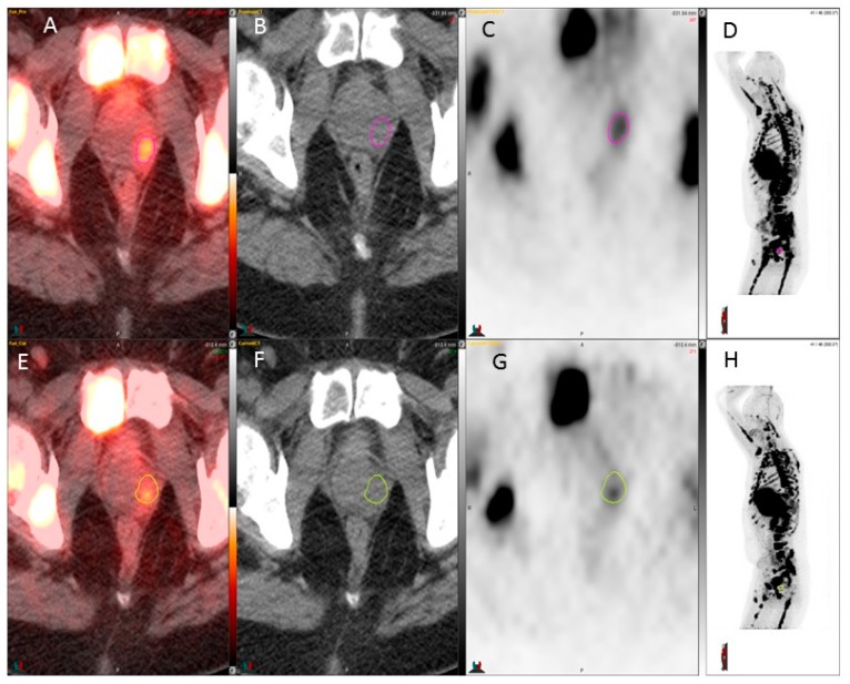
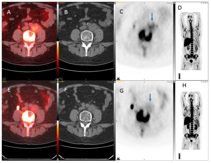
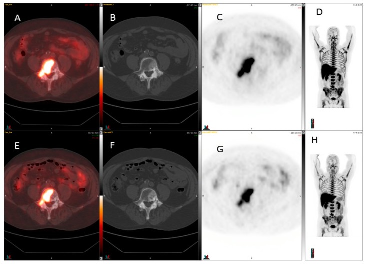
Fluorothymidine is a thymidine analog labeled with fluorine-18 fluorothymidine for positron emission tomography (18F-FLT-PET) imaging. Thymidine is a nucleic acid that is used to build DNA. Fluorine-18 fluorothymidine (18F-FLT) utilizes the same metabolic pathway as does thymidine but has a very low incidence of being incorporated into the DNA (<1%). 18F-FLT-PET could have a role in the evaluation of response to targeted therapy. We present here a pilot study where we investigated cellular metabolism and proliferation in patients with prostate cancer before and after targeted therapy. Seven patients with Stage IV prostate adenocarcinoma, candidates for targeted therapy inhibiting the hepatocyte growth factor/tyrosine-protein kinase Met (HGF/C-MET) pathway, were included in this study. The HGF/C-MET pathway is implicated in prostate cancer progression, and an evaluation of the inhibition of this pathway could be valuable. 18F-FLT was performed at baseline and within four weeks post-therapy. Tumor response was assessed semi-quantitatively and using visual response criteria. The range of SUVmax for 18F-FLT at baseline in the prostate varied from 2.5 to 4.2. This study demonstrated that 18F-FLT with positron emission tomography/computerized tomography (18F-FLT PET/CT) had only limited applications in the early response evaluation of prostate cancer. 18F-FLT PET/CT may have some utility in the assessment of response in lymph node disease. However, 18F-FLT PET/CT was not found to be useful in the evaluation of the prostate bed, metastatic skeletal disease, and liver disease.
Keywords: fluorine-18 fluorothymidine (18F-FLT); molecular imaging; positron emission tomography/computerized tomography (PET/CT); prostate cancer.
Conflict of interest statement
The authors declare no conflict of interest.
Figures
Similar articles
-
Molecular Imaging with 3'-deoxy-3'[(18)F]-Fluorothymidine (18F-FLT) PET/CT for Early Response to Targeted Therapies in Sarcomas: A Pilot Study.Diagnostics (Basel). 2020 Feb 25;10(3):125. doi: 10.3390/diagnostics10030125. Diagnostics (Basel). 2020. PMID: 32106426 Free PMC article.
-
More advantages in detecting bone and soft tissue metastases from prostate cancer using 18F-PSMA PET/CT.Hell J Nucl Med. 2019 Jan-Apr;22(1):6-9. doi: 10.1967/s002449910952. Epub 2019 Mar 7. Hell J Nucl Med. 2019. PMID: 30843003
-
Imaging peritoneal metastasis of gastric cancer with 18F-fluorothymidine positron emission tomography/computed tomography: a proof-of-concept study.Br J Radiol. 2018 Sep;91(1089):20180259. doi: 10.1259/bjr.20180259. Epub 2018 Jun 27. Br J Radiol. 2018. PMID: 29916721 Free PMC article.
-
3'-Deoxy-3'-[18F]fluorothymidine.2004 Oct 1 [updated 2005 Jan 15]. In: Molecular Imaging and Contrast Agent Database (MICAD) [Internet]. Bethesda (MD): National Center for Biotechnology Information (US); 2004–2013. 2004 Oct 1 [updated 2005 Jan 15]. In: Molecular Imaging and Contrast Agent Database (MICAD) [Internet]. Bethesda (MD): National Center for Biotechnology Information (US); 2004–2013. PMID: 20641574 Free Books & Documents. Review.
-
PET imaging of proliferation with pyrimidines.J Nucl Med. 2013 Jun;54(6):903-12. doi: 10.2967/jnumed.112.112201. Epub 2013 May 14. J Nucl Med. 2013. PMID: 23674576 Review.
Cited by
-
Molecular imaging of metastatic atrial angiosarcoma with positron emission tomography (PET) tracer 3'-deoxy-3'[(18)F]-fluorothymidine, [(18)F]-FLT imaging and early response evaluation.BMJ Case Rep. 2019 May 6;12(5):e218979. doi: 10.1136/bcr-2016-218979. BMJ Case Rep. 2019. PMID: 31061188 Free PMC article.
-
Deciphering Tumor Response: The Role of Fluoro-18-d-Glucose Uptake in Evaluating Targeted Therapies with Tyrosine Kinase Inhibitors.J Clin Med. 2024 May 31;13(11):3269. doi: 10.3390/jcm13113269. J Clin Med. 2024. PMID: 38892979 Free PMC article.
-
Molecular Imaging with 3'-deoxy-3'[(18)F]-Fluorothymidine (18F-FLT) PET/CT for Early Response to Targeted Therapies in Sarcomas: A Pilot Study.Diagnostics (Basel). 2020 Feb 25;10(3):125. doi: 10.3390/diagnostics10030125. Diagnostics (Basel). 2020. PMID: 32106426 Free PMC article.
References
LinkOut - more resources
Full Text Sources
Other Literature Sources
Miscellaneous
