. 2017 Aug;27(4):238-250.
doi: 10.1089/nat.2016.0654. Epub 2017 Apr 4.

Dual-Phase Iontophoresis for the Delivery of Antisense Oligonucleotides

Affiliations

Dual-Phase Iontophoresis for the Delivery of Antisense Oligonucleotides

Daniel J Gibson et al. Nucleic Acid Ther. 2017 Aug.

Abstract

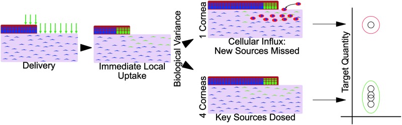
In support of ongoing research in the study of corneal and skin wound healing, we sought to improve on previously published results by using iontophoresis to deliver RNA interference-based oligonucleotides. By using a electromechanics-based approach, we were able to devise a two-phase solution that separated the buffering solution from the antisense oligonucleotide (ASO) solution. The separation was obtained by making the drug solution a higher density than the buffer, leading it to sink directly onto the tissue surface. This change immediately decreased the distance that the ASO would have to travel before delivery. The changes enabled delivery into ex vivo skin and corneas in 10 or fewer minutes and into in vivo corneas in 5 min. In vivo studies demonstrated short-term bioavailability of at least 24 h, a lack of chemical or thermal injury, a lack of interference in the healing of a corneal injury, and an antisense effect till at least day 7, but not day 14. The only side-effect observed was postdelivery edema that was not present when the vehicle alone was iontophoresed. This suggests that electro-osmotic flow from the delivery chamber was not the mechanism, but that the delivered solute likely increased the tissue's osmolarity. These results support the continued development and utilization of this ASO delivery approach in research-grade oligonucleotides to probe molecular biological pathways and in support of testing therapeutic ASOs in the skin and cornea.

Keywords: antisense; biological variance; cornea; delivery; iontophoresis; skin.


Conflict of interest statement

D.J.G.: Patent: U.S. 8,838,229, issued: yes, licensed: no, royalties: no, Grant: Trainee on a NEI T32 Vision Training grant (T32-EY007132). S.S.T.: Patent: U.S. 8,838,229, issued: yes, licensed: no, royalties: no, Grant: Unrestricted grant from Research to Prevent Blindness. G.S.S.: Patent: U.S. 8,838,229, issued: yes, licensed: no, royalties: no, Grant: PI on a R01 Regulation of Stromal Wound Healing (R01-EY05587). All three authors are co-inventors on a patent (U.S. 8,838,229), which has been issued, but has neither been licensed nor generated any royalties.

Figures

<b>FIG. 1.</b>
FIG. 1.
A pilot study guided by the initial literature review. (A) Corneal opacification when following the current literature's suggested methods. The animal was not allowed to leave general anesthesia and was immediately euthanized. (B) Ex vivo testing with Nile Blue (as a pH indicator). (C) The migrating cylindrical front of solvent with pH >8.0 would explain the observed opacification pattern.
<b>FIG. 2.</b>
FIG. 2.
Physical considerations enable substantial improvement of iontophoresis. The physical model of the iontophoresis setup as described in the literature (left) and as is proposed for improved performance and safety (right).
<b>FIG. 3.</b>
FIG. 3.
The ex vivo rabbit eye setup. (A) A typical phototherapeutic keratectomy wound in the center of an ex vivo cornea, 6.0 mm in diameter, 125 μm deep. (B) For both the intact and wounded models, the apparatus was the same.
<b>FIG. 4.</b>
FIG. 4.
In vitro testing of single- versus dual-phase arrangements. The graphic depicts the gross arrangement (not to scale) of the single- and dual-phase systems. PBS, phosphate-buffered saline; TAE, Tris Acetate EDTA.
<b>FIG. 5.</b>
FIG. 5.
Ex vivo results in intact rabbit corneas. The pink on the background is believed to be excess oligonucleotide, since it was not stained in the control (subset panels at the bottom) and the degree of staining was roughly consistent with the amount of oligonucleotide in the stroma (none was seen for the 1.5, 2.0, or 3.0 mA samples). The black boxes roughly correlate to the higher magnification fields above them. The scale bars represent 100 μm.
<b>FIG. 6.</b>
FIG. 6.
Quantifying the number of ASO+ Cells. (A) Macrophotographs of postdelivery fluorescence with the epithelial cell transfection rates noted. (B) Confocal micrographs of whole mounted epithelium and stroma. (C) The gating scheme used for the fluorescence-assisted cell sorting analysis. ASO, antisense oligonucleotide.
<b>FIG. 7.</b>
FIG. 7.
Ex vivo results in an excimer-ablated rabbit cornea. The scale bars represent 75 μm. Ep, epithelium; Str, stroma; En, endothelium.
<b>FIG. 8.</b>
FIG. 8.
Ex vivo results in an intact mouse skin and cornea. The 5-FAM ASO as seen before, during, and after delivery by macrophotography and fluorescence microscopy. SC, stratum corneum; EPI, epidermis; Derm, dermis; Epi, epithelium; Str, stroma; Endo, endothelium.
<b>FIG. 9.</b>
FIG. 9.
Ex vivo results in an intact pig skin. Testing delivery on regular and highly keratinized pig skin.
<b>FIG. 10.</b>
FIG. 10.
Ex vivo results in a disrupted pig skin. A model for scenarios with the underlying dermis as the drug target and debridement being acceptable (chronic wounds). Also, delivery into a sutured surgical incision.
<b>FIG. 11.</b>
FIG. 11.
In vivo delivery of ASOs into an intact cornea. The 5-FAM ASO could be immediately visualized either in native light (yellow) or via blue-excited green fluorescence. Green is nuclease-labile ASO, whereas red is nuclease-resistant ASO detected by immunostaining. All corneal sections are depicted as epithelium up, endothelium down. The scale bars represent 100 μm. The 1° antibody withheld control was equivalently exposed, but it had no strong substrate development (not shown).
<b>FIG. 12.</b>
FIG. 12.
The early physiological response to CTGF ASO delivered by iontophoresis. (A) Edema in corneas treated with iontophoresis of an ASO versus the carrier solution. (B) The postsurgery edema time course. (C) The epithelial closure time course. Note: (B, C) are from the same group of corneas. For the ASO treated, n = 7; for the nonintervention (No Tx), n = 3. (D) A typical corneal wound closure time course where closure by days 3 or 4 is typical.
<b>FIG. 13.</b>
FIG. 13.
The statistical distribution of CTGF in healing corneas and CTGF reduction by an ASO. (A) The internally normalized levels of CTGF mRNA in the epithelium and stroma 1 and 2 days after injury. (B) The log10-transformed mRNA data. The red circle represents the potential “outlier,” whereas the green represents the otherwise closely aggregated samples. (C) The nontransformed results from the CTGF ELISA and (D) the log10-transformed ELISA data.
<b>FIG. 14.</b>
FIG. 14.
The hypothetical source for biological variance and its consequences for RNA interference therapies. After a single dose, ASOs are found within the dosed cells. Afterward, stochastic events can lead to a splitting of the study population. In healing corneas, an influx of CTGF+ neutrophils has been observed and would not be affected by the initial dose. The anticipated consequences of such an occurrence are consistent with the observed results in both CTGF mRNA and protein in healing corneas.

Similar articles

Cited by

References

    1. Sriram S, Robinson P, Pi L, Lewin AS. and Schultz G. (2013). Triple combination of siRNAs targeting TGFbeta1, TGFbetaR2, and CTGF enhances reduction of collagen I and smooth muscle actin in corneal fibroblasts. Invest Ophthalmol Vis Sci 54:8214–8223 - PMC - PubMed
    1. Sisco M, Kryger ZB, O'Shaughnessy KD, Kim PS, Schultz GS, Ding XZ, Roy NK, Dean NM. and Mustoe TA. (2008). Antisense inhibition of connective tissue growth factor (CTGF/CCN2) mRNA limits hypertrophic scarring without affecting wound healing in vivo. Wound Repair Regen 16:661–673 - PubMed
    1. Rae JL. and Blankenship JE. (1973). Bioelectric measurements in the frog lens. Exp Eye Res 15:209–217 - PubMed
    1. Araie M. and Maurice DM. (1985). A reevaluation of corneal endothelial permeability to fluorescein. Exp Eye Res 41:383–390 - PubMed
    1. Koretz JF, Bertasso AM, Neider MW, True-Gabelt BA. and Kaufman PL. (1987). Slit-lamp studies of the rhesus monkey eye: II. Changes in crystalline lens shape, thickness and position during accommodation and aging. Exp Eye Res 45:317–326 - PubMed

Substances