Review
. 2017 Jun 12;10(1):293-320.
doi: 10.1146/annurev-anchem-061516-045205. Epub 2017 Mar 24.

Beyond Antibodies as Binding Partners: The Role of Antibody Mimetics in Bioanalysis

Affiliations
Review

Beyond Antibodies as Binding Partners: The Role of Antibody Mimetics in Bioanalysis

Xiaowen Yu et al. Annu Rev Anal Chem (Palo Alto Calif). .

Abstract

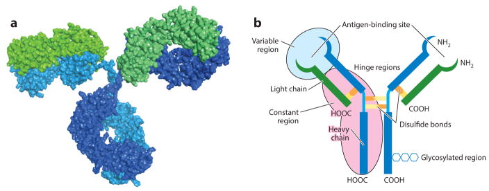
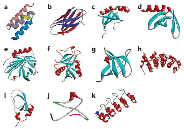
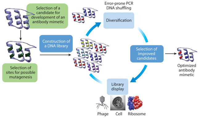
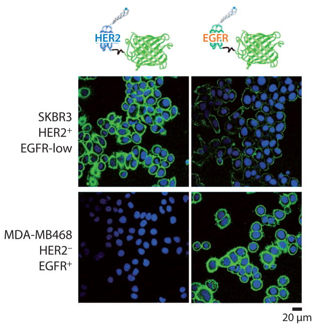
The emergence of novel binding proteins or antibody mimetics capable of binding to ligand analytes in a manner analogous to that of the antigen-antibody interaction has spurred increased interest in the biotechnology and bioanalytical communities. The goal is to produce antibody mimetics designed to outperform antibodies with regard to binding affinities, cellular and tumor penetration, large-scale production, and temperature and pH stability. The generation of antibody mimetics with tailored characteristics involves the identification of a naturally occurring protein scaffold as a template that binds to a desired ligand. This scaffold is then engineered to create a superior binder by first creating a library that is then subjected to a series of selection steps. Antibody mimetics have been successfully used in the development of binding assays for the detection of analytes in biological samples, as well as in separation methods, cancer therapy, targeted drug delivery, and in vivo imaging. This review describes recent advances in the field of antibody mimetics and their applications in bioanalytical chemistry, specifically in diagnostics and other analytical methods.

Keywords: antibodies; antibody mimetics; biosensors; diagnostics; molecular imaging; protein scaffolds.


Figures

Figure 1
Figure 1
(a) The X-ray crystal structure of an antibody. Heavy chains (constant fraction) and light chains (antigen-binding fraction) are shaded in blue/turquoise and green, respectively. (b) Schematic of an antibody with important regions highlighted.
Figure 2
Figure 2
X-Ray crystal structures of antibody mimetics. (a) An affibody molecule, with the hydroxylamine-susceptible asparagine-glycine residues highlighted in yellow (Protein Data Bank: 1LP1); (b) an adnectin molecule (Protein Data Bank: 3RZW); (c) an affimer molecule (Protein Data Bank: 1NB5); (d ) an affitin molecule (Protein Data Bank: 4CJ2); (e) an anticalin molecule (Protein Data Bank: 4GH7); ( f ) an atrimer molecule, with the flexible loops involved in binding colored in orange (Protein Data Bank: 1TN3); ( g) a fynomer molecule (Protein Data Bank: 1M27); (h) an armadillo repeat protein molecule (Protein Data Bank: 4DB9); (i ) a Kunitz domain inhibitor molecule (Protein Data Bank: 1ZR0); ( j) a knottin molecule (Protein Data Bank: 2IT7); (k) a designed ankyrin repeat protein molecule (Protein Data Bank: 2Q4J).
Figure 3
Figure 3
Directed evolution cycle for isolating novel antibody mimetics. Once the parent binding moiety is decided and the sites have been identified for mutagenesis, a new library is built and displayed using one of the display techniques. The selected candidates are then matured via error-prone shuffling. This process is repeated until the desired binding protein is developed. Abbreviation: PCR, polymerase chain reaction.
Figure 4
Figure 4
The use of affiprobes to stain cells with different HER2 and EGFR phenotype. The images were taken by a confocal microscope. Adapted with permission from Reference . Copyright 2010, John Wiley & Sons. Abbreviations: EGFR, epidermal growth factor receptor; HER2, human epidermal growth factor receptor 2.
Figure 5
Figure 5
In vivo imaging of nude mice bearing tumors with fluorescence-labeled affibodies Eaff682 and Haff800. The EGFR-overexpressing A431 tumor is located on the left side, whereas the HER2-overexpressing SKOV3 tumor is located on the right side. Tumors are indicated by white arrows. Reprinted with permission from Reference . Copyright 2010, Elsevier. Abbreviations: EGFR, epidermal growth factor receptor; HER2, human epidermal growth factor receptor 2.
Figure 6
Figure 6
(a) Applications of monobodies (adnectins) for in vitro and in vivo imaging. (i ) Schematic of the FN3 monobody library screening for SFK targeting binders; (ii ) biosensing mechanism of the SFK-targeting monobody; (iii ) SFK activation dynamics in living cells. Reproduced with permission from Reference . Copyright 2011, Macmillan Publishers. (iv) Differential interference contrast image (left) and ratio image (right) of PDGF-B–stimulated NIH 3T3 MEF cells microinjected with the SFK-targeting monobody. Scale bars are 20 μm. (v) Ratio image of a PTK1 cell microinjected with the biosensor. Scale bar is 20 μm. (b) In vivo targeting of the E1h monobody. Optical images of PC3 tumors after injection of E1h-Cy5.5 into mice. Nude mice were pretreated with nontagged E1h (+) or without (−). Panel reproduced with permission from Reference under the terms of the Creative Commons Attribution 4.0 International license, http://creativecommons.org/licenses/by/4.0. Abbreviations: KD, knockdown; MEF, mouse embryonic fibroblast; PBS, phosphate-buffered saline; PDGF-B, platelet-derived growth factor B; SFK, Src family kinase; SH, Src homology.
Figure 7
Figure 7
(a) Redox charge transfer resistance (Rct) plotted against a concentration of C-reactive protein (CRP) on a logarithmic scale for the P7i22 affimer interface. (b) The variance of the real part of impedance with log CRP concentration. Adapted with permission from Reference . Copyright 2012, American Chemical Society.
Figure 8
Figure 8
Binding curves of CDK2 on a PDEA-immobilized adlayer. (a) Signal dependence on the aqueous concentration of CDK2 target. (b) Linear range of CDK2 binding; error bars indicate assay reproducibility. Adapted with permission from Reference . Copyright 2009, American Chemical Society. Abbreviations: CDK2, cyclin-dependent protein kinase 2; PDEA, 2-(2-pyridinyldithio) ethane amine; SPR, surface plasmon resonance.
Figure 9
Figure 9
Peptide aptamers bind recombinant anterior gradient-2 (AGR2) in an ELISA. (a) AGR2 titration using peptide capture. (b) AGR2 capture using peptide titration. (c) AGR2 binding activity when adsorbed directly onto the solid phase. Adapted with permission from Reference . Copyright 2007, American Chemical Society. Abbreviations: ELISA, enzyme-linked immunosorbent assay; RLU, relative light units.
Figure 10
Figure 10
(a) Detection of autochthonous medulloblastoma in genetically engineered mice. (i ) Biphotonic images of (left) tumor-free wild-type mouse and (right) mouse with medulloblastoma. (ii ) Images of brains from the same mice following necropsy confirm the signal from the tumor. (iii ) Histologic confirmation of medulloblastoma tumor. (iv) Confocal microscopy images from the same brain of mouse with medulloblastoma. Reproduced with permission from Reference . Copyright 2016, American Association for Cancer Research. (b) The AF680-EETI 2.5F dye illuminates mouse medulloblastoma in vivo. Mouse with (left) and without (right) tumor. Reproduced with permission from Reference . (c) Coronal micropositron emission tomography images of U87MG tumor-bearing mice at 0.5, 1, and 2 h postinjection of the 18F-FP-2.5D dye (top) with cyclic RGD pentapeptides as a blocking agent (bottom). Red arrows indicate tumors. Reproduced with permission from Reference under the terms of the Creative Commons Attribution 4.0 International license, http://creativecommons.org/licenses/by/4.0.
Figure 11
Figure 11
Micro single-photon emission computed tomography/computed tomography scans of [99mTc(CO)3]+-G3 DARPin-His6 at 1 h in SCID mice bearing (a) HER2-positive human breast tumor (arrow) and (b) HER2- negative human breast tumor (arrow). Reprinted with permission from Reference under the terms of the Creative Commons Attribution 4.0 International license, http://creativecommons.org/licenses/by/4.0. Abbreviations: DARPin, designed ankyrin repeat protein; HER2, human epidermal growth factor receptor 2; SCID, severe combined immunodeficient.

References

    1. Pier GB, Lyczak JB, Wetzler LM. Immunology, Infection, and Immunity. Washington, DC: ASM; 2004.
    1. Woof JM, Burton DR. Human antibody-Fc receptor interactions illuminated by crystal structures. Nat Rev Immunol. 2004;4:89–99. - PubMed
    1. Litman GW, Rast JP, Shamblott MJ, Haire RN, Hulst M, et al. Phylogenetic diversification of immunoglobulin genes and the antibody repertoire. Mol Biol Evol. 1993;10:60–72. - PubMed
    1. Kuhn P, Fühner V, Unkauf T, Moreira GM, Frenzel A, et al. Recombinant antibodies for di- agnostics and therapy against pathogens and toxins generated by phage display. Proteom Clin Appl. 2016;10:922–48. - PMC - PubMed
    1. Waldmann H. Prospects for the application of antibodies in medicine. Methods Mol Med. 2000;40:63–72. - PubMed

Publication types