Review
. 2017 May 22:46:357-377.
doi: 10.1146/annurev-biophys-062215-011256. Epub 2017 Mar 22.

Single-Molecule Studies of Telomeres and Telomerase

Affiliations
Review

Single-Molecule Studies of Telomeres and Telomerase

Joseph W Parks et al. Annu Rev Biophys. .

Abstract

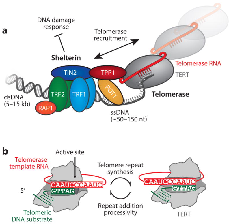
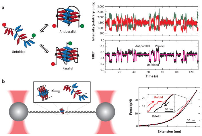
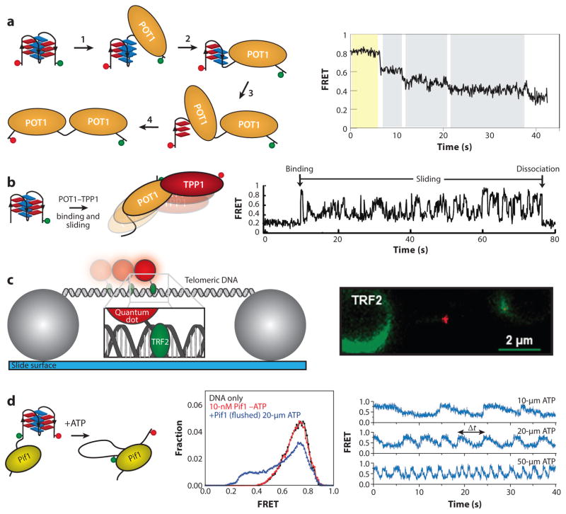
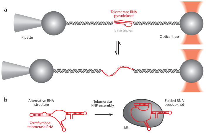
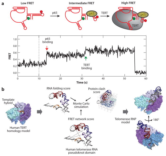
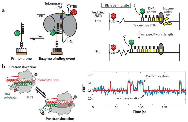
Telomeres are specialized chromatin structures that protect chromosome ends from dangerous processing events. In most tissues, telomeres shorten with each round of cell division, placing a finite limit on cell growth. In rapidly dividing cells, including the majority of human cancers, cells bypass this growth limit through telomerase-catalyzed maintenance of telomere length. The dynamic properties of telomeres and telomerase render them difficult to study using ensemble biochemical and structural techniques. This review describes single-molecule approaches to studying how individual components of telomeres and telomerase contribute to function. Single-molecule methods provide a window into the complex nature of telomeres and telomerase by permitting researchers to directly visualize and manipulate the individual protein, DNA, and RNA molecules required for telomere function. The work reviewed in this article highlights how single-molecule techniques have been utilized to investigate the function of telomeres and telomerase.

Keywords: single-molecule biophysics; telomerase; telomere.


Figures

Figure 1
Figure 1
(a) Schematic diagram of shelterin and telomerase at the telomere. The shelterin complex binds to double-stranded telomeric DNA through TRF1 and TRF2 homodimers. TIN2 recruits the POT1–TPP1 heterodimer to the TRF complex. POT1 binds to the single-stranded, guanine-rich tail. These proteins prevent the DNA-damage response mechanisms from identifying telomeres as double-stranded breaks. An interaction between TPP1 and the N-terminal domain of telomerase regulates recruitment of telomerase to the telomeres. (b) The telomerase catalytic cycle. Telomerase catalysis begins with the 3′ end of the DNA substrate ( green) bound in the active site of the telomerase ribonucleoprotein (RNP) complex. The DNA primer aligns with the RNA template (red ) through Watson-Crick base pairing, providing the telomerase reverse transcriptase (TERT, gray) with an RNA–DNA hybrid substrate. Upon completion of a single telomere DNA repeat, the product–template hybrid must dissociate, realign, and reenter the TERT active site to support repeat addition processivity. Abbreviations: dsDNA, double-stranded DNA; ssDNA, single-stranded DNA.
Figure 2
Figure 2
Single-molecule observation and mechanical manipulation of G-quadruplexes (GQs). (a) A doubly dye-labeled DNA construct is used in a single-molecule Förster resonance energy transfer (smFRET) experiment to measure the folding dynamics of a GQ. The smFRET signature reveals dynamic interconversion of the GQ between multiple structural states after passing through an unfolded intermediate. Panel a adapted with permission from Reference . (b) Optical tweezers are used to mechanically unfold a telomere GQ. Upon relaxation of the force, the single-stranded DNA can refold into the native GQ state. Analysis of the unfolding/refolding forces provides information about the energy landscape of folding. Panel b adapted with permission from Reference .
Figure 3
Figure 3
Telomere-associated proteins interact dynamically with telomere DNA. (a) A single-molecule Förster resonance energy transfer (smFRET) assay is used to probe the state of the telomere DNA quadruplex in the presence of POT1. The stepwise unfolding of a quadruplex by POT1 is observed as a series of consecutive decreases in FRET throughout the single-molecule trace. (b) Using the same experimental setup as in panel a, the heterodimeric POT1–TPP1 complex displays a dynamic sliding behavior once bound to the DNA substrate. Panels a and b adapted with permission from Reference . (c) A double-stranded telomere DNA tightrope is stretched between two immobilized beads. TRF2 protein conjugated to a quantum dot is added to solution, and single-molecule observation of dynamic TRF2 association with telomere DNA is made in real time. Comparison of telomere versus non-telomere DNA provides kinetic parameters about TRF2 diffusion along telomere DNA. Panel c adapted with permission from Reference . (d ) A schematic depiction of an smFRET assay for measuring quadruplex unfolding by the helicase Pif1. Unwinding of the quadruplex by Pif1 is ATP dependent and reversible. The sawtooth pattern represents binding by Pif1, followed by the repetitive unfolding of the quadruplex by the DNA reeling action of a single Pif1 enzyme. Panel d adapted with permission from Reference .
Figure 4
Figure 4
Folding dynamics of the telomerase RNA (TR) pseudoknot. (a) Using optical tweezers, the human telomerase pseudoknot was stretched to induce unfolding of the pseudoknot through base-pair shearing. Refolding of the pseudoknot was observed as the force is relaxed. Panel a adapted with permission from Reference . (b) Tetrahymena telomerase RNA pseudoknot folding during assembly into an active ribonucleoprotein (RNP) complex was monitored by single-molecule Förster resonance energy transfer (smFRET) (69). This result is consistent with models for an alternative RNA structure in the absence of telomerase proteins derived from biochemical and structural analyses (13, 19). Abbreviation: TERT, telomerase reverse transcriptase.
Figure 5
Figure 5
Assembly of the telomerase ribonucleoprotein (RNP) visualized by single-molecule Förster resonance energy transfer (smFRET). (a) In Tetrahymena thermophila telomerase, an smFRET pair was used to measure conformational changes in telomerase RNA (TR) during RNP assembly. Sequential binding of p65 and then telomerase reverse transcriptase (TERT) induces stepwise folding of TR visualized by consecutive increases in the FRET signal. Panel a adapted with permission from Reference . (b) A schematic pipeline for molecular modeling of the human telomerase complex. A homology model was generated for human TERT and used as a rigid modeling scaffold. smFRET distance constraints were used to guide Rosetta modeling of the RNA pseudoknot domain within the RNP. Convergent models of the RNP complex were produced, providing insight into human telomerase RNP architecture. Panel b adapted with permission from Reference .
Figure 6
Figure 6
Intramolecular telomerase dynamics. (a) Single-molecule Förster resonance energy transfer (smFRET) assay measuring internal movement of telomerase RNA throughout the catalytic cycle. RNA sequences flanking the template exhibit reciprocal compression and expansion motion during telomere DNA synthesis. Panel a adapted with permission from Reference . (b) DNA substrate dynamics in human telomerase observed by smFRET. Upon completion of one round of telomere repeat synthesis, the 3′ end of the DNA primer exhibits rapid dynamics between the two possible base-pairing registers with the RNA template. Kinetic analysis of these single-molecule data suggests that template hybrid realignment is much faster than the rate-limiting step of telomerase translocation required during repeat addition processivity. Panel b adapted with permission from Reference . Abbreviations: TBE, template boundary element; TERT, telomerase reverse transcriptase; TRE, telomeric repeat elongation.
Figure 7
Figure 7
Real-time telomerase activity assay at the single-molecule level. Human telomerase immobilized on a surface can bind to and extend a telomere DNA primer. As telomerase extends the primer processively, the telomere DNA repeats are extruded out of the telomerase complex and bound by a fluorescently labeled peptide nucleic acid (PNA) probe. Consecutive binding of multiple PNA probes generates a stepping pattern in the single-molecule fluorescence trace, providing real-time kinetic parameters governing the telomerase catalytic process. Adapted with permission from Reference . Abbreviation: dNTP, deoxynucleotide.

References

    1. Akiyama BM, Parks JW, Stone MD. The telomerase essential N-terminal domain promotes DNA synthesis by stabilizing short RNA–DNA hybrids. Nucleic Acids Res. 2015;43:5537–49. - PMC - PubMed
    1. Alves D, Li H, Codrington R, Orte A, Ren X, et al. Single-molecule analysis of human telomerase monomer. Nat Chem Biol. 2008;4:287–89. - PubMed
    1. Amiard S, Doudeau M, Pinte S, Poulet A, Lenain C, et al. A topological mechanism for TRF2-enhanced strand invasion. Nat Struct Mol Biol. 2007;14:147–54. - PubMed
    1. An N, Fleming AM, Burrows CJ. Interactions of the human telomere sequence with the nanocavity of the α-hemolysin ion channel reveal structure-dependent electrical signatures for hybrid folds. J Am Chem Soc. 2013;135:8562–70. - PMC - PubMed
    1. An N, Fleming AM, Middleton EG, Burrows CJ. Single-molecule investigation of G-quadruplex folds of the human telomere sequence in a protein nanocavity. PNAS. 2014;111:14325–31. - PMC - PubMed