Review
. 2017 Jul;24(4):314-321.
doi: 10.1097/MOH.0000000000000347.

Hematopoietic stem cells under pressure

Affiliations
Review

Hematopoietic stem cells under pressure

Miguel Ganuza et al. Curr Opin Hematol. 2017 Jul.

Abstract

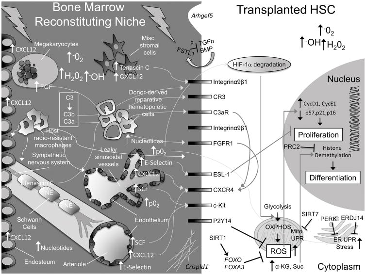
Purpose of review: Hematopoietic stem cells (HSCs) and progenitors are tasked with maintaining hematopoietic homeostasis in the face of numerous insults and challenges, including infection, inflammation, and exsanguination. HSCs possess the remarkable ability to reconstitute the entire hematopoietic system of an organism whose own hematopoietic system has been ablated. This ability is exploited routinely in the clinic via HSC transplantation (HSCT). Here, we focus on the physiological and molecular bottlenecks overcome by HSCs during transplantation.

Recent findings: During transplantation, HSCs encounter a damaged bone marrow niche, characterized molecularly by increases in oxygen concentrations and an altered cytokine milieu. New mechanisms and pathways have been recently implicated during HSCT, including transplanted HSC-dependent secretion of conditioning molecules that facilitate engraftment and pathways that protect HSCs from perturbed organelle homeostasis.

Summary: Better understanding the molecular processes HSCs employ to withstand the stress of transplant will illuminate novel targets for further improving conditioning regimens and engraftment during HSCT.


Conflict of interest statement

Conflicts of interest.

Authors declare no conflicts of interest.

Figures

Figure 1
Figure 1. Summary of molecular alterations driven by classical pre-transplant conditioning regimens in HSCs and the bone marrow niche
Here, we present a schematic to highlight some of the gross physical and molecular changes that occur in the bone marrow niche and within HSCs. For simplicity, not every known cellular component of the niche is pictured. In the niche, C3 is cleaved to C3b and C3a, which interact with HSC CR3 and C3aR receptors and stimulate homing by increasing, among other things, CXCR4. Megakaryocytes, which are attracted to the endosteum from sinusoidal vessels by increasing endosteal-CXCL12, also upregulate CXCR4 on HSCs via increased secretion of FGF. Schwann cells and stromal cells release TENASCIN C, which stimulates HSC migration and adhesion. Endothelial cells upregulate E-SELECTIN, CXCL12 and SCF. Sinusoidal vessels are damaged and leaky, resulting in an increase in O2 partial pressure (p02) and BM ROS levels. This contributes to H1F-1α degradation in HSCs, promoting their transition from glycolysis to oxidative respiration (OXPHOS), which further increases intracellular ROS levels. FOXOs, FOXA3, and signaling downstream of P2Y14 help HSCs cope with rising ROS levels. SIRT1 activates FOXOs. SIRT7 inhibits the increase in the mitochondrial unfolded protein response (UPR). Increased ROS stimulates HSC division and an ER-UPR. PERK and ERDJ14 counteract this effect in transplanted HSCs. Free nucleotide levels rise in the BM and are sensed by purinergic receptors, like P2Y14, which regulates ROS. Increasing intracellular α-Ketoglutarate (α-KG) promotes HSC differentiation via Histone demethylation. PRC2 complex counteracts this effect by promoting Histone methylation. Transplanted HSCs condition the reconstituting niche by secreting FSTL1 and extracellular matrix components (via Crispld1) and (very likely) additional factors (e.g. IL-8). Transplanted hematopoietic cells facilitate recovery of the conditioned niche. Figure Key: the bone marrow space is depicted on a dark gray background, the HSC intracellular space is light gray, and the HSC nucleus is dark gray. Major cell types are labeled in white font, major changes in the bone marrow space are labeled in white font, and major changes in the HSC are labeled in black font.

References

    1. Bernitz JM, Kim HS, MacArthur B, Sieburg H, Moore K. Hematopoietic Stem Cells Count and Remember Self-Renewal Divisions. Cell. 2016;167(5):1296–309. e10. Epub 2016/11/15. This study describes a population of dormant HSCs that persist throughout adulthood and also presents evidence that HSCs retain memory of their proliferative history and that long term HSCs are exceptionally rare in the aged HSC pool. - PMC - PubMed
    1. Gratwohl A, Baldomero H, Aljurf M, Pasquini MC, Bouzas LF, Yoshimi A, et al. Hematopoietic stem cell transplantation: a global perspective. JAMA. 2010;303(16):1617–24. Epub 2010/04/29. - PMC - PubMed
    1. Current Uses and Outcomes of Hematopoietic Cell Transplantation (HCT): CIBMTR Summary Slides, [database on the Internet]. 2016.

    1. Sasine JP, Yeo KT, Chute JP. Concise Review: Paracrine Functions of Vascular Niche Cells in Regulating Hematopoietic Stem Cell Fate. Stem Cells Transl Med. 2016 Epub 2016/09/15. - PMC - PubMed
    1. Olson TS, Caselli A, Otsuru S, Hofmann TJ, Williams R, Paolucci P, et al. Megakaryocytes promote murine osteoblastic HSC niche expansion and stem cell engraftment after radioablative conditioning. Blood. 2013;121(26):5238–49. Epub 2013/05/15. - PMC - PubMed

Substances