. 2017 Apr 4;13(4):e1006597.
doi: 10.1371/journal.pgen.1006597. eCollection 2017 Apr.

Bovine and murine models highlight novel roles for SLC25A46 in mitochondrial dynamics and metabolism, with implications for human and animal health

Affiliations

Bovine and murine models highlight novel roles for SLC25A46 in mitochondrial dynamics and metabolism, with implications for human and animal health

Amandine Duchesne et al. PLoS Genet. .

Abstract

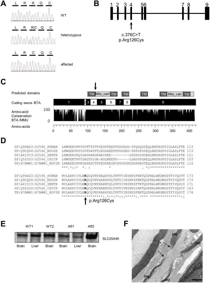
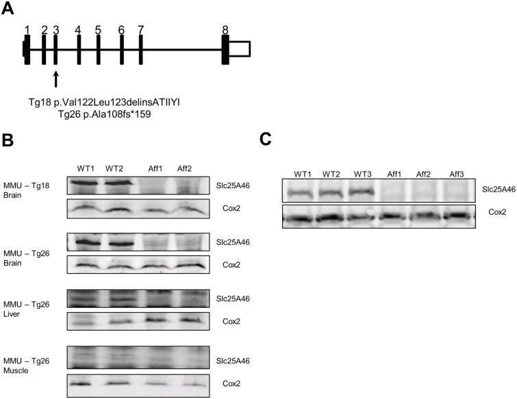
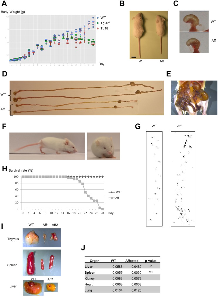
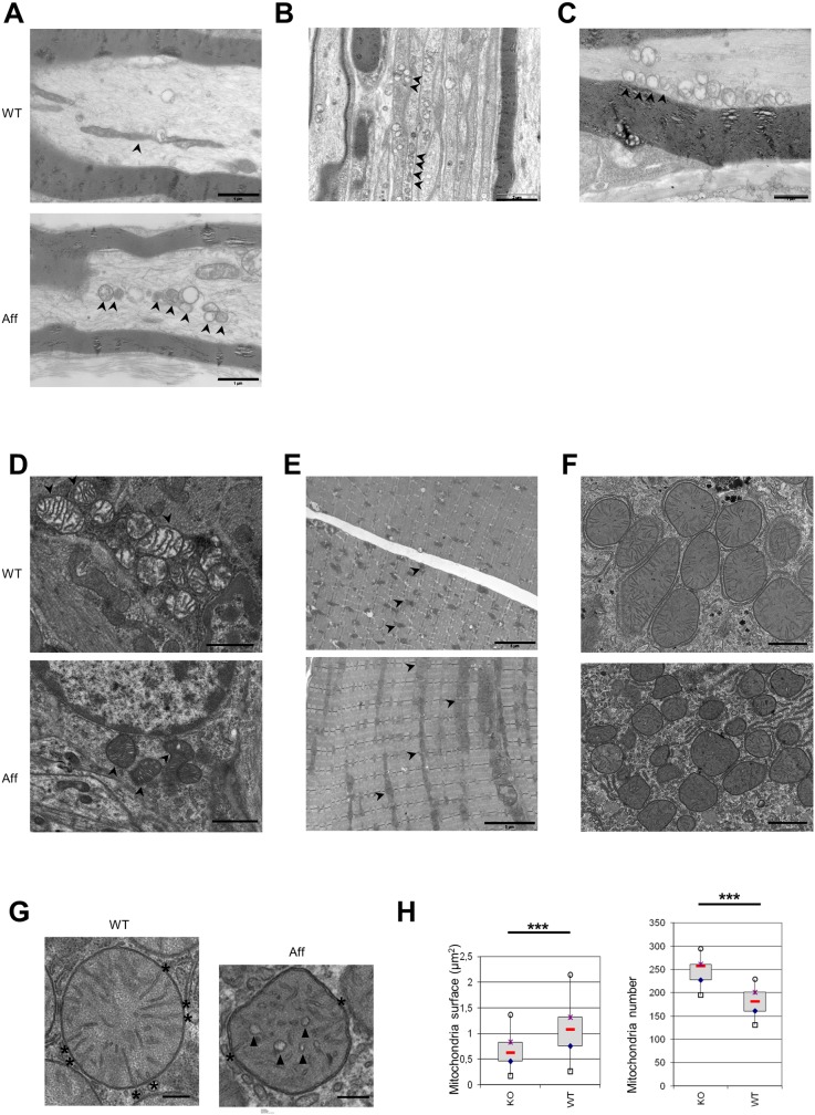
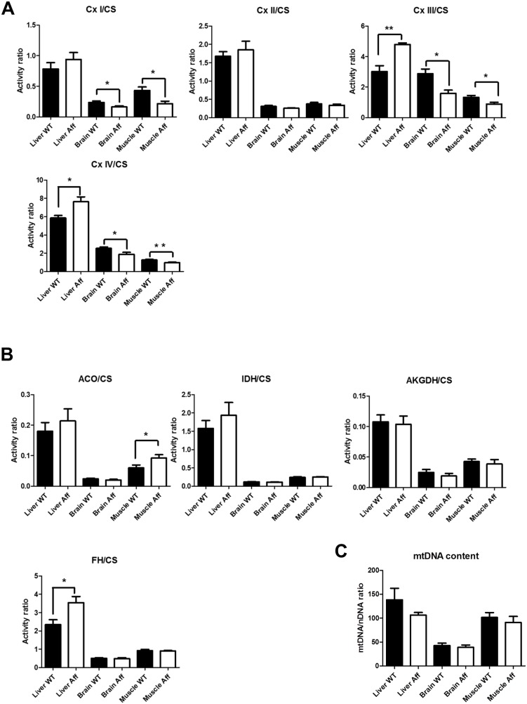
Neuropathies are neurodegenerative diseases affecting humans and other mammals. Many genetic causes have been identified so far, including mutations of genes encoding proteins involved in mitochondrial dynamics. Recently, the "Turning calves syndrome", a novel sensorimotor polyneuropathy was described in the French Rouge-des-Prés cattle breed. In the present study, we determined that this hereditary disease resulted from a single nucleotide substitution in SLC25A46, a gene encoding a protein of the mitochondrial carrier family. This mutation caused an apparent damaging amino-acid substitution. To better understand the function of this protein, we knocked out the Slc25a46 gene in a mouse model. This alteration affected not only the nervous system but also altered general metabolism, resulting in premature mortality. Based on optic microscopy examination, electron microscopy and on biochemical, metabolic and proteomic analyses, we showed that the Slc25a46 disruption caused a fusion/fission imbalance and an abnormal mitochondrial architecture that disturbed mitochondrial metabolism. These data extended the range of phenotypes associated with Slc25a46 dysfunction. Moreover, this Slc25a46 knock-out mouse model should be useful to further elucidate the role of SLC25A46 in mitochondrial dynamics.


Conflict of interest statement

The authors have declared that no competing interests exist.

Figures

Fig 1
Fig 1. Identification of the causal mutation for bovine axonopathy.
(A) Sanger sequence traces for the causal mutation in the bovine SLC25A46 gene done on a wild-type (WT), a heterozygous carrier and an affected animal. (B) Schematic diagram of the SLC25A46 gene in cattle, located on chromosome 7, with the position of the mutation indicated (arrow). (C) Schematic diagram of coding exons from SLC25A46 gene in cattle (protein with 419 amino acids) with the predicted functional domains of the protein. Conservation between bovine and murine protein sequences is reported (mutation in cattle is indicated by an arrow). TM, Transmembrane; Mito_carr, mitochondrial carrier; BTA, Bos taurus (bovine); MMU, Mus musculus (mouse) (D) Based on protein alignment, the affected amino acid was highly conserved in vertebrates and located in a conserved region from the protein. XENTR, Xenopus tropicalis; CHICK, chicken; BOVIN, bovine. (E) Proteins were extracted with a Mitochondria Isolation kit from bovine WT and affected brain and liver tissues. Samples were analyzed by immunoblotting with antibody against human mitochondrial protein SLC25A46, targeting the N-term domain of the protein. (F) Radial nerve (proximal part). Electron micrograph. Longitudinal section from a calf homozygous for the mutation. Note the enlarged node of Ranvier, between the two arrows. Note the uneven myelin sheath (m) on each side of the node of Ranvier (scale bar = 2 μm).
Fig 2
Fig 2. Construction of two mouse lines with disruption of Slc25a46.
(A) Schematic diagram of the Slc25a46 gene in mice, located on chromosome 18, with positions of mutations in Tg18 and Tg26 lines (arrows). (B) Total proteins were extracted from brain, muscle and liver of WT, Tg18 homozygous and Tg26 homozygous mice. Samples were analyzed by immunoblotting, with antibodies against mitochondrial proteins Slc25a46 and Cox2 (internal loading control). (C) Proteins were extracted with a Mitochondria Isolation kit from brains of WT and Tg18 mice. Samples were analyzed by immunoblotting with antibody against the mitochondrial proteins Slc25a46 and Cox2. WT, Wild-Type; Aff, affected. Note: entire Western Blots with SLC25A46 antibody are shown in S5 Fig.
Fig 3
Fig 3. Representative phenotypes in homozygous mice from Tg18 and Tg26 lines (with disruption of the Slc25a46 gene).
(A) Growth curve (birth to 22 days pn) for WT (blue circles), Tg18 homozygous (red circles) and Tg26 homozygous (green circles) mice. Homozygous mice from the two knockout lines stopped gaining weight at ~2 weeks pn. (B) Note size difference between a WT and an affected mouse at 3 weeks post-natal (pn). (C) Representative image of stomach filled with milk for 2 weeks old WT and Tg-/- mice. (D) Gastrointestinal tract (stomach to colon) in WT and affected mice. (E). Representative hemorrhages in the intestinal tract from the oldest affected mice. (F) Representative image of unsteady gait of affected mice. (G) Footprint analyses of WT and Tg-/- mice. The soles of the limbs were labeled with ink. Mice walked on paper in a 10-cm lane surrounded by walls. The ataxic gait is clearly evidenced in the Tg-/- mice, as well as a rapid weakness that prevents them to walk as long as the WT mice. (H) Survival rate curve for WT and Tg-/- animals. Only Tg-/- mice that died prematurely were recorded (27 Tg-/- mice) as well as 40 WT mice. For ethical reasons, most Tg-/- mice were euthanized as soon as they displayed poor health. None of the Tg-/- mice were capable of survival beyond 27 days pn. (I) Thymus, spleen and liver from WT (27 days pn), and affected mice (27 and 22 days pn); note the decreased size of these organs in affected mice. (J) Ratio between organ weight and body weight in 3 weeks old WT (n = 3) and affected mice (n = 4). Liver and spleen were smaller in affected animals. ** p = 0.01, *** p = 0.001, Student test. WT, Wild-Type; Aff, affected; dpn, days post-natal.
Fig 4
Fig 4. Phenotyping of Tg-/- mice.
(A) Brain. HES staining. Coronal section from Tg-/- mouse displaying one vacuolated neuron in the lateral vestibular nucleus, indicated by an arrow (scale bar = 100 μm) (B) Nerve root from the lumbar spinal cord. Electron micrograph. Longitudinal section from Tg-/- mouse; note the macrophage containing lipid debris, indicated by an arrow (scale bar = 2 μm). (C) Sciatic nerve, distal part. Electron micrograph. Transversal sections from WT and Tg-/- mice. Axon diameter and myelin sheath are comparable (scale bar = 10 μm). (D) Optic nerves. Electron micrograph. Longitudinal sections from WT and Tg-/- mice. There are no significant quantitative or qualitative differences between mitochondria of WT and Tg-/- mice (scale bar = 1 μm). Arrowheads indicate mitochondria.
Fig 5
Fig 5. Characterization of ultrastructural abnormalities in bovine and murine models with SLC25A46 mutations.
(A) Distal sciatic nerve. Electron micrograph. Longitudinal sections from a WT mouse, displaying intra-axonal, randomly distributed, elongated normal mitochondria and from an Tg-/- mouse, displaying numerous aggregated intra-axonal mitochondria of various shapes; most of them presenting abnormal cristae and membranes (scale bar 1 = μm). (B) Proximal sciatic nerve. Electron micrograph. Longitudinal sections from a Tg-/- mouse, displaying numerous aggregated intra-axonal mitochondria of various shapes in myelinated and non-myelinated axons; most of them presenting abnormal cristae and membranes (scale bar 2 = μm). (C) Radial nerve (proximal part). Electron micrograph. Longitudinal section from a calf homozygous for the mutation. Abnormally aggregated, small and round mitochondria are located at the periphery of the axon; most have abnormal cristae and membranes (scale bar = 1 μm). (D) Intestinal enteric plexus. Electron micrograph. Sections from a WT mouse, displaying normal mitochondria and from Tg-/- mouse displaying smaller dark mitochondria with vesicular-like abnormal cristae (scale bar = 1 μm). (E) Quadriceps femoris muscle. Electron micrograph. Sections from WT and Tg-/- mouse showing more abundant and aggregated mitochondria in Tg-/- mouse (scale bar = 5 μm). (F) Liver. Electron micrograph. Section from a WT mouse, with round mitochondria and from an Tg-/- mouse, with numerous, dark and smaller mitochondria (scale bar = 1 μm). (G) Liver. Electron micrograph. Mitochondria in a WT mouse have numerous organized cristae (radiating from the inner mitochondrial membrane to the center of the mitochondria). Mitochondria from Tg-/- mouse display disorganized, vesicular-like cristae, rarely attached to the inner mitochondrial membrane (scale bar = 250 nm). Stars indicate cristae and inner mitochondrial membrane contact points. Arrowheads indicate vesicular-like cristae. (H) Mitochondrial phenotype shown in (G) were quantified from the liver of two WT and two Tg-/- mice (10 independent images per individual for mitochondrial number and three independent images per individual for mitochondrial area). Mitochondrial surface refers to 2D-area of each cut mitochondria on the electron microscopy images. Mitochondrial number refers to number of cut mitochondria on electron microscopy images per cell. *** p = 0.001; Student test. WT, Wild Type; Aff, affected. Arrowheads indicated mitochondria in panels (A) to (E).
Fig 6
Fig 6. Analysis of the mitochondrial metabolism and mitochondrial DNA (mtDNA) in WT and Tg-/- mice in liver, brain and muscle, three tissues which have mitochondrial morphology abnormalities in homozygous mutant mice.
(A) Analysis of the respiratory chain complex activities: complexes I, II, III, IV. Since mitochondria number varied in WT and affected tissues, all activities were normalized with Citrate Synthase activity (estimate of mitochondria number). Cx, complex; CS, citrate synthase. (B) Analysis of the activity for some enzymes involved in Krebs cycle. Since mitochondria number varied in WT and affected tissues, all activities were normalized with Citrate Synthase activity. ACO, aconitase; IDH, isocitrate dehydrogenase; AKGDH, α-ketoglutarate dehydrogenase; FH, fumarate hydratase; CS, citrate synthase. (C) mtDNA content was estimated by qPCR. * p = 0.05, ** p = 0.01, Student test. WT, Wild-Type; Aff, affected.

Similar articles

Cited by

References

    1. Zick M, Rabl R, Reichert AS. Cristae formation-linking ultrastructure and function of mitochondria. Biochim Biophys Acta—Mol Cell Res [Internet]. Elsevier B.V.; 2009;1793(1):5–19. - PubMed
    1. Nunnari J, Suomalainen A. Mitochondria: In sickness and in health. Cell [Internet]. Elsevier Inc.; 2012;148(6):1145–59. 10.1016/j.cell.2012.02.035 - DOI - PMC - PubMed
    1. Kühlbrandt W. Structure and function of mitochondrial membrane protein complexes. BMC Biol [Internet]. BMC Biology; 2015;13(1):89. - PMC - PubMed
    1. Lin MT, Beal MFF. Mitochondrial dysfunction and oxidative stress in neurodegenerative diseases. Nature [Internet]. 2006;443(7113):787–95. 10.1038/nature05292 - DOI - PubMed
    1. Westermann B. Mitochondrial fusion and fission in cell life and death. Nat Rev Mol Cell Biol [Internet]. Nature Publishing Group; 2010;11(12):872–84. 10.1038/nrm3013 - DOI - PubMed

Publication types

Substances