LRG1 promotes proliferation and inhibits apoptosis in colorectal cancer cells via RUNX1 activation
- PMID: 28376129
- PMCID: PMC5380360
- DOI: 10.1371/journal.pone.0175122
LRG1 promotes proliferation and inhibits apoptosis in colorectal cancer cells via RUNX1 activation
Abstract
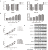
Leucine-rich-alpha-2-glycoprotein 1 (LRG1) has been shown to be involved in various human malignancies. Whether it plays a role in colorectal cancer (CRC) development remains unclear. Here, we investigated whether and through what mechanism LRG1 functions in human CRC cells. The plasma level of LRG1 was significantly increased in CRC patients, but it was remarkably decreased in patients with resected colorectal cancers. Meanwhile, both mRNA and protein levels of LRG1 were remarkable overexpressed in CRC tissues than normal tissues. The knockdown of LRG1 significantly inhibited cell proliferation, induced cell cycle arrest at the G0/G1 phase, and promoted apoptosis in SW480 and HCT116 cells in vitro. In addition, LRG1 silencing led to the downregulation of the levels of key cell cycle factors, such as cyclin D1, B, and E and anti-apoptotic B-cell lymphoma-2(Bcl-2). However, it up-regulated the expression of pro-apoptotic Bax and cleaved caspase-3. Furthermore, RUNX1 could be induced by LRG1 in a concentration-dependent manner, while the knockdown of RUNX1 blocked the promotion of the proliferation and inhibition of apoptosis induced by LRG1. Collectively, these findings indicate that LRG1 plays a crucial role in the proliferation and apoptosis of CRC by regulating RUNX1 expression. Thus, LRG1 may be a potential detection biomarker as well as a marker for monitoring recurrence and therapeutic target for CRC.
Conflict of interest statement
Figures


Similar articles
-
LRG1 modulates epithelial-mesenchymal transition and angiogenesis in colorectal cancer via HIF-1α activation.J Exp Clin Cancer Res. 2016 Feb 9;35:29. doi: 10.1186/s13046-016-0306-2. J Exp Clin Cancer Res. 2016. PMID: 26856989 Free PMC article.
-
Stable knockdown of LRG1 by RNA interference inhibits growth and promotes apoptosis of glioblastoma cells in vitro and in vivo.Tumour Biol. 2015 Jun;36(6):4271-8. doi: 10.1007/s13277-015-3065-3. Epub 2015 Jan 15. Tumour Biol. 2015. PMID: 25589464
-
Colorectal cancer-associated fibroblasts promote metastasis by up-regulating LRG1 through stromal IL-6/STAT3 signaling.Cell Death Dis. 2021 Dec 20;13(1):16. doi: 10.1038/s41419-021-04461-6. Cell Death Dis. 2021. PMID: 34930899 Free PMC article.
-
LRG1 in cancer: mechanisms, potential therapeutic target, and use in clinical diagnosis.Mol Biol Rep. 2025 Jun 10;52(1):577. doi: 10.1007/s11033-025-10669-y. Mol Biol Rep. 2025. PMID: 40493117 Review.
-
LRG1: an emerging player in disease pathogenesis.J Biomed Sci. 2022 Jan 21;29(1):6. doi: 10.1186/s12929-022-00790-6. J Biomed Sci. 2022. PMID: 35062948 Free PMC article. Review.
Cited by
-
Hsa_circ_0040809 and hsa_circ_0000467 promote colorectal cancer cells progression and construction of a circRNA-miRNA-mRNA network.Front Genet. 2022 Oct 20;13:993727. doi: 10.3389/fgene.2022.993727. eCollection 2022. Front Genet. 2022. PMID: 36339002 Free PMC article.
-
LRG1 is an adipokine that mediates obesity-induced hepatosteatosis and insulin resistance.J Clin Invest. 2021 Dec 15;131(24):e148545. doi: 10.1172/JCI148545. J Clin Invest. 2021. PMID: 34730111 Free PMC article.
-
Research Progress on Leucine-Rich Alpha-2 Glycoprotein 1: A Review.Front Pharmacol. 2022 Jan 5;12:809225. doi: 10.3389/fphar.2021.809225. eCollection 2021. Front Pharmacol. 2022. PMID: 35095520 Free PMC article. Review.
-
RUNX1 promotes tumour metastasis by activating the Wnt/β-catenin signalling pathway and EMT in colorectal cancer.J Exp Clin Cancer Res. 2019 Aug 1;38(1):334. doi: 10.1186/s13046-019-1330-9. J Exp Clin Cancer Res. 2019. PMID: 31370857 Free PMC article.
-
Label-free quantitative proteomics reveals aberrant expression levels of LRG, C9, FN, A1AT and AGP1 in the plasma of patients with colorectal cancer.Clin Proteomics. 2023 Apr 6;20(1):15. doi: 10.1186/s12014-023-09407-y. Clin Proteomics. 2023. PMID: 37024778 Free PMC article.
References
-
- IARC, Globocan 2012. World Health Organization. Paris, 2014 URL: http://globocan.iarc.fr/Default.aspx.
-
- Duffy MJ. Carcinoembryonic antigen as a marker for colorectal cancer: Is it clinically useful? Clinical Chemistry. 2001; 47: 624–30. - PubMed
-
- Haupt H, Baudner S. Isolation and characterization of an unknown, leucinerich 3.1-S- alpha2-glycoprotein from human serum. Hoppe Seyler Z Physiol Chem.1977;358(6): 639–46. - PubMed
MeSH terms
Substances
LinkOut - more resources
Full Text Sources
Other Literature Sources
Medical
Research Materials
Miscellaneous

