. 2017 Sep 1;109(9):djx016.
doi: 10.1093/jnci/djx016.

Evidence for a Mesothelial Origin of Body Cavity Effusion Lymphomas

Affiliations

Evidence for a Mesothelial Origin of Body Cavity Effusion Lymphomas

David Sanchez-Martin et al. J Natl Cancer Inst. .

Abstract

Background: Primary effusion lymphoma (PEL) is a Kaposi's sarcoma herpes virus (KSHV)-induced lymphoma that typically arises in body cavities of HIV-infected patients. PEL cells are often co-infected with Epstein-Barr virus (EBV). "PEL-like" lymphoma is a KSHV-unrelated lymphoma that arises in body cavities of HIV-negative patients. "PEL-like" lymphoma is sometimes EBV positive. The derivation of PEL/"PEL-like" cells is unclear.

Methods: Mesothelial cells were cultured from body cavity effusions of 23 patients. Cell proliferation, cytokine secretion, marker phenotypes, KSHV/EBV infection, and clonality were evaluated by standard methods. Gene expression was measured by quantitative polymerase chain reaction and immunoblotting. A mouse model of PEL (3 mice/group) was used to evaluate tumorigenicity.

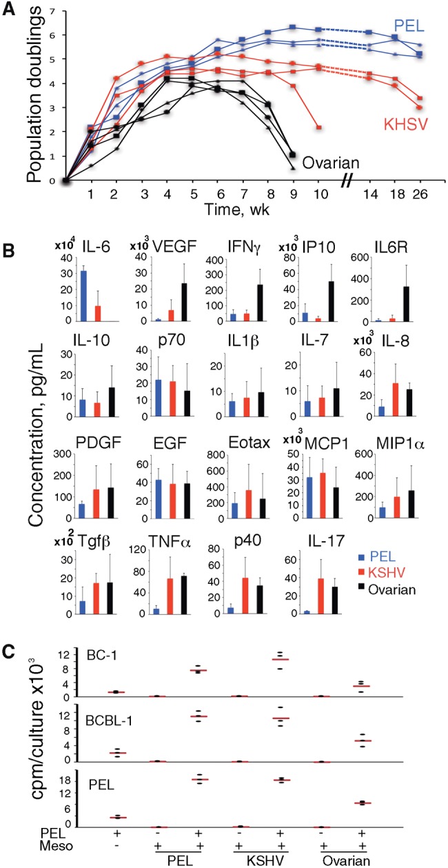
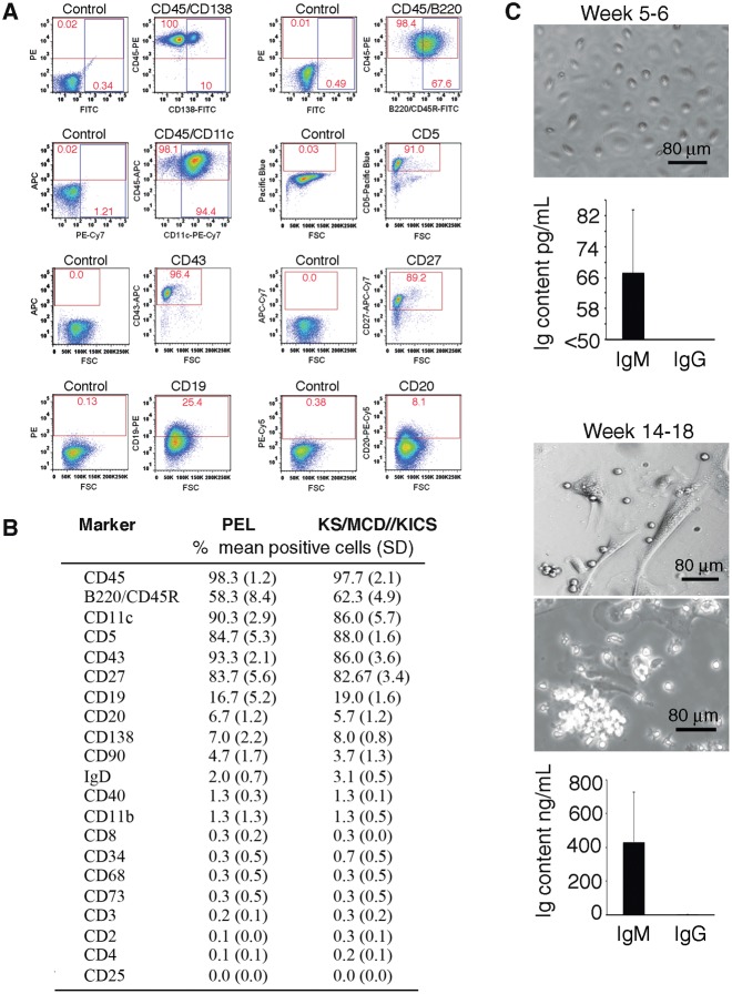
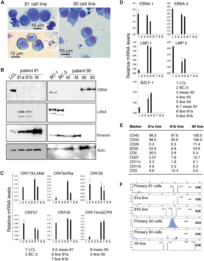
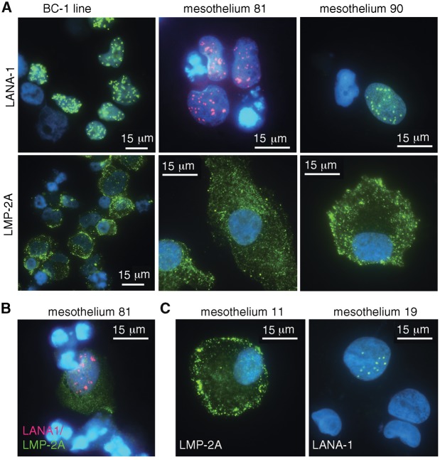
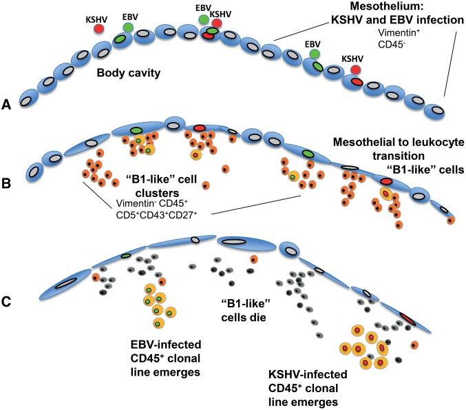
Results: We found that the mesothelia derived from six effusions of HIV-infected patients with PEL or other KSHV-associated diseases contained rare KSHV + or EBV + mesothelial cells. After extended culture (16-17 weeks), some mesothelial cells underwent a trans-differentiation process, generating lymphoid-type CD45 + /B220 + , CD5 + , CD27 + , CD43 + , CD11c + , and CD3 - cells resembling "B1-cells," most commonly found in mouse body cavities. These "B1-like" cells were short lived. However, long-term KSHV + EBV - and EBV + KSHV - clonal cell lines emerged from mesothelial cultures from two patients that were clonally distinct from the monoclonal or polyclonal B-cell populations found in the patients' original effusions.

Conclusions: Mesothelial-to-lymphoid transformation is a newly identified in vitro process that generates "B1-like" cells and is associated with the emergence of long-lived KSHV or EBV-infected cell lines in KSHV-infected patients. These results identify mesothelial cultures as a source of PEL cells and lymphoid cells in humans.


Figures

Figure 1.
Figure 1.
Derivation of mesothelial cells from effusions. A) Proliferative capacity of primary mesothelial cells over time in culture. Mesothelial cells from primary effusion lymphoma (PEL): PEL effusion; Kaposi's sarcoma herpes virus (KSHV): KSHV effusion; ovarian: ovarian carcinoma effusion. B) Cytokines in mesothelial conditioned medium (n = 3/group) at week 3 or 4. Bar graphs: group means (±SEM). C) Proliferation of PEL cells alone, mesothelia alone, or coculture of PEL cells over mesothelia (PEL+/Meso+). Dot plot: data from triplicate cultures from a representative experiment (three performed); the horizontal red line reflects the means. EGF = endothelial growth factor; IL = interleukin; KHSV = Kaposi's sarcoma herpes virus; PDGF = platelet-derived growth factor; PEL = primary effusion lymphoma; TNF = tumor necrosis factor; VEGF = vascular endothelial growth factor.
Figure 2.
Figure 2.
Emergence of lymphoid-type cells from mesothelial cell cultures. A) Mesothelial cell culture imaged at weeks 4, 16, 17, and 19; phase contrast microscopy. Arrows: lymphoid-type cells; asterisks: “senescent” mesothelial cells; area limited by the red dotted line is magnified in the inset. B) Sessile bodies and lymphoid-like cell in an Azurite+ mesothelial culture. C) Cytospun cells from culture supernatant of mesothelial cells at week 17. Giemsa staining showing small lymphoid-like (lymph), plasmablast/plasma cell-like (plasm), and mesothelial-like (meso) cells.
Figure 3.
Figure 3.
Characterization of lymphoid-like cells from mesothelial cell cultures. A and B) Nonadherent cells recovered from mesothelial cell cultures analyzed by flow cytometry. A) Representative profiles from antibody and control Ig staining; percent cells in quadrants are shown. B) Cumulative results; cultures were derived from effusions of primary effusion lymphoma (PEL) patients (n = 3) and patients with KS, KSHV-associated inflammatory cytokine syndrome (KICS), or KSHV-associated plasmablastic multicentric Castleman disease (KSHV-MCD; n = 4); percent mean positive cells (±SD) shown. C) Immunoglobulin (Ig)M and IgG content in supernatants of mesothelial cell cultures at weeks 5 to 6 and 14 to 18. Top: representative mesothelial cell monolayers at supernatant harvest. Bottom: bar graphs showing the means (n = 5; ±SD; error bars). Ig = immunoglobulin; KICS = KSHV-associated inflammatory cytokine syndrome; KS = Kaposi's sarcoma; MCD = multicentric Castleman disease; PE = primary effusion.
Figure 4.
Figure 4.
Emergence of cell lines from mesothelial cultures. A) Anaplastic morphology of cytospun 81a, 81b, and 90 cells; Giemsa staining. B) Immunoblotting detection of EBV nuclear antigen (EBNA1), latency-associated nuclear antigen (LANA1), Vimentin and actin in cell lysates from patient 81 (left): 81a and 81b lines (passage 3) and mesothelial cells (M, week 4); and patient 90 (right): mesothelial cells (M, weeks 3 and 4); and 90 line (passage 1 and 4). LCL: EBV+ lymphoblastoid cell line; BC-1 (EBV+/KSHV+) and BC-3 (EBV-/KSHV+): PEL lines. Relative KSHV (C) and EBV (D) mRNA levels in mesothelial cells (Meso) from patients 81 and 90 (weeks 2–4) and cell lines 81a, 81b, and 90 (passage 1 to 3). E) Flow cytometric analysis of 81a, 81b, and 90 cell lines. Results reflect percent positive cells. F) Analysis of clonality in primary cells from patient (81 and 90) effusion and cell lines (81a, 81b, and 90) derived from mesothelial culture. EBNA = EBV nuclear antigen; EBV = Epstein-Barr virus; KSHV = Kaposi's sarcoma herpes virus; LANA = latency-associated nuclear antigen; LCL = lymphoblastoid cell line; LMP = latent membrane protein; ORF = open reading frame.
Figure 5.
Figure 5.
Kaposi's sarcoma herpes virus (KSHV) and Epstein-Barr virus (EBV) infect primary mesothelial cells. A–C) KSHV and EBV infection in primary mesothelial cells from effusions of patients with primary effusion lymphoma (PEL; mesothelia 81 and 11) and KSHV-associated diseases but no PEL (mesothelia 90 and 19). Immunofluorescence detection of KSHV-latency-associated nuclear antigen (LANA)–1 (green or red) and EBV-latent membrane protein–2A (green); nuclear DAPI: blue. LANA = latency-associated nuclear antigen; LMP = latent membrane protein.
Figure 6.
Figure 6.
A model for primary effusion lymphoma origin. A) Mesothelial cells lining body cavities become infected with endogenous Kaposi's sarcoma herpes virus (KSHV), Epstein-Barr virus (EBV), or both viruses. B) Vimentin+CD45- senescent mesothelial cells undergo mesothelial-to-lymphoid transition (MLT), giving rise to Vimentin-CD45+CD5+CD43+CD27+ “B1-like” lymphoid cells that cluster onto the mesothelium and then detach from it. C) Clonal population of CD45+, KSHV+, and EBV+ leukocytes emerge concurrent with the death of “B1-like” leukocytes. EBV = Epstein-Barr virus; KSHV = Kaposi's sarcoma herpes virus; MLT = mesothelial-to-lymphoid transition.

Similar articles

Cited by

References

    1. Knowles DM, Inghirami G, Ubriaco A, et al.Molecular genetic analysis of three AIDS-associated neoplasms of uncertain lineage demonstrates their B-cell derivation and the possible pathogenetic role of the Epstein-Barr virus. Blood. 1989;73(3):792–799. - PubMed
    1. Cesarman E, Chang Y, Moore PS, et al.Kaposi's sarcoma-associated herpesvirus-like DNA sequences in AIDS-related body-cavity-based lymphomas. N Engl J Med. 1995;332(18):1186–1191. - PubMed
    1. Chadburn A, Hyjek EM, Tam W, et al.Immunophenotypic analysis of the Kaposi sarcoma herpesvirus (KSHV; HHV-8)-infected B cells in HIV+ multicentric Castleman disease (MCD). Histopathology. 2008;53(5):513–524. - PubMed
    1. Di Bartolo DL, Hyjek E, Keller S, et al.Role of defective Oct-2 and OCA-B expression in immunoglobulin production and Kaposi's sarcoma-associated herpesvirus lytic reactivation in primary effusion lymphoma. J Virol. 2009;83(9):4308–4315. - PMC - PubMed
    1. Ascoli V, Calabro ML, Giannakakis K, et al.Kaposi's sarcoma-associated herpesvirus/human herpesvirus 8-associated polyclonal body cavity effusions that mimic primary effusion lymphomas. Int J Cancer. 2006;119(7):1746–1748; author reply 1749–1750. - PubMed

MeSH terms