. 2017 Mar 1;109(3):1-13.
doi: 10.1093/jnci/djw229.

Rad51 Degradation: Role in Oncolytic Virus-Poly(ADP-Ribose) Polymerase Inhibitor Combination Therapy in Glioblastoma

Affiliations

Rad51 Degradation: Role in Oncolytic Virus-Poly(ADP-Ribose) Polymerase Inhibitor Combination Therapy in Glioblastoma

Jianfang Ning et al. J Natl Cancer Inst. .

Abstract

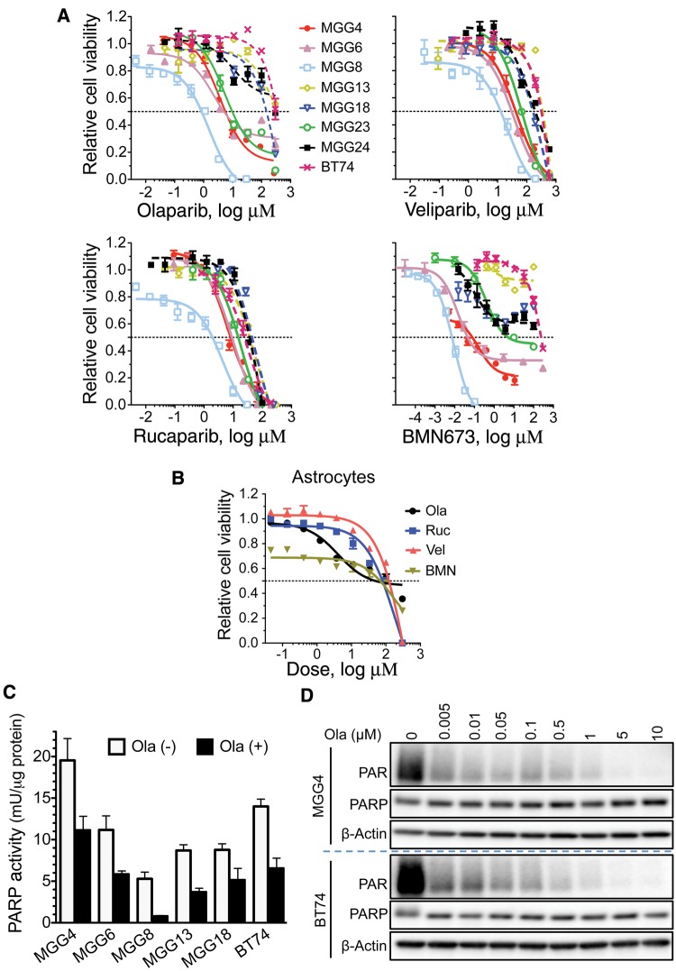
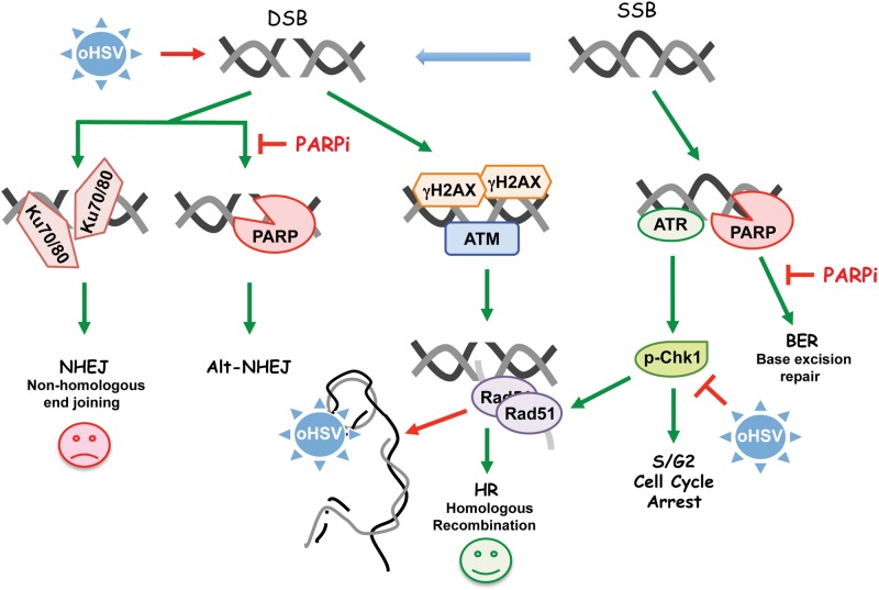
Background: Clinical success of poly(ADP-ribose) polymerase inhibitors (PARP i ) has been limited to repair-deficient cancers and by resistance. Oncolytic herpes simplex viruses (oHSVs) selectively kill cancer cells, irrespective of mutation, and manipulate DNA damage responses (DDR). Here, we explore potential synthetic lethal-like interactions between oHSV and PARP i .

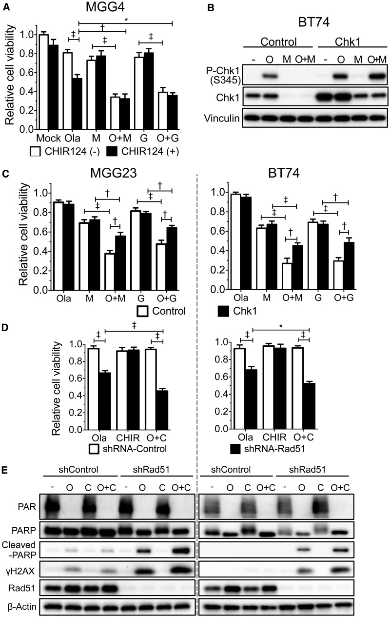
Methods: The efficacy of combining PARP i , oHSV MG18L, and G47Δ in killing patient-derived glioblastoma stem cells (GSCs) was assessed using cell viability assays and Chou-Talalay synergy analysis. Effects on DDR pathways, apoptosis, and cell cycle after manipulation with pharmacological inhibitors and lentivirus-mediated knockdown or overexpression were examined by immunoblotting and FACS. In vivo efficacy was evaluated in two GSC-derived orthotopic xenograft models (n = 7-8 per group). All statistical tests were two-sided.

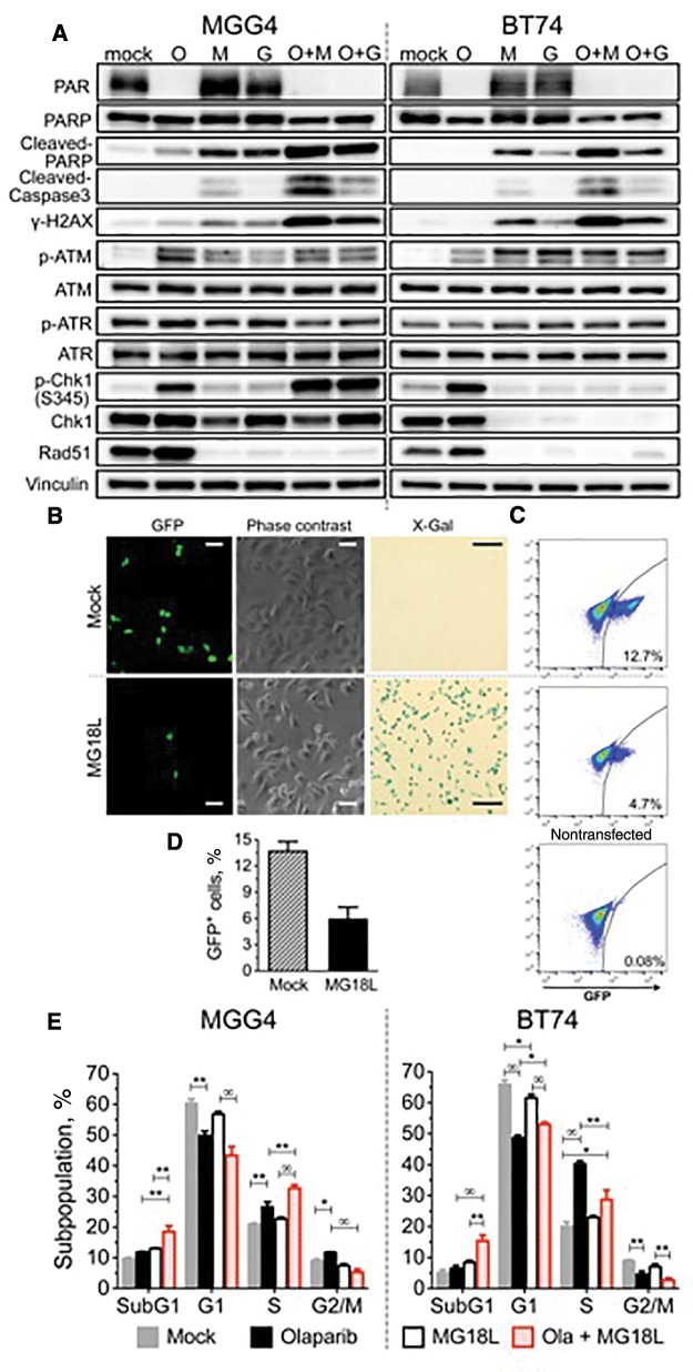
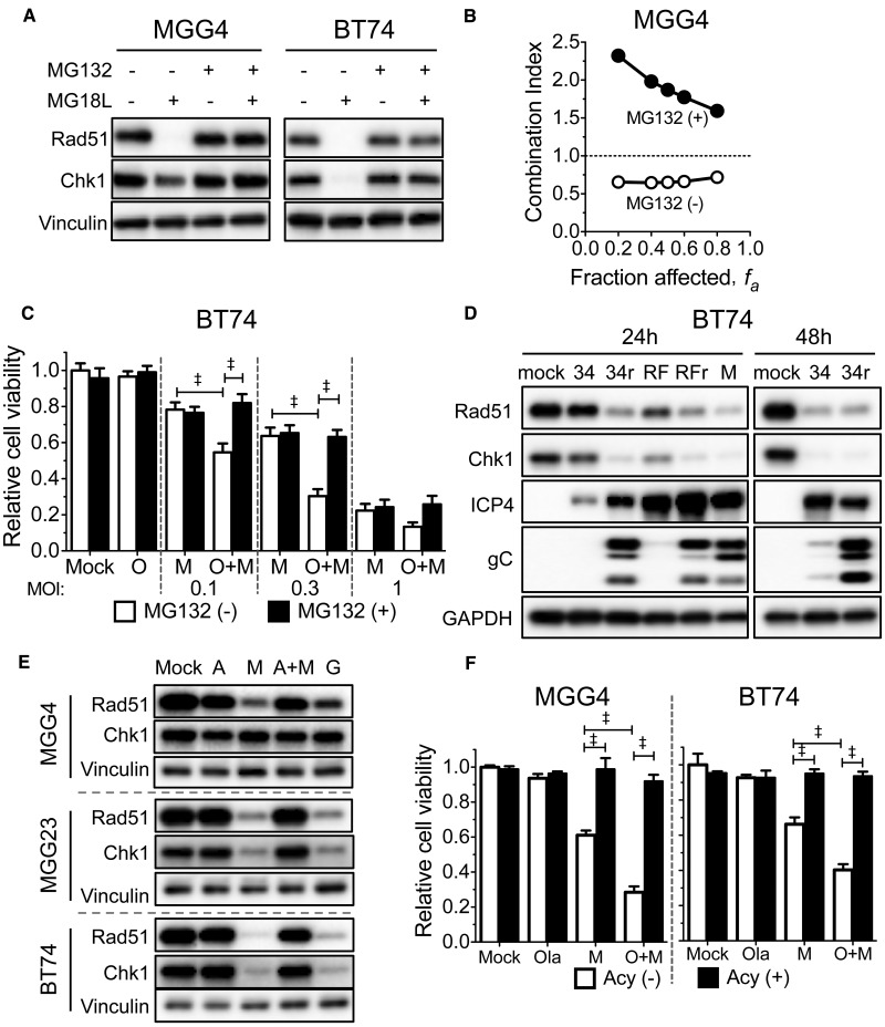
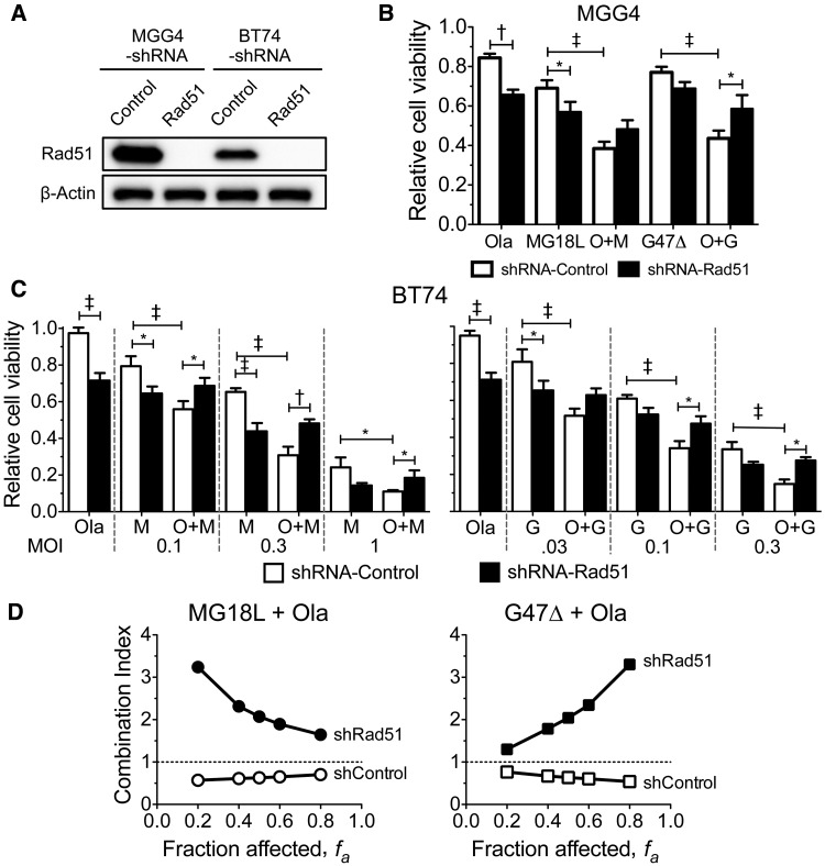
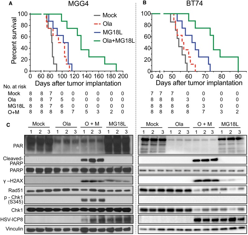
Results: GSCs are differentially sensitive to PARP i despite uniform inhibition of PARP activity. oHSV sensitized GSCs to PARP i , irrespective of their PARP i sensitivity through selective proteasomal degradation of key DDR proteins; Rad51, mediating the combination effects; and Chk1. Rad51 degradation required HSV DNA replication. This synthetic lethal-like interaction increased DNA damage, apoptosis, and cell death in vitro and in vivo. Combined treatment of mice bearing PARP i -sensitive or -resistant GSC-derived brain tumors greatly extended median survival compared to either agent alone (vs olaparib: P ≤.001; vs MG18L: P = .005; median survival for sensitive of 83 [95% CI = 77 to 86], 94 [95% CI = 75 to 107], 102 [95% CI = 85 to 110], and 131 [95% CI = 108 to 170] days and for resistant of 54 [95% CI = 52 to 58], 56 [95% CI = 52 to 61], 62 [95% CI = 56 to 72], and 75 [95% CI = 64 to 90] days for mock, PARPi, oHSV, and combination, respectively).

Conclusions: The unique oHSV property to target multiple components of DDR generates cancer selective sensitivity to PARP i . This combination of oHSV with PARP i is a new anticancer strategy that overcomes the clinical barriers of PARP i resistance and DNA repair proficiency and is applicable not only to glioblastoma, an invariably lethal tumor, but also to other tumor types.


Figures

Figure 1.
Figure 1.
Effect of poly(ADP-ribose) polymerase inhibitors (PARPis) on glioblastoma stem cell (GSC) cytotoxicity and PARP activity. A) Dose response curves for PARPis. GSCs were plated at 5000 cells/well, except MGG24 and BT74 at 8000 cells/well, and treated the next day with indicated PARPis (olaparib, veliparib, rucaparib, and BMN673) at different doses for six days, followed by MTS assay for cell viability. Nonlinear regression curves (log(inhibitor) vs response) were plotted. B) Dose response curves for PARPis on normal human astrocytes. Cells were plated at 3000 cells/well and treated as in (A). C) PARP activity, as measured by PARP Assay Kit, was inhibited in all GSCs after olaparib treatment (Ola (+), 30 μM) for 24 hours. Data are represented as mean ± SD. D) PARylated proteins (PAR), a measure of PARP activity, were detected by immunoblotting after treatment with indicated doses of olaparib for 24 hours in MGG4 and BT74. β-actin is loading control. Ola = olaparib; PARP = poly(ADP-ribose) polymerase.
Figure 2.
Figure 2.
The interaction of olaparib with MG18L and G47Δ in killing poly(ADP-ribose) polymerase inhibitor (PARPi)–sensitive and –resistant glioblastoma stem cells (GSCs). A) Dose response curves for MG18L (left) and G47Δ (right) in the indicated GSCs, determined as in Figure 1A. Combination of olaparib and MG18L or G47Δ in MGG4 (B), MGG23 (C), BT74 (D), MGG24 (E), and normal astrocytes (F). Left: The fixed dose of MG18L was MOI = 0.04, 0.001, 0.05, and 0.05 for (B), (C), (D), and (E), respectively, indicated with blue arrow. Middle: The fixed dose of olaparib was 1, 1, 10, and 10 µM for (B), (C), (D), and (E), respectively, indicated with brown arrow. Right: B and C) Interaction between olaparib (Ola) and MG18L or G47Δ in MGG4 and MGG23, as determined by the Chou-Talalay median effect method (19,40). Combination index < 1, = 1, and > 1 indicates synergistic, additive, and antagonistic interactions, respectively. Right: D and E) Combination of olaparib and G47Δ in BT74 (Ola = 10 μM) and MGG24 (Ola = 5 μM). Increasing virus dose is statistically significantly different from the previous dose, with or without olaparib (P < .0001). *P = .004; †P < .001; ‡P < .0001 (multiple comparisons test, Tukey). F) Combination of olaparib (10 μM, Ola (+)) and MG18L or G47Δ (0.1 MOI) in astrocytes. Cell viability was determined by MTS assay after six-day treatment and represented as mean ± SD. All statistical tests were two-sided. MOI = multiplicity of infection; Ola = olaparib; PARP = poly(ADP-ribose) polymerase.
Figure 3.
Figure 3.
Effect of oncolytic herpes simplex virus, poly(ADP-ribose) polymerase inhibitor (PARPi), and combination on DNA damage responses and cell cycle. A) PARPi-sensitive MGG4 (left) and PARPi-resistant BT74 (right) cells were treated with olaparib (O; 10 µM for MGG4 and 30 µM for BT74) or vehicle (mock) for 48 hours and then mock-infected or infected with MG18L (M) or G47Δ (G; MOI = 1) or in combination (O + M, O + G). Cells were harvested for immunoblotting at 30 hours after infection. β-actin as loading control. B) Effect of MG18L on homologous recombination. DR-GFP-transduced U2OS cells were transfected with a plasmid expressing I-SceI, and 24 hours later infected with MG18L (1MOI) or mock (PBS), followed 24 hours later by analysis of GFP-positive cells with fluorescence (left) and phase-contrast microscopy (middle). X-gal staining performed at 16 hours after MG18L infection (right), showing that almost all the cells were infected. Scale bars = 1 μm for GFP and phase contrast and 5 μm for X-gal. C) Representative fluorescence-activated cell sorting (FACS) analysis of GFP-positive cells (gated right quadrant, percent positive) from mock (top), MG18L infected (middle), and control nontransfected (lower). Total of 1X 10⁵ cells were analyzed for each sample. D) Quantification of GFP-positive cells analyzed by FACS as in (C) from three independent experiments. Data represented as mean ± SD; P = .002 (two-sided unpaired t test). E) Cell cycle analysis of treated MGG4 (left) and BT74 (right). Cells were treated as indicated with olaparib (3 μM for MGG4 and 30 μM for BT74) and/or MG18L (MOI = 0.5) and cell cycle phases determined after 24 hours by FACS. Values are the mean of three independent experiments and represented as mean ± SD. *P < .01; **P < .001; ∞P< .0001. P values of .01 or greater are not indicated (multiple comparisons test, Tukey). In MGG4: mock vs olaparib for G2/M, P = .004. In BT74: mock vs MG18L and olaparib vs Ola+MG18L for G1, P = .005; mock vs Ola+MG18L for S, P = .002. All statistical tests were two-sided. Ola = olaparib; PARP = poly(ADP-ribose) polymerase.
Figure 4.
Figure 4.
The role of Rad51 and Chk1 degradation and viral DNA replication on synergy between oncolytic herpes simplex viruses (oHSV) and olaparib. A) MG132 blocks MG18L-induced degradation of Rad51 and Chk1. MGG4 and BT74 cells were treated with MG18L (MOI = 1) and/or MG132 (1 μM) as indicated for 30 hours before harvesting for immunoblot analysis and probed with antibodies to Rad51, Chk1, and vinculin as loading control. B) Requirement for proteasomal activity on synergy. MGG4 cells were treated with olaparib, MG18L, or the combination in the absence (open circles) or presence of 0.05 μM MG132 (+MG132, filled circles) for six days. Cell viability was measured by MTS assay, and the combination index determined. Combination Index < 1, = 1, and > 1 indicates synergistic, additive, and antagonistic interactions, respectively. C) BT74 cells were treated with olaparib (O; 10 μM), MG18L (M; at the doses indicated), or combination (O + M), in the absence (MG132 (-)) or presence of MG132 (0.1 μM; MG132 (+)) for six days, followed by MTS assay for cell viability, represented as mean ± SD. ‡P < .0001 (multiple comparisons tests, Tukey) between indicated pairs. D) BT74 cells were mock-infected or infected with HSV ICP0 mutants 7134 (34) and ICP0-RING finger domain mutant virus KOS RFm (RF), or rescued wild-type HSV 7134R (34r) and KOS RFr (RFr), or MG18L (M) with MOI = 10, except 7134R at 48 hours (MOI = 1). Cells were harvested for immunoblotting at 24 hours or 48 hours after infection. Membranes were probed with antibodies to Rad51, Chk1, ICP4, gC, and GAPDH as loading control. E) Acyclovir treatment blocks MG18L-induced Rad51 and Chk1 degradation. Glioblastoma stem cells (MGG4, MG23, BT74) were treated with MG18L (M, A + M; MOI = 1), G207 (G; MOI = 1), and/or acyclovir (A, A + M; 10 μM), for 30 hours before harvesting. Membranes were probed with antibodies to Rad51, Chk1, and vinculin as loading control. F) Acyclovir abrogates the combination effect of oHSV with poly(ADP-ribose) polymerase inhibitor. MGG4 (left) and BT74 (right) cells were treated with olaparib (O; 1 μM for MGG4 and 10 μM for BT74), MG18L (M; 0.05 MOI for MGG4 and 0.3 MOI for BT74), or combination (O + M) in the absence (Acy (-)) or presence of acyclovir (5 μM; A, Acy (+)) for six days, followed by MTS assay for cell viability, represented as mean ± SD. ‡P < .0001 (multiple comparisons test, Tukey) between indicated pairs. All statistical tests were two-sided.
Figure 5.
Figure 5.
Role of Rad51 knockdown on synergy between olaparib and oncolytic herpes simplex viruses. A) MGG4 or BT74 cells transduced with lentivirus expressing Rad51- or control-shRNA were grown under puromycin selection for seven days prior to immunoblot analysis. β-actin is loading control. B) MGG4 cells transduced with shRNA-control or shRNA-Rad51 were treated with olaparib (Ola; 1 μM), MG18L (0.04 MOI), G47Δ (0.04 MOI), or the combination (O + M, O + G) for six days, and cell viability was measured by MTS assay and represented as mean ± SD. *P < .01; †P < .001; ‡P < .0001 (multiple comparisons test, Tukey) between indicated pairs. MG18L shRNA-control vs shRNA-Rad51 (P = .02) and O + G shRNA-control vs shRNA-Rad51 (P = .02). C) BT74 cells transduced with shRNA-control or shRNA-Rad51 were treated with olaparib (Ola; 10 μM), MG18L (left; M), G47Δ (right; G), or combination (O + M, O + G) at the MOIs indicated for six days, and cell viability was measured by MTS assay and represented as mean ± SD. *P < .05; †P < .001; ‡P < .0001 (multiple comparisons test, Tukey) between indicated pairs. The combination of O + G statistically significantly increased viability with shRNA-Rad51 vs shRNA-control; P = .05, .01, .02 for MOIs of .03, .1, .3, respectively. The combination of O + M statistically significantly increased viability with shRNA-Rad51 vs shRNA-control; P = .02, .0005, .02 for MOIs of 0.1, 0.3, 1, respectively. M (1 MOI, shRNA-control) vs O + M (1 MOI, shRNA-control), P = .01; M (0.1 MOI, shRNA-control) vs M (0.1 MOI, shRNA-Rad51), P = .004; and G (0.03 MOI, shRNA-control) vs G (0.03 MOI, shRNA-Rad51), P = .002. D) Rad51 shRNA abrogates synergy. Interaction of olaparib and MG18L (left) or G47Δ (right) in MGG4 transduced with shRNA-control (shcontrol) or shRNA-Rad51 (shRad51). Combination index < 1, = 1, and > 1 indicates synergistic, additive, and antagonistic interactions, respectively. All statistical tests were two-sided. Ola = olaparib.
Figure 6.
Figure 6.
The role of Chk1 on poly(ADP-ribose) polymerase (PARP) inhibitor sensitivity of glioblastoma stem cells and in combination with oncolytic herpes simplex virus (oHSV). A) Chk1 inhibitor CHIR-124 sensitizes MGG4 to olaparib but not combination with oHSV. Olaparib (Ola; 1μM), MG18L (M; 0.04 MOI), G47Δ (G; 0.04 MOI), or the combination (O + M and O + G), with (CHIR124 (+), 0.1 μM) or without CHIR-124 (CHIR-124 (-)) for six days, and viability was measured by MTS assay. With CHIR-124, Ola was statistically significantly different from O + G (P = .003). B) Immunoblot analysis of Chk1-overexpressing BT74 (BT74-Chk1) or control-transduced (BT74-control) cells treated with mock (-), olaparib (O; 30 μM), MG18L (M; MOI = 1), or combination (O + M) and harvested after 30 hours. C) Chk1 overexpression reduces combination effect. Left: MGG23 transduced with control or Chk1 (Fig S6D) and treated with olaparib (O; 2 μM), MG18L (M; 0.002 MOI), G47Δ (G; 0.1 MOI), or combination (O + M, O + G). Right: Transduced BT74 treated with olaparib (10 μM), MG18L (0.3 MOI), G47Δ (0.1 MOI). MTS assay was performed for cell viability after six-day treatment. D) MGG23 (left) and BT74 (right), transduced with shRNA-control or shRNA-Rad51, were treated with olaparib (Ola; 2, 10 μM) or CHIR-124 (CHIR; 0.01, 1.5 μM), respectively, or combination (O + C) for six days before MTS assay. For BT74, in the presence of shRNA-Rad51, O + C was statistically significantly different from Ola (P = .001). E) Immunoblot analysis of shRNA-control or shRNA-Rad51 transduced MGG23 (left) treated with mock (-), olaparib (O; 10 μM), CHIR-124 (C; 0.1 μM), or combination (O + C) or BT74 (right) treated with olaparib (30 μM) or CHIR124 (0.2 μM). Membranes were probed with antibodies to PAR, PARP, cleaved PARP, γH2AX, Rad51, and β-actin as loading control. Cell viability is represented as mean ± SD. *P < .01; †P < .001; ‡P < .0001 (multiple comparisons test, Tukey) between indicated pairs. All statistical tests were two-sided.
Figure 7.
Figure 7.
Efficacy of combination therapy in glioblastoma stem cell–derived intracerebral tumors. A) Mice implanted intracerebrally with 2 x 10⁵ MGG4 cells were treated with olaparib (Ola; 50 mg/kg) in 10% DMSO/10% 2-hydroxyl-propyl-β-cyclodextrine/PBS or vehicle, administered intraperitoneally starting on day 17 postimplantation with six cycles of five-day on and two-day off, and MG18L (1 X 10⁶ pfu) or PBS intratumorally injected on day 19. Mock vs olaparib (P = .03) or vs MG18L (P = .003); olaparib + MG18L vs MG18L (P = .005) or vs olaparib (P = .001); log-rank test. B) Mice implanted intracerebrally with 1 x 10⁵ BT74 cells were treated with olaparib or vehicle administered intraperitoneally daily for 26 days starting on day 9 postimplantation, and MG18L or PBS intratumorally injected on day 11. Mock vs MG18L (P = .009); olaparib + MG18L vs MG18L (P = .005) or vs olaparib (P = .0004); log-rank test. C) DNA damage responses induced by olaparib (Ola), MG18L, or combination (O + M) in vivo. Intracerebral tumors were established with MGG4 (left) and treated with olaparib (Ola) or vehicle (mock) intraperitoneally starting at day 61 after implantation for five days and/or MG18L (2 X 10⁶ pfu) injected on day 63, and harvested on day 65. BT74 tumors (right) were treated similarly, except with olaparib starting on day 42, MG18L injected on day 44, and death on day 46. Lysates from tumor-bearing hemispheres from individual mice (1, 2, 3) were electrophoresed and probed with antibodies to PAR, poly(ADP-ribose) polymerase (PARP), cleaved PARP, γH2AX, Rad51, p-Chk1 (S345), Chk1, HSV-ICP8, and vinculin as loading control. All statistical tests were two-sided. Ola = olaparib; PARP = poly(ADP-ribose) polymerase.
Figure 8.
Figure 8.
Model for combination therapy of poly(ADP-ribose) polymerase inhibitors (PARPi) and inducing synthetic lethality in glioblastoma stem cells. Oncolytic herpes simplex virus (oHSV) induces DSBs (detected by γH2AX) that activate ATM to recruit repair proteins. oHSV DNA replication promotes the degradation of Rad51 and Chk1, blocking homologous recombination and disturbing cell cycle processes. DNA single strand breaks (SSBs) activate ATR, which phosphorylates Chk1. PARP binds to DNA breaks to facilitate BER and Alt-NHEJ and blocks Ku protein binding for NHEJ. Therefore, PARPis inhibit BER and facilitate SSB conversion to DSB, and inhibit Alt-NHEJ and facilitate NHEJ, which is detrimental. Red perpendicular mark indicates inhibition (degradation), green arrow indicates activation, and red arrow indicates detrimental activity. BER = base excision repair; DSB = DNA double-strand break; HR = homologous recombination; NHEJ = nonhomologous end joining; oHSV = oncolytic herpes simplex virus; PARPi = poly(ADP-ribose) polymerase inhibitor; SSB = DNA single-strand break.

Similar articles

Cited by

References

    1. O'Connor MJ. Targeting the DNA damage response in cancer. Mol Cell. 2015;60(4):547–560. - PubMed
    1. Scott CL, Swisher EM, Kaufmann SH.. Poly (ADP-ribose) polymerase inhibitors: Recent advances and future development. J Clin Oncol. 2015;33(12):1397–1406. - PMC - PubMed
    1. Dietlein F, Thelen L, Reinhardt HC.. Cancer-specific defects in DNA repair pathways as targets for personalized therapeutic approaches. Trends Genet. 2014;30(8):326–339. - PubMed
    1. Lord CJ, Tutt AN, Ashworth A.. Synthetic lethality and cancer therapy: Lessons learned from the development of PARP inhibitors. Annu Rev Med. 2015;66:455–470. - PubMed
    1. Smith S, Weller SK.. HSV-I and the cellular DNA damage response. Future Virol. 2015;10(4):383–397. - PMC - PubMed

MeSH terms