

Alterations in endo-lysosomal function induce similar hepatic lipid profiles in rodent models of drug-induced phospholipidosis and Sandhoff disease
- PMID: 28377426
- PMCID: PMC5496029
- DOI: 10.1194/jlr.M073395
Alterations in endo-lysosomal function induce similar hepatic lipid profiles in rodent models of drug-induced phospholipidosis and Sandhoff disease
Abstract
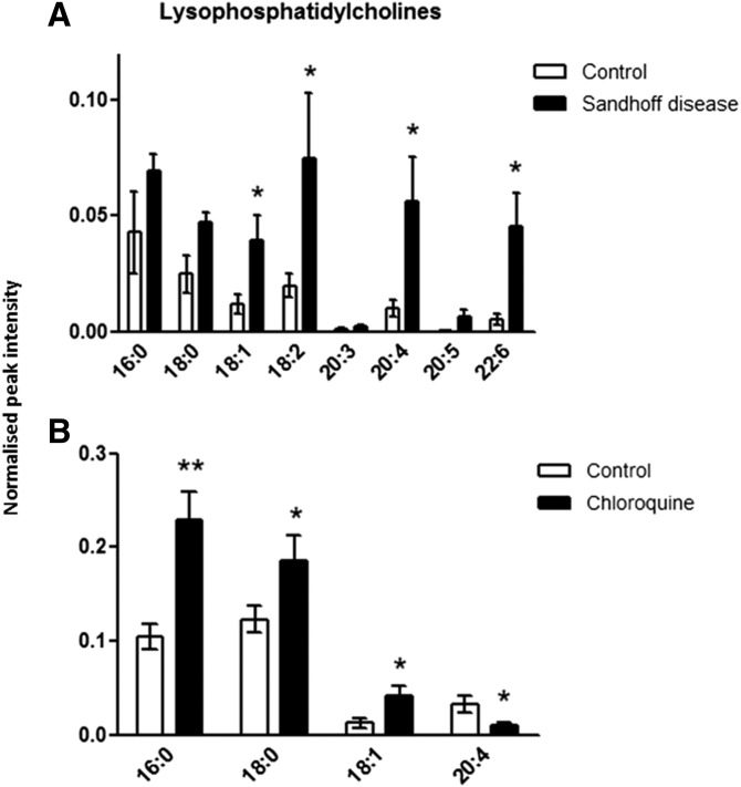
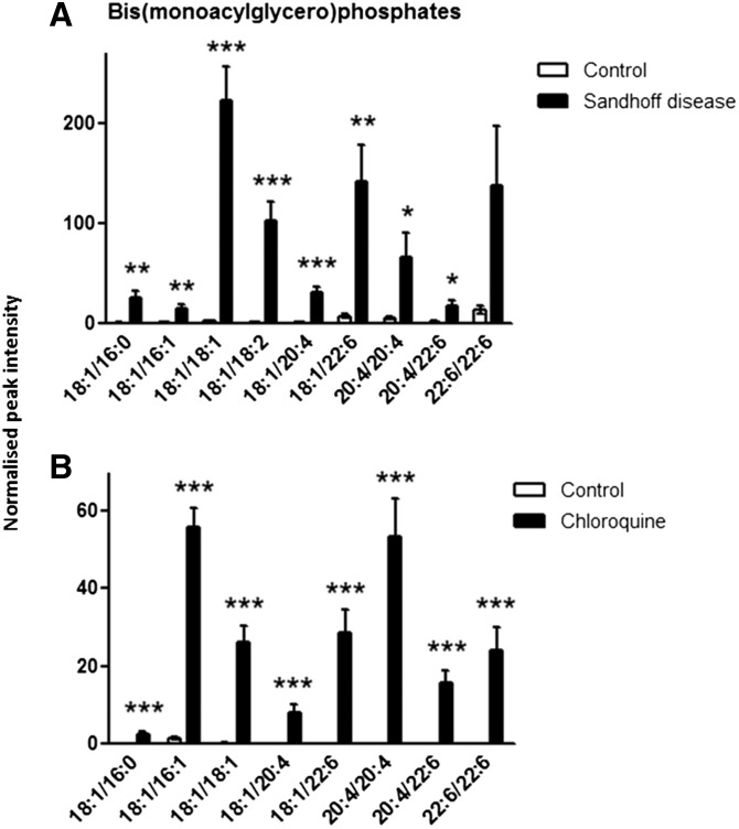
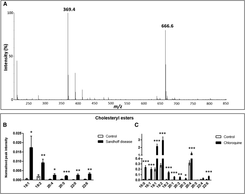
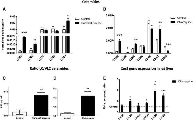
Drug-induced phospholipidosis (DIPL) is characterized by an increase in the phospholipid content of the cell and the accumulation of drugs and lipids inside the lysosomes of affected tissues, including in the liver. Although of uncertain pathological significance for patients, the condition remains a major impediment for the clinical development of new drugs. Human Sandhoff disease (SD) is caused by inherited defects of the β subunit of lysosomal β-hexosaminidases (Hex) A and B, leading to a large array of symptoms, including neurodegeneration and ultimately death by the age of 4 in its most common form. The substrates of Hex A and B, gangliosides GM2 and GA2, accumulate inside the lysosomes of the CNS and in peripheral organs. Given that both DIPL and SD are associated with lysosomes and lipid metabolism in general, we measured the hepatic lipid profiles in rodent models of these two conditions using untargeted LC/MS to examine potential commonalities. Both model systems shared a number of perturbed lipid pathways, notably those involving metabolism of cholesteryl esters, lysophosphatidylcholines, bis(monoacylglycero)phosphates, and ceramides. We report here profound alterations in lipid metabolism in the SD liver. In addition, DIPL induced a wide range of lipid changes not previously observed in the liver, highlighting similarities with those detected in the model of SD and raising concerns that these lipid changes may be associated with underlying pathology associated with lysosomal storage disorders.
Keywords: ceramides; lipidomics; lysophospholipid; lysosome; mass spectrometry; storage diseases; toxicology.
Copyright © 2017 by the American Society for Biochemistry and Molecular Biology, Inc.
Figures
References
-
- Nelson A. A., and Fitzhugh O. G.. 1948. Chloroquine (SN-7618) pathologic changes observed in rats which for 2 years had been fed various proportions. Arch. Pathol. (Chic). 45: 454–462. - PubMed
-
- Kodavanti U. P., and Mehendale H.. 1990. Cationic amphiphilic drugs and phospholipid storage disorder. Pharmacol. Rev. 42: 327–354. - PubMed
-
- Reasor M. J., and Kacew S.. 2001. Drug-induced phospholipidosis: are there functional consequences? Exp. Biol. Med. (Maywood) 226: 825–830. - PubMed
-
- Dake M. D., Madison J. M., Montgomery C. K., Shellito J. E., Hinchcliffe W., Winkler M. L., and Bainton D. F.. 1985. Electron microscopic demonstration of lysosomal inclusion bodies in lung, liver, lymph nodes, and blood leukocytes of patients with amiodarone pulmonary toxicity. Am. J. Med. 78: 506–512. - PubMed
-
- Nonoyama T., and Fukuda R.. 2008. Drug-induced phospholipidosis -pathological aspects and its prediction. J. Toxicol. Pathol. 21: 9–24.
Publication types
MeSH terms
Substances
Grants and funding
LinkOut - more resources
Full Text Sources
Other Literature Sources
Molecular Biology Databases
Miscellaneous
