. 2017 Apr 4;8(2):e00040-17.
doi: 10.1128/mBio.00040-17.

Bactericidal Permeability-Increasing Proteins Shape Host-Microbe Interactions

Affiliations

Bactericidal Permeability-Increasing Proteins Shape Host-Microbe Interactions

Fangmin Chen et al. mBio. .

Abstract

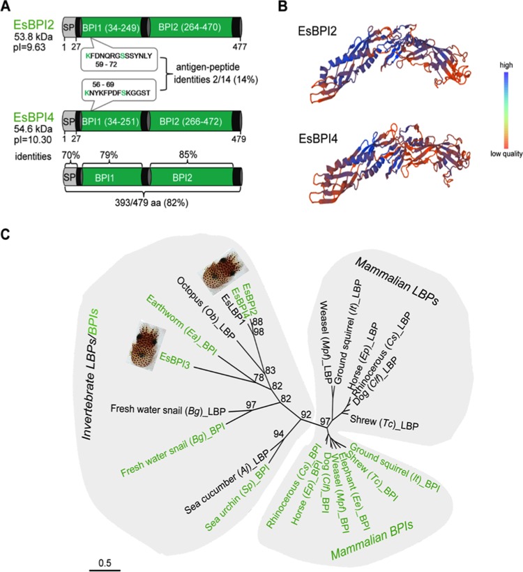
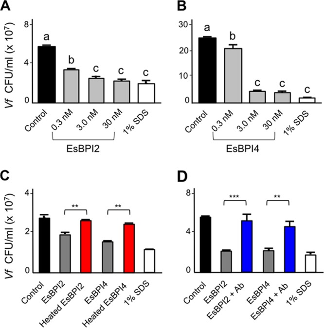
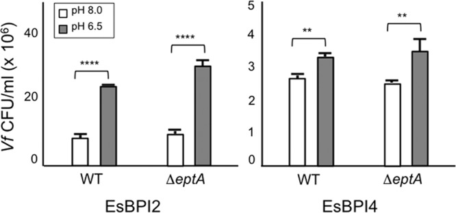
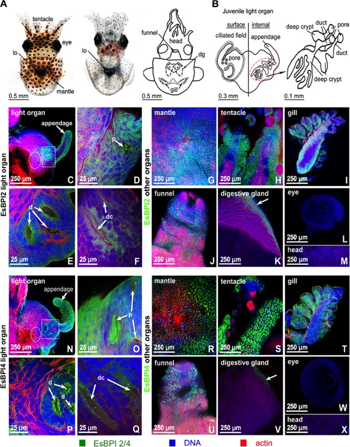
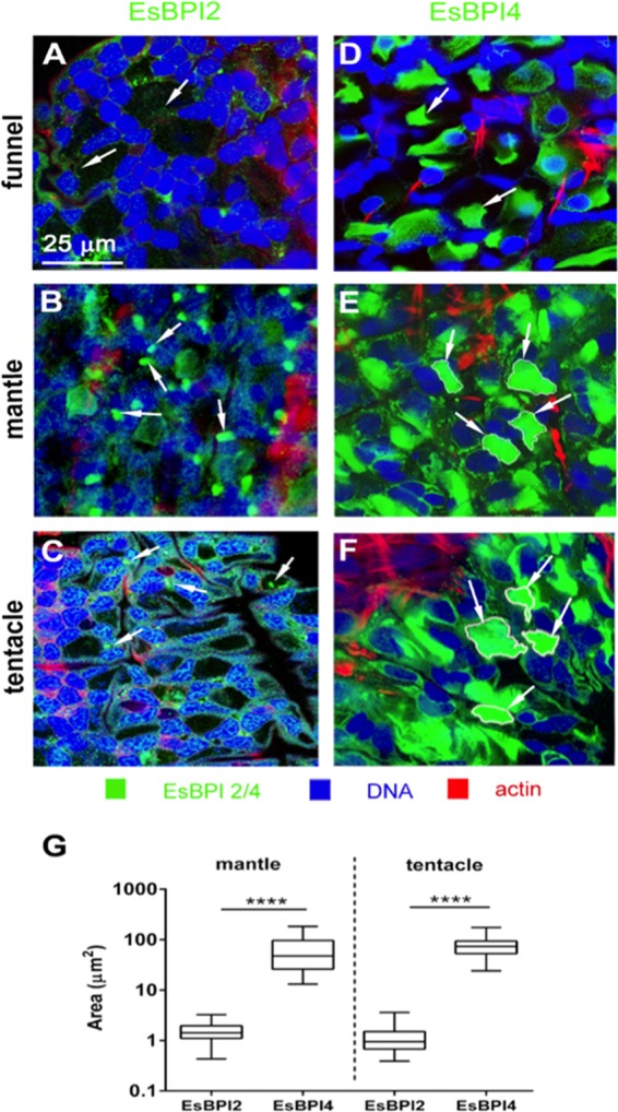
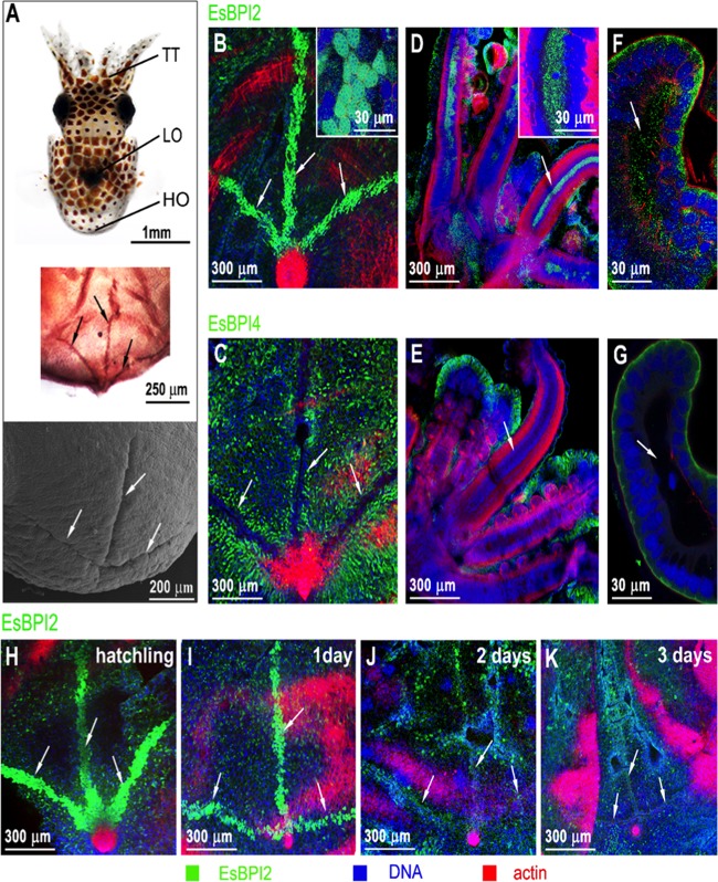
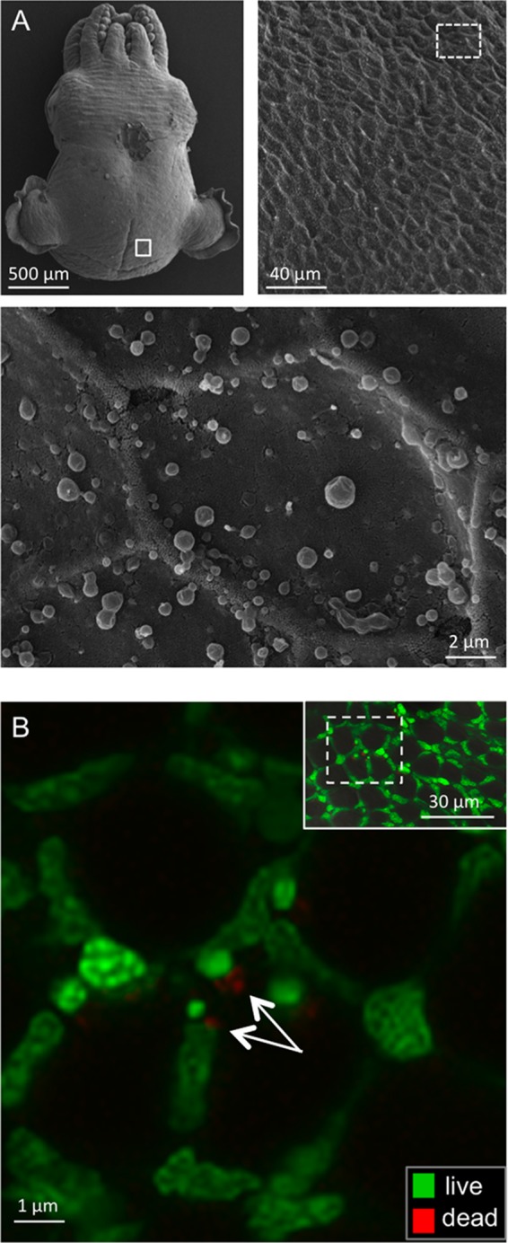
We characterized bactericidal permeability-increasing proteins (BPIs) of the squid Euprymna scolopes, EsBPI2 and EsBPI4. They have molecular characteristics typical of other animal BPIs, are closely related to one another, and nest phylogenetically among invertebrate BPIs. Purified EsBPIs had antimicrobial activity against the squid's symbiont, Vibrio fischeri, which colonizes light organ crypt epithelia. Activity of both proteins was abrogated by heat treatment and coincubation with specific antibodies. Pretreatment under acidic conditions similar to those during symbiosis initiation rendered V. fischeri more resistant to the antimicrobial activity of the proteins. Immunocytochemistry localized EsBPIs to the symbiotic organ and other epithelial surfaces interacting with ambient seawater. The proteins differed in intracellular distribution. Further, whereas EsBPI4 was restricted to epithelia, EsBPI2 also occurred in blood and in a transient juvenile organ that mediates hatching. The data provide evidence that these BPIs play different defensive roles early in the life of E. scolopes, modulating interactions with the symbiont.IMPORTANCE This study describes new functions for bactericidal permeability-increasing proteins (BPIs), members of the lipopolysaccharide-binding protein (LBP)/BPI protein family. The data provide evidence that these proteins play a dual role in the modulation of symbiotic bacteria. In the squid-vibrio model, these proteins both control the symbiont populations in the light organ tissues where symbiont cells occur in dense monoculture and, concomitantly, inhibit the symbiont from colonizing other epithelial surfaces of the animal.

Keywords: Vibrio fischeri; antimicrobial peptides; bioinformatics; confocal microscopy; symbiosis.


Figures

FIG 1
FIG 1
Characteristics of the EsBPI2 and EsBPI4. (A) Domain structure with full-sequence and domain alignment (SP, signal peptide; BPI1 and BPI2, bactericidal permeability-increasing domains). The boxes show the locations and sequences of the variable regions used for production of antibodies to EsBPI2 and EsBPI4. aa, amino acids; pI, isoelectric point; kDa, kilodaltons. (B) Model of EsBPI2/4 showing the characteristic “boomerang” shape predicted from human BPI templates (SWISS-MODEL template ID 1ewf.1.A and 1bp1.1.A, respectively). The color gradient indicates the structure quality by local QMEAN4 score (46). (C) An unrooted LBP/BPI phylogenetic tree produced by the maximum likelihood method (see Table S2 in the supplemental material for the species used and associated accession numbers). The numbers are aLRT values, which approximate likelihood ratio test scores (48) as percentages, and represent the level of confidence associated with the branch at a given node. The bar shows branch length distances as the number of amino acid substitutions per site.
FIG 2
FIG 2
Bactericidal activity of EsBPI2 and EsBPI4 against V. fischeri. (A and B) Survival of wild-type V. fischeri strain ES114 (Vf) following 1-h incubation with different concentrations of EsBPI2 (A) or EsBPI4 (B) compared to incubation with a chemical cell-permeant (SDS). Values are shown as means plus standard errors of the means (sem) (error bars) for three biological replicates, each with three technical replicates. Bars labeled with different letters are statistically significantly different (P < 0.001). (C and D) Effect of a pretreatment with either heat (70°C for 10 min; red) (C) or preincubation with the cognate antibody (Ab; blue) (D) on the killing of V. fischeri by a 30 nM concentration of either EsBPI2 or EsBPI4 after 1 h of incubation. Values are shown as means plus sem (error bars) for three biological replicates, each with three technical replicates. Pairwise comparisons of viability (CFU/milliliter) for untreated protein/heat-denatured protein (C) and untreated protein/preadsorbed with its cognate antibody protein (D) for each protein were tested by a one-way ANOVA, followed by a Tukey’s multiple-comparison test and are indicated as follows: **, P < 0.01; ***, P < 0.001. For each protein, heat or antibody treatment resulted in an activity not significantly different from that of the control (black).
FIG 3
FIG 3
Effect of low-pH pretreatment on V. fischeri susceptibility to EsBPI2 and EsBPI4. Survival of either wild-type (WT) or an eptA mutant derivative of V. fischeri strain ES114 exposed to EsBPI2/4. The two strains were cultured for 3 h in SWT adjusted to either pH 8.0 or 6.5 with 20 mM Tris-HCl prior to a 1-h incubation with one of the EsBPIs. Values are shown as means plus sem (error bars) for three biological replicates, each with three technical replicates. Significant differences in viability (CFU/milliliter) between the two strains with different pH treatments were tested by a two-way ANOVA and are indicated as follows: **, P < 0.01; ****, P < 0.0001.
FIG 4
FIG 4
Localization of EsBPI2 and EsBPI4 in juvenile squid tissues. Proteins were probed with rabbit anti-EsBPI2 or anti-EsBPI4 antibodies, which were detected with FITC-conjugated goat anti-rabbit secondary antibody (EsBPI2/4 [green]), and counterstained with rhodamine-phalloidin (actin cytoskeleton [red]) and TOTO-3 (blue) (nuclei). (A) Dorsal and ventral views of the juvenile squid, and diagram to illustrate internal regions depicted in the following panels of the figure. In the diagram, the digestive gland (dg) is shown. lo, light organ. (B) Enlargement of the light organ showing the surface in contact with the environment (left) and the internal structure (right) where symbionts colonize. (Right) Anatomical structures of the three crypts indicated by the dashed red circle. (C to F) EsBPI2 ICC labeling at the top surface of the light organ (C), in the pore (p) (white arrow) (D), in the ducts (d) (white arrows) (area of white dashed box in panel C) (E), and in the deep crypts (dc) (deep crypt lumen indicated by white arrows) (white dashed circle in panel C, which is tissue superficial to the deep crypts) (F). (G to M) Localization of EsBPI2 ICC labeling in other organs examined. The white arrow points to the epithelial edge of the digestive gland. (N to Q) EsBPI4 ICC labeling on the top surface of the light organ (N), in the pores (p) (white arrows) (O), in the ducts (d) (white arrows) (white dashed box area in panel N) (P), and in the deep crypts (dc) (white arrows indicate deep crypt lumen) (white dashed circle in panel N, which is tissue superficial to the deep crypts) (Q). (R to X) Localization of EsBPI4 ICC labeling in other organs examined. The white arrow points to the epithelial edge of the digestive gland.
FIG 5
FIG 5
Subcellular localization of EsBPI2 and EsBPI4 by ICC. (See the legend to Fig. 4 for ICC methods.) (A to C) Typical punctate cytosolic ICC labeling of EsBPI2 (white arrows); (D to F) large stores of EsBPI4 in the cytosol (white arrows). (G) Size of labeled areas within cells. The area of cytosolic ICC labeling of EsBPI2 and EsBPI4 was determined by examining 30 regions in each tissue for three different animals. Values that are significantly different (P < 0.0001) are indicated by a bar and four asterisks.
FIG 6
FIG 6
EsBPI2 is unique in the blood and Hoyle organ. (See the legend to Fig. 4 for ICC methods.) (A) Hoyle organ location. (Top) Position of the Hoyle organ (HO) in the body, relative to the tentacles (TT) and light organ (LO). (Middle) Low-magnification light micrograph of the posterior end of the animal showing the trifurcated branching of the HO. (Bottom) Low-magnification scanning electron micrograph, showing the surface view of the HO branches. (B) ICC labeling of EsBPI2 in the HO. (C) Exclusion of EsBPI4 from the HO. (D and E) EsBPI2 ICC labeling of blood sinuses of the tentacles (D) and lack of EsBPI4 ICC labeling of these sites (E). (F and G) EsBPI2 ICC labeling of blood sinuses of the juvenile light-organ superficial appendages (F) and lack of EsBPI4 ICC labeling of these sites (G). Insets in panels B and D show higher-magnification images of ICC labeling of EsBPI2 in the Hoyle organ and blood sinus of the tentacle, respectively. (H to K) The typical loss of the HO in early squid development (29, 31) and concomitant loss of EsBPI2 labeling.
FIG 7
FIG 7
Detection of microbes associating with the outer mantle surface of E. scolopes. (A) Scanning electron microscopy (SEM). (Top, right and left) Low-magnification images of the animal showing the region examined for microbes (white boxes). (Bottom) High-magnification image of the epithelial surface (n = 4) (the whole skin of the animal was examined) with no microbes detected. (The irregular, round globules on the surface are mucous secretions from the skin.) (B) Confocal micrographs of a live, anesthetized animal that had been washed with filter-sterilized seawater three times for 5 min each time and then labeled for detection of live (green) or dead (red) bacterial cells. Images were taken in the same surface region as for SEM. (Inset, top right) Low-magnification view to show region (box outlined with white dashed line) examined at higher magnification. White arrows indicate possible vestiges of dead bacterial cells (n = 5).

References

    1. Baron OL, Deleury E, Reichhart JM, Coustau C. 2016. The LBP/BPI multigenic family in invertebrates: evolutionary history and evidences of specialization in mollusks. Dev Comp Immunol 57:20–30. doi:10.1016/j.dci.2015.11.006. - DOI - PubMed
    1. Krasity BC, Troll JV, Weiss JP, McFall-Ngai MJ. 2011. LBP/BPI proteins and their relatives: conservation over evolution and roles in mutualism. Biochem Soc Trans 39:1039–1044. doi:10.1042/BST0391039. - DOI - PMC - PubMed
    1. Schultz H, Hume J, Zhang DS, Gioannini TL, Weiss JP. 2007. A novel role for the bactericidal/permeability increasing protein in interactions of Gram-negative bacterial outer membrane blebs with dendritic cells. J Immunol 179:2477–2484. doi:10.4049/jimmunol.179.4.2477. - DOI - PubMed
    1. Bingle CD, Craven CJ. 2004. Meet the relatives: a family of BPI- and LBP-related proteins. Trends Immunol 25:53–55. doi:10.1016/j.it.2003.11.007. - DOI - PubMed
    1. Chiang SC, Veldhuizen EJ, Barnes FA, Craven CJ, Haagsman HP, Bingle CD. 2011. Identification and characterisation of the BPI/LBP/PLUNC-like gene repertoire in chickens reveals the absence of a LBP gene. Dev Comp Immunol 35:285–295. doi:10.1016/j.dci.2010.09.013. - DOI - PMC - PubMed

MeSH terms

Substances

LinkOut - more resources