. 2017 Apr 4;8(2):e00321-17.
doi: 10.1128/mBio.00321-17.

Catabolism of the Last Two Steroid Rings in Mycobacterium tuberculosis and Other Bacteria

Affiliations

Catabolism of the Last Two Steroid Rings in Mycobacterium tuberculosis and Other Bacteria

Adam M Crowe et al. mBio. .

Abstract

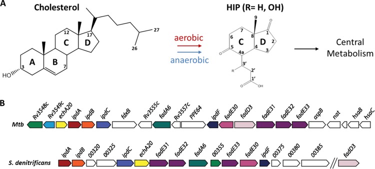
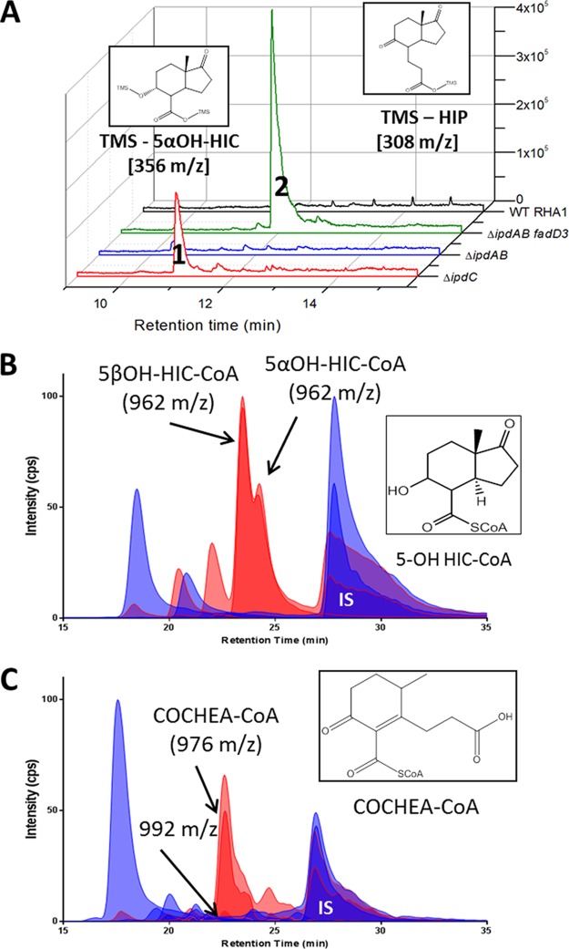
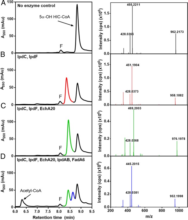
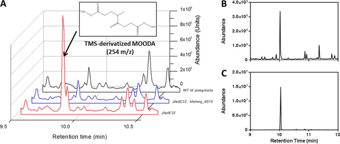
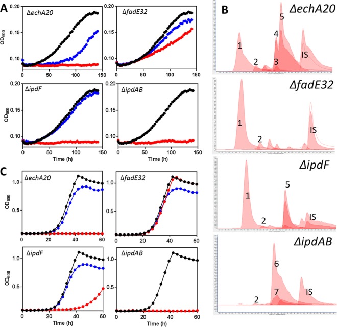
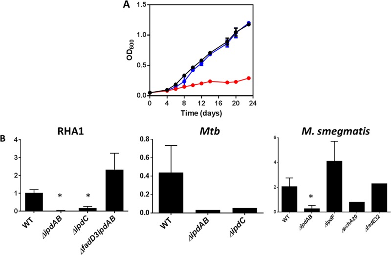
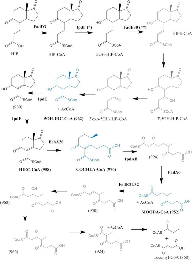
Most mycolic acid-containing actinobacteria and some proteobacteria use steroids as growth substrates, but the catabolism of the last two steroid rings has yet to be elucidated. In Mycobacterium tuberculosis, this pathway includes virulence determinants and has been proposed to be encoded by the KstR2-regulated genes, which include a predicted coenzyme A (CoA) transferase gene (ipdAB) and an acyl-CoA reductase gene (ipdC). In the presence of cholesterol, ΔipdC and ΔipdAB mutants of either M. tuberculosis or Rhodococcus jostii strain RHA1 accumulated previously undescribed metabolites: 3aα-H-4α(carboxyl-CoA)-5-hydroxy-7aβ-methylhexahydro-1-indanone (5-OH HIC-CoA) and (R)-2-(2-carboxyethyl)-3-methyl-6-oxocyclohex-1-ene-1-carboxyl-CoA (COCHEA-CoA), respectively. A ΔfadE32 mutant of Mycobacterium smegmatis accumulated 4-methyl-5-oxo-octanedioic acid (MOODA). Incubation of synthetic 5-OH HIC-CoA with purified IpdF, IpdC, and enoyl-CoA hydratase 20 (EchA20), a crotonase superfamily member, yielded COCHEA-CoA and, upon further incubation with IpdAB and a CoA thiolase, yielded MOODA-CoA. Based on these studies, we propose a pathway for the final steps of steroid catabolism in which the 5-member ring is hydrolyzed by EchA20, followed by hydrolysis of the 6-member ring by IpdAB. Metabolites accumulated by ΔipdF and ΔechA20 mutants support the model. The conservation of these genes in known steroid-degrading bacteria suggests that the pathway is shared. This pathway further predicts that cholesterol catabolism yields four propionyl-CoAs, four acetyl-CoAs, one pyruvate, and one succinyl-CoA. Finally, a ΔipdAB M. tuberculosis mutant did not survive in macrophages and displayed severely depleted CoASH levels that correlated with a cholesterol-dependent toxicity. Our results together with the developed tools provide a basis for further elucidating bacterial steroid catabolism and virulence determinants in M. tuberculosis.IMPORTANCE Bacteria are the only known steroid degraders, but the pathway responsible for degrading the last two steroid rings has yet to be elucidated. In Mycobacterium tuberculosis, this pathway includes virulence determinants. Using a series of mutants in M. tuberculosis and related bacteria, we identified a number of novel CoA thioesters as pathway intermediates. Analysis of the metabolites combined with enzymological studies establishes how the last two steroid rings are hydrolytically opened by enzymes encoded by the KstR2 regulon. Our results provide experimental evidence for novel ring-degrading enzymes, significantly advance our understanding of bacterial steroid catabolism, and identify a previously uncharacterized cholesterol-dependent toxicity that may facilitate the development of novel tuberculosis therapeutics.

Keywords: CoA thioester; Mycobacterium tuberculosis; catabolism; cholesterol; ring opening.


Figures

FIG 1
FIG 1
HIP catabolic genes in representative actinobacteria and proteobacteria. (A) The aerobic catabolism and anaerobic catabolism of cholesterol and other steroids appear to converge at HIP-CoA. (B) The HIP catabolic gene clusters of M. tuberculosis (Mtb) H37Rv and S. denitrificans DSMZ18526. Homologous genes are colored the same. For gene annotation, see Table 1.
FIG 2
FIG 2
Growth of ΔipdAB M. tuberculosis. WT M. tuberculosis Erdman (black), ΔipdAB M. tuberculosis (red), or ΔipdAB::ipdAB M. tuberculosis (blue) cells were grown on (A) 0.5 mM cholesterol, (B) on 0.2% glycerol, or (C) in phorbol myristate acetate (PMA)-differentiated THP-1 cells (MΦ). Data represent the mean from biological triplicates.
FIG 3
FIG 3
Growth of ΔipdC R. jostii RHA1. WT RHA1::pTipQC2 (black), ΔipdC RHA1::pTipQC2 (red), or ΔipdC RHA1::pTipRv3553 (blue) cells were grown on (A) 1 mM cholesterol or (B) 10 mM sodium pyruvate. OD600, optical density at 600 nm.
FIG 4
FIG 4
Accumulation of cholesterol-derived metabolites from ΔipdAB and ΔipdC strains. (A) GC-MS traces of culture supernatants of ΔipdC, ΔipdAB, ΔfadD3 ipdAB, and WT R. jostii RHA1 incubated with cholesterol. Peaks 1 and 2 correspond to TMS-5α-OH HIC and TMS-HIP, respectively. (B and C) CoA metabolome of cholesterol-incubated cells of (B) WT (blue) and ΔipdC (red) RHA1 or (C) WT (blue) and ΔipdAB (red) M. tuberculosis. The major unique peaks in the ΔipdC and ΔipdAB metabolomes correspond to 5αOH-HIC-CoA and COCHEA-CoA, respectively (inset). Lighter-shaded curves in panels B and C are based on the 768→261 transition observed in free CoASH as well as CoA thioesters subjected to in-source fragmentation.
FIG 5
FIG 5
LC-MS analyses of the transformation of 5-OH HIC-CoA by purified enzymes. The left panels show HPLC traces of reaction mixtures containing 100 μM 5-OH HIC-CoA, 125 μM NAD+, 50 μM CoASH, 5 μM flavin mononucleotide (FMN) (10 mM phosphate [pH 7.5]) and (A) no enzyme (control), (B) IpdFMtb and IpdCDoc21, (C) IpdFMtb, IpdCDoc21, and EchA20RHA1, or (D) IpdFMtb, IpdCDoc21, EchA20RHA1, IpdABRHA1, and FadA6Mtb. LC-MS analyses of the reaction products identified the major HPLC peaks. The major peaks are color coded with fragmentation patterns in the right-hand panels and correspond to (peak 1) 5α-OH HIC-CoA (962 m/z), (peak 2) HIEC-CoA (958 m/z), (peak 3) COCHEA-CoA (976 m/z), and (peak 4) MOODA-CoA (952 m/z). Other LC peaks correspond to acetyl-CoA (810 m/z) and FMN (labeled “F”).
FIG 6
FIG 6
Cholesterol-derived metabolite of ΔfadE32 M. smegmatis. (A) GC-MS traces of culture supernatants of cholesterol-grown ΔfadE32 (red), ΔfadE32::msmeg_6015 (blue), and WT (black) M. smegmatis. The major metabolite observed in the mutant was MOODA (inset). (B) GC-MS trace of the product following hydrolysis of a metabolite of 952 m/z in 1 M NaOH. (C) MOODA purified from ΔfadE32 M. smegmatis incubated with cholesterol.
FIG 7
FIG 7
Characterization of KstR2 regulon mutants of M. smegmatis. Growth of ΔechA20, ΔfadE32, ΔipdF, and ΔipdAB M. smegmatis mutants on (A) 1.5 mM HIP or (C) 1 mM HIP plus 0.2% glycerol. Curves show WT (black), KstR2 regulon mutants (red), and corresponding complements (blue) and are the means from three biological replicates. (B) CoA metabolomes of mutants. The numbers correspond to CoASH (1), acetyl-CoA (2), HIEC-CoA (3), 5β-OH HIC-CoA (4), 5α-OH HIC-CoA (5), COCHEA-CoA (6), and unknown CoA thioester of 992 m/z (7). IS, p-coumaroyl-CoA internal standard. Lighter-shaded curves indicate the 768→261 transition (Fig. 4).
FIG 8
FIG 8
Cholesterol-dependent toxicity. (A) Growth of WT (black), ΔipdAB (red), and ΔipdAB::ipdAB (blue) M. tuberculosis grown on 7H9 medium containing 0.5 mM cholesterol and 0.2% glycerol. The data represent the average from biological triplicates. (B) The relative abundance of CoASH (768→261) was normalized to the internal standard (p-coumaroyl-CoA [914→407]) in KstR2 regulon mutants. *, P < 0.05 compared to the WT strain. Error bars represent standard deviations. The numbers of replicates were as follows: 5, 5, 5, and 3 for WT, ΔipdAB, ΔipdC, and ΔfadD3 ΔipdAB R. jostii RHA1, respectively; 2, 1, and 1 for WT, ΔipdAB, and ΔipdC M. tuberculosis (Mtb), respectively; and 4, 5, 1, 4, and 1 for WT, ΔipdAB, ΔechA20, ΔipdF, and ΔfadE32 M. smegmatis, respectively.
FIG 9
FIG 9
Proposed HIP catabolic pathway. NMR-confirmed metabolites are in blue. Metabolites for which MS data were obtained are in black. Other metabolites are in gray. *, the current study established that IpdF has this activity, but its physiological relevance is unclear. **, the role of FadE30 assigned previously (24).

References

    1. Bergstrand LH, Cardenas E, Holert J, Van Hamme JD, Mohn WW. 2016. Delineation of steroid-degrading microorganisms through comparative genomic analysis. mBio 7:e00166-16. doi: 10.1128/mBio.00166-16. - DOI - PMC - PubMed
    1. van der Geize R, Yam K, Heuser T, Wilbrink MH, Hara H, Anderton MC, Sim E, Dijkhuizen L, Davies JE, Mohn WW, Eltis LD. 2007. A gene cluster encoding cholesterol catabolism in a soil actinomycete provides insight into Mycobacterium tuberculosis survival in macrophages. Proc Natl Acad Sci U S A 104:1947–1952. doi: 10.1073/pnas.0605728104. - DOI - PMC - PubMed
    1. Horinouchi M, Hayashi T, Kudo T. 2012. Steroid degradation in Comamonas testosteroni. J Steroid Biochem Mol Biol 129:4–14. doi: 10.1016/j.jsbmb.2010.10.008. - DOI - PubMed
    1. Yang FC, Chen YL, Tang SL, Yu CP, Wang PH, Ismail W, Wang CH, Ding JY, Yang CY, Yang CY, Chiang YR. 2016. Integrated multi-omics analyses reveal the biochemical mechanisms and phylogenetic relevance of anaerobic androgen biodegradation in the environment. ISME J 10:1967–1983. doi: 10.1038/ismej.2015.255. - DOI - PMC - PubMed
    1. Donova MV, Egorova OV. 2012. Microbial steroid transformations: current state and prospects. Appl Microbiol Biotechnol 94:1423–1447. doi: 10.1007/s00253-012-4078-0. - DOI - PubMed

Publication types

MeSH terms

Grants and funding

LinkOut - more resources