. 2017 Apr 4;8(2):e00368-17.
doi: 10.1128/mBio.00368-17.

Topoisomerase II Inhibitors Induce DNA Damage-Dependent Interferon Responses Circumventing Ebola Virus Immune Evasion

Affiliations

Topoisomerase II Inhibitors Induce DNA Damage-Dependent Interferon Responses Circumventing Ebola Virus Immune Evasion

Priya Luthra et al. mBio. .

Abstract

Ebola virus (EBOV) protein VP35 inhibits production of interferon alpha/beta (IFN) by blocking RIG-I-like receptor signaling pathways, thereby promoting virus replication and pathogenesis. A high-throughput screening assay, developed to identify compounds that either inhibit or bypass VP35 IFN-antagonist function, identified five DNA intercalators as reproducible hits from a library of bioactive compounds. Four, including doxorubicin and daunorubicin, are anthracycline antibiotics that inhibit topoisomerase II and are used clinically as chemotherapeutic drugs. These compounds were demonstrated to induce IFN responses in an ATM kinase-dependent manner and to also trigger the DNA-sensing cGAS-STING pathway of IFN induction. These compounds also suppress EBOV replication in vitro and induce IFN in the presence of IFN-antagonist proteins from multiple negative-sense RNA viruses. These findings provide new insights into signaling pathways activated by important chemotherapy drugs and identify a novel therapeutic approach for IFN induction that may be exploited to inhibit RNA virus replication.IMPORTANCE Ebola virus and other emerging RNA viruses are significant but unpredictable public health threats. Therapeutic approaches with broad-spectrum activity could provide an attractive response to such infections. We describe a novel assay that can identify small molecules that overcome Ebola virus-encoded innate immune evasion mechanisms. This assay identified as hits cancer chemotherapeutic drugs, including doxorubicin. Follow-up studies provide new insight into how doxorubicin induces interferon (IFN) responses, revealing activation of both the DNA damage response kinase ATM and the DNA sensor cGAS and its partner signaling protein STING. The studies further demonstrate that the ATM and cGAS-STING pathways of IFN induction are a point of vulnerability not only for Ebola virus but for other RNA viruses as well, because viral innate immune antagonists consistently fail to block these signals. These studies thereby define a novel avenue for therapeutic intervention against emerging RNA viruses.

Keywords: ATM signaling; DNA damage; Ebola virus; cGAS-STING pathway; innate immune responses.


Figures

FIG 1
FIG 1
Establishing a high-throughput screening (HTS) assay to identify inhibitors of VP35. (A) Schematic for high-throughput screening assay of VP35 function. Stable VP35 cells were dispensed in 384-well plates using an automated dispenser. Two hours later, cells were treated with SeV (negative control) or SeV plus doxorubicin (positive control). Compound addition was done via pin tool transfer. Twenty hours posttreatment, a luciferase assay was performed. (B) Results of HTS. A total of 2,080 bioactive compounds were screened (8 screening plates). Each screening plate was run in duplicate (indicated by A or B). Data points indicate relative luciferase units (RLU) for each sample. Controls were as described for panel A. The overall Z factor for the screen was greater than 0.5, and the signal-to-background ratio (S/B) was greater than 100. (C) Z values for each 384-well plate in the pilot screen are plotted. (D) The 5 hits identified by the pilot screen that had a Z score greater than 5 in both replicates are listed along with the average Z score for the two replicates. See also Fig. S1 in the supplemental material.
FIG 2
FIG 2
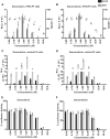
IFN induction and cytotoxicity of doxorubicin and daunorubicin in reporter cell lines. (A to D) (A and B) Dose response in VP35 cells for activation of the IFN-β reporter luciferase reporter gene (bars) and for cytotoxicity (triangles) of doxorubicin (A) or daunorubicin (B). (C and D) Dose response in control-FF cells for activation of the IFN-β reporter luciferase reporter gene by doxorubicin (C) and daunorubicin (D). (E and F) The effect of doxorubicin (E) and daunorubicin (F) on expression of a constitutively expressed firefly luciferase gene. Percent luciferase activity is relative to that with no drug treatment. Data represent means ± standard deviations and are representative of three independent experiments. RLU, relative luciferase units.
FIG 3
FIG 3
Induction of an IFN response by doxorubicin and daunorubicin is not cell type specific. Dose response for activation of the IFN-β reporter gene (bars) and for cytotoxicity (triangles) of doxorubicin (A) or daunorubicin (B) in A549 cells transfected with empty vector (vector) or VP35. Reverse transcription-quantitative polymerase chain reaction (qRT-PCR) was performed for endogenous IFN-β (C) or ISG54 (D) mRNA levels in A549 cells transfected with empty vector (vector) or VP35 and treated with doxorubicin. The RNA was isolated 12 h after treatment with the indicated concentrations of drug, and levels were normalized to levels of β-actin mRNA. Data represent means ± standard deviations and are representative of three independent experiments.
FIG 4
FIG 4
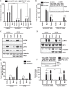
An ATM-dependent IFN response that is not blocked by VP35 is stimulated by doxorubicin and daunorubicin. (A) An IFN-β promoter assay was performed as described above except that cells (control or VP35) were treated with DMSO, ATM kinase inhibitor Ku55933 (10 μM), or mirin (10 μM) for 2 h before doxorubicin (1 μM) or daunorubicin (1 μM) treatment or SeV infection. ****, P value < 0.0001 (one-way analysis of variance followed by Tukey’s test). (B) IFN-β promoter reporter gene assays were performed as described above except that cells were transfected with scrambled shRNA (sh Scrm.) or ATM-specific shRNA (sh ATM) plasmids. ****, P value < 0.0001 (one-way analysis of variance followed by Tukey’s test). A Western blot for ATM and VP35 is shown in the inset. M, mock treated (medium + DMSO); S, SeV infected; D, doxorubicin (1 μM) treated. (C) Phospho-ATM (S1981), phospho-p53 (S15), total ATM, total p53, and VP35 levels were assessed by Western blotting in HEK293T cells transfected with empty vector or VP35 and mock treated (mock), treated with doxorubicin (Doxo), or infected with SeV (SeV) at 4 h posttreatment. β-Tubulin served as a loading control. (D) Phospho-IRF-3 (p-IRF-3) and total IRF-3 (IRF-3) levels were assessed in HEK293T cells transfected with FLAG-IRF-3 plasmid and either empty vector (vector) or VP35 plasmid. The cells were either mock treated or treated with doxorubicin or infected with SeV for 8 h. β-Tubulin served as a loading control. Total IRF-3 levels were assessed by using anti-FLAG, p-IRF-3 levels were assessed by using anti-p-IRF-3 (Ser396), and VP35 levels were assessed by using anti-VP35 antibodies. (E) NF-κB firefly luciferase reporter gene activity in mock- or VP35-transfected cells that were mock treated (medium + DMSO), treated with doxorubicin, or infected with SeV in the presence or absence of ATM kinase inhibitor Ku55933. Cells treated with 50 ng/ml of TNF-α for 2 h served as a known NF-κB activation control. Fold induction is relative to the mock-treated, vector control. Data represent means ± standard deviations and are representative of three independent experiments. **, P value < 0.01. (F) IFN-β reporter gene assays were performed as described above in control cells or VP35 cells but in the presence of scrambled siRNA (scrm.) or Top2A-specific siRNA (Top2a). ***, P value < 0.001 (one-way analysis of variance followed by Tukey’s test). The inset shows Western blotting assays to detect Top2A and β-tubulin. M, mock treated; D, doxorubicin treated; S, SeV infected.
FIG 5
FIG 5
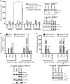
cGAS and STING enhance IFN induction by doxorubicin. (A) IFN-β reporter gene assays were performed as described above in control cells or cell lines with stable expression of STING. These were transfected with empty vector, cGAS-wt, NTase mutant cGAS (cGAS-NTM), or DNA binding cGAS mutant (cGAS-DBM). Some cells were also transfected with VP35 plasmid, as indicated. The next day, cells were mock treated, treated with doxorubicin (Doxo), or infected with SeV. Twenty hours later, reporter gene activity was measured. The Western blot indicates expression of STING, cGAS, VP35, and β-tubulin as a loading control. (B and C) IFN-β reporter control cells or cells stably expressing STING and wt-cGAS were transduced with empty vector or VP35-expressing lentiviruses. Three days later, cells were pretreated with ATM kinase inhibitor Ku55933 (10 μM) for 2 h (B) or transfected with scrambled short hairpin RNA (sh scrnm.) or ATM-specific short hairpin RNA plasmid (sh ATM) to knock down ATM expression (C) and mock treated (medium + DMSO), treated with doxorubicin (Doxo, 3 μM), induced with c-di-GMP (20 μg), or infected with SeV. Twenty hours later, IFN-β reporter activation was measured by luciferase assay. The Western blots show expression of STING, cGAS, ATM, VP35, and β-tubulin. ****, P value < 0.0001 (one-way analysis of variance followed by Tukey’s test). Error bars represent means ± standard deviations, and values are representative of three independent experiments. See also Fig. S2 and S3 in the supplemental material.
FIG 6
FIG 6
Effect of doxorubicin in vitro. (A) The toxicity of doxorubicin was evaluated in A549 cells using the CellTiter-Glo assay at 48 h after treatment with the drug. (B) Titers of an Ebola virus that expresses GFP (EBOV) after infection at a multiplicity of 2 of A549 cells in the presence of DMSO or doxorubicin (10 μM) (Doxo). The cells were pretreated with doxorubicin prior to infection for 1 h, and doxorubicin was added back to the medium after the infection. The error bars indicate the standard deviations from three independent replicates. **, P value < 0.01 (Student’s two-tailed t test). Data represent means ± standard deviations from two independent experiments (each performed in triplicate). (C and D) qRT-PCR for endogenous IFN-β (C) and ISG54 (D) mRNA levels normalized to β-actin mRNA at indicated postinfection time points. The error bars indicate the standard deviations from three independent replicates. ***, P value < 0.001; ****, P value < 0.0001 (Student’s two-tailed t test). hpi, hours postinfection.
FIG 7
FIG 7
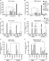
Doxorubicin bypasses multiple RNA virus IFN antagonists. IFN-β promoter (A) or ISG54 promoter (B) firefly luciferase reporter gene assays were performed. The empty vector (vector) or expression plasmids for the indicated viral IFN antagonists were transfected. The next day, cells were mock treated, treated with doxorubicin, or infected with SeV. Eighteen hours later, luciferase activity was determined. Fold induction was determined by setting the mock-treated (medium + DMSO) empty-vector controls to 1. Error bars indicate standard deviations from three independent replicates. Experiments similar to those described for panels A and B were performed to detect IFN-β promoter (C) or ISG54 promoter (D) reporter gene activity but with ATM kinase inhibitor pretreatment. An experiment similar to that described for panel C was performed using either the control-FF cells (E) or the cGAS-wt-STING-FF stable IFN-β reporter cells described in the legend to Fig. 5B (F). Data represent means ± standard deviations and are representative of three independent experiments. Error bars indicate standard deviations from three independent replicates. **, P value < 0.01; ***, P value < 0.001; ****, P value < 0.0001 (one-way analysis of variance followed by Tukey’s test). See also Fig. S4 in the supplemental material.
FIG 8
FIG 8
Proposed model for activation of IFN by doxorubicin bypassing IFN antagonism by Ebola virus VP35 protein. Ebola virus VP35 antagonizes IFN responses triggered by RIG-I-like receptors (RLR), which include RIG-I and melanoma differentiation-associated protein 5 (MDA5). RLR detect cytoplasmic double-stranded RNAs (dsRNAs) or RNAs with 5′ triphosphate (5′ pppdsRNA), products of RNA virus replication. The activation of RLR is further facilitated by protein kinase R activator (PACT). Upon activation, RLR signal through the mitochondrial antiviral signaling protein (MAVS) to activate kinases IκB kinase ε (IKKε) and TBK1. These kinases phosphorylate IFN regulatory factor 3 (IRF-3) or IRF-7, which then accumulates in the nucleus and promotes expression of type I IFNs. Doxorubicin treatment results in IFN induction by two independent pathways: the DNA damage repair response pathway involving ATM and DNA sensor machinery cGAS-STING. The DNA damage leads to activation of ATM that triggers activation of an IRF-3 and/or NF-κB response, thus leading to IFN activation. Furthermore, damaged DNA can also be detected by a cytoplasmic DNA sensor, cGAS, which through the STING–TBK1–IRF-3 axis leads to activation of IFN responses. Interestingly, these DNA-mediated IFN activation pathways are not subverted by the presence of Ebola virus VP35 protein. Thus, these data suggest novel avenues for developing antiviral therapeutics.

Similar articles

Cited by

References

    1. Mahanty S, Bray M. 2004. Pathogenesis of filoviral haemorrhagic fevers. Lancet Infect Dis 4:487–498. doi:10.1016/S1473-3099(04)01103-X. - DOI - PubMed
    1. Wong G, Qiu X, Olinger GG, Kobinger GP. 2014. Post-exposure therapy of filovirus infections. Trends Microbiol 22:456–463. doi:10.1016/j.tim.2014.04.002. - DOI - PubMed
    1. Mateo M, Reid SP, Leung LW, Basler CF, Volchkov VE. 2010. Ebolavirus VP24 binding to karyopherins is required for inhibition of interferon signaling. J Virol 84:1169–1175. doi:10.1128/JVI.01372-09. - DOI - PMC - PubMed
    1. Reid SP, Leung LW, Hartman AL, Martinez O, Shaw ML, Carbonnelle C, Volchkov VE, Nichol ST, Basler CF. 2006. Ebola virus VP24 binds karyopherin alpha1 and blocks STAT1 nuclear accumulation. J Virol 80:5156–5167. doi:10.1128/JVI.02349-05. - DOI - PMC - PubMed
    1. Reid SP, Valmas C, Martinez O, Sanchez FM, Basler CF. 2007. Ebola virus VP24 proteins inhibit the interaction of NPI-1 subfamily karyopherin alpha proteins with activated STAT1. J Virol 81:13469–13477. doi:10.1128/JVI.01097-07. - DOI - PMC - PubMed

Publication types