. 2017 Apr 4;7(1):623.
doi: 10.1038/s41598-017-00755-y.

Flow Cytometric Measurement of Blood Cells with BCR-ABL1 Fusion Protein in Chronic Myeloid Leukemia

Affiliations

Flow Cytometric Measurement of Blood Cells with BCR-ABL1 Fusion Protein in Chronic Myeloid Leukemia

Liza Löf et al. Sci Rep. .

Abstract

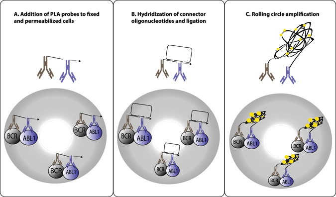
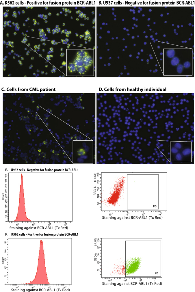
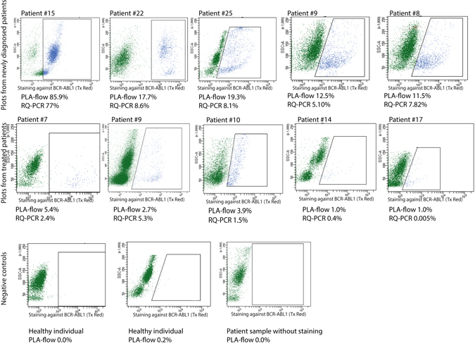
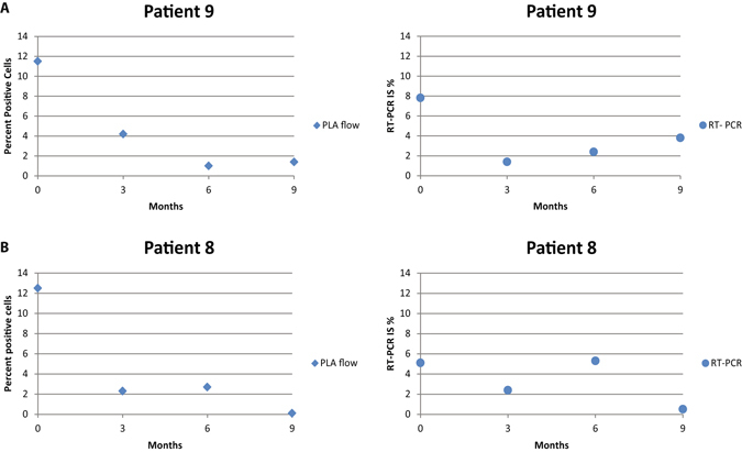
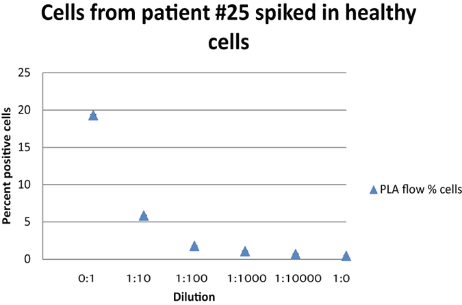
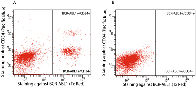
Chronic myeloid leukemia (CML) is characterized in the majority of cases by a t(9;22)(q34;q11) translocation, also called the Philadelphia chromosome, giving rise to the BCR-ABL1 fusion protein. Current treatment with tyrosine kinase inhibitors is directed against the constitutively active ABL1 domain of the fusion protein, and minimal residual disease (MRD) after therapy is monitored by real-time quantitative PCR (RQ-PCR) of the fusion transcript. Here, we describe a novel approach to detect and enumerate cells positive for the BCR-ABL1 fusion protein by combining the in situ proximity ligation assay with flow cytometry as readout (PLA-flow). By targeting of the BCR and ABL1 parts of the fusion protein with one antibody each, and creating strong fluorescent signals through rolling circle amplification, PLA-flow allowed sensitive detection of cells positive for the BCR-ABL1 fusion at frequencies as low as one in 10,000. Importantly, the flow cytometric results correlated strongly to those of RQ-PCR, both in diagnostic testing and for MRD measurements over time. In summary, we believe this flow cytometry-based method can serve as an attractive approach for routine measurement of cells harboring BCR-ABL1 fusions, also allowing simultaneously assessment of other cell surface markers as well as sensitive longitudinal follow-up.


Conflict of interest statement

UL is founder and holds stock in Olink Bioscience, having rights to the in situ PLA technique.

Figures

Figure 1
Figure 1
Detection of cells expressing BCR-ABL1 with PLA-flow. The assay employs a pair of oligonucleotide-conjugated antibodies (PLA probes) with affinity for the BCR and ABL1 domains of the fusion protein in fixed and permeabilized cells (A). The oligonucleotides on PLA probes remaining in close proximity after washes serve as templates for hybridization of two additional DNA oligonucleotides, guiding their ligation into DNA circles (B). After ligation, the DNA circles are locally amplified by rolling circle amplification (RCA) to generate RCA products that are visualized and detected by addition of complementary fluorophore-labeled oligonucleotides (C), followed by detection of labeled cells via flow cytometry.
Figure 2
Figure 2
In situ PLA detection by fluorescence microscopy of BCR-ABL1 fusion protein. (A) Detection of the BCR-ABL1 positive cell line K562, (B) Detection of the BCR-ABL1 negative cell line U937, (C) cells from a patient treated for CML, (D) nucleated blood cells from a healthy individual. Signals were recorded using (E) PLA-flow for detection of BCR-ABL1 in the BCR-ABL1 negative cell line U937 compared to (F) the BCR-ABL1 positive cell line K562. Experiments were repeated more than three times.
Figure 3
Figure 3
PLA-flow detection of BCR-ABL1 positive cells from patients diagnosed with CML and normal controls. The top row dot-plots are examples from analysis with PLA-flow of newly diagnosed patients, while the middle row represents patients previously treated or under treatment. At the time for sample collection, patients 7 and 9 were treated with imatinib, patient 10 did not receive any treatment and patients 14 and 17 were treated with dasatinib. The cells were gated first for side and forward scatter and then a positive gate was placed around cells that were considered positive compared to a negative control. The negative control used for gating was a patient sample that was not subjected to any PLA probes. The bottom row displays examples of the negative controls, i.e. two samples from healthy individuals and one sample from a patient where no PLA probes were added. The patient samples were also analyzed with RQ-PCR to estimate BCR-ABL1 transcript levels. Results from both analyses are indicated below each plot where applicable.
Figure 4
Figure 4
Long-term follow-up of newly diagnosed patients. Two CML patients were followed from the time of initial diagnosis, by measuring BCR-ABL1 positive cells using PLA-flow and BCR-ABL1 transcript levels using RQ-PCR, every 3 months over a period of 9 months. (A and B) Display results for patient 1 and 2, respectively, analyzed with PLA-flow (diamonds) and RQ-PCR (circles).
Figure 5
Figure 5
Demonstration of the sensitivity of PLA-flow. Blood from a newly diagnosed CML patient was spiked in blood from a healthy individual in a ten-fold dilution series. Samples were analyzed with PLA-flow to detect positive cells expressing the fusion protein BCR-ABL1. The X-axis indicates the dilutions, while the Y-axis displays percentages of cells expressing BCR-ABL1. Error bars represent standard deviation (SD).
Figure 6
Figure 6
Correlation of BCR-ABL1 fusion protein positive cells as determined by PLA-flow and BCR-ABL1 fusion transcript levels as determined by RQ-PCR. A total of 81 samples from 36 different patients were analyzed and compared; seven samples (indicated with red circles) represented samples analyzed before initiation of therapy, whereas the remaining samples were collected at follow-up. Two correlations were performed, one for follow-up samples (ρ = 0.7) and one for diagnostic samples (ρ = 0.93). Patient numbers and numbers of samples investigated for each patient is indicated with different colors/symbols.
Figure 7
Figure 7
Simultaneous staining of CML cells for BCR-ABL1 and for CD34. (A) The image displays simultaneous immunofluorescence staining for CD34 during BCR-ABL1 PLA-flow in a newly diagnosed CML patient. Three distinct populations were observed: A major population negative for both BCR-ABL1 and CD34, one subpopulation (13.8%) expressing BCR-ABL1 but not CD34, and one subpopulation (1.8%) positive for both CD34 and BCR-ABL1. (B) The same experiment was performed on a healthy individual as a negative control. The X-axes represent intensity of staining against BCR-ABL1 and the Y-axes reflect the CD34 staining.

References

    1. Groffen J, et al. Philadelphia Chromosomal Breakpoints Are Clustured within a limited region, bcr, on chromosome 22. Cell. 1984;36:93–99. doi: 10.1016/0092-8674(84)90077-1. - DOI - PubMed
    1. Bartram CR, d. K A, Hagemeijer A, van Agthoven T, Geurts van Kessel A, Bootsma D, et al. Translocation of c-abl oncogene correlates with the presence of a Philadelphia chromosome in chronic myelocytic leukaemia. Nature. 1983;306:277–280. doi: 10.1038/306277a0. - DOI - PubMed
    1. de Klein A, et al. A cellular oncogene is translocated to the Philadelphia chromosome in chronic myelocytic leukaemia. Nature. 1982;300:765–767. doi: 10.1038/300765a0. - DOI - PubMed
    1. Ben-Neriah Y, Daley G, Mes-Masson A, Witte O, Baltimore D. The chronic myelogenous leukemia-Specific P210 protein Is the product of the bcr/abl Hybrid Gene. Science. 1986;233:212–214. doi: 10.1126/science.3460176. - DOI - PubMed
    1. Rowley J. A new consistent chromosomal abnormality in chronic myelogenous leukaemia identifed by quinacrine fluorescence and giemsa staining. Nature. 1973;243:290–293. doi: 10.1038/243290a0. - DOI - PubMed

Publication types

MeSH terms