. 2017 Apr 4;7(1):618.
doi: 10.1038/s41598-017-00747-y.

The 5'UTR in human adenoviruses: leader diversity in late gene expression

Affiliations

The 5'UTR in human adenoviruses: leader diversity in late gene expression

Mirja Ramke et al. Sci Rep. .

Abstract

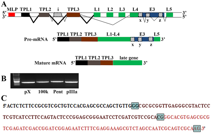
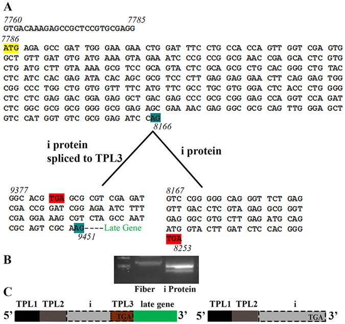
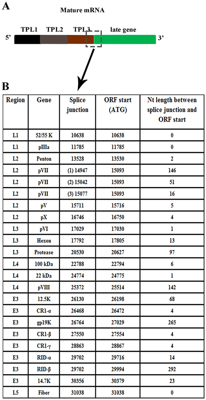
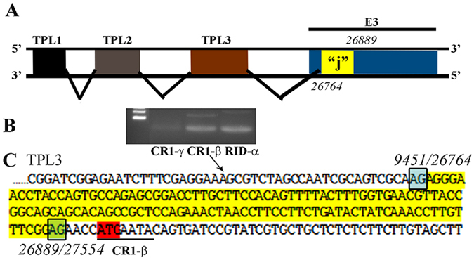
Human adenoviruses (HAdVs) shut down host cellular cap-dependent mRNA translation while initiating the translation of viral late mRNAs in a cap-independent manner. HAdV 5' untranslated regions (5'UTRs) are crucial for cap-independent initiation, and influence mRNA localization and stability. However, HAdV translational regulation remains relatively uncharacterized. The HAdV tripartite leader (TPL), composed of three introns (TPL 1-3), is critical to the translation of HAdV late mRNA. Herein, we annotated and analyzed 72 HAdV genotypes for the HAdV TPL and another previously described leader, the i-leader. Using HAdV species D, type 37 (HAdV-D37), we show by reverse transcription PCR and Sanger sequencing that mRNAs of the HAdV-D37 E3 transcription unit are spliced to the TPL. We also identified a polycistronic mRNA for RID-α and RID-β. Analysis of the i-leader revealed a potential open reading frame within the leader sequence and the termination of this potential protein in TPL3. A potential new leader embedded within the E3 region was also detected and tentatively named the j-leader. These results suggest an underappreciated complexity of post-transcriptional regulation, and the importance of HAdV 5'UTRs for precisely coordinated viral protein expression along the path from genotype to phenotype.


Conflict of interest statement

The authors declare that they have no competing interests.

Figures

Figure 1
Figure 1
Organization of the human adenovirus late transcription unit. (A) Schematic of the major late transcription unit of HAdV-C2adapted from 4, chosen because of prior experimental evidence for the shown leader sequences. Major late promoter (MLP: red; Late gene family L1-L5: green; E3 transcription unit: blue; tripartite leader (TPL) 1, 2, 3, and i, x, y, and z-leaders: grey). The thickness of the angled lines indicates the approximate abundance of the splice events in the referenced paper. Common splicing events between TPL3 and L3, y, and L5 were omitted for simplicity. (B) To examine splicing of the tripartite leader of HAdV-D37 during natural infection, human A549 cells were infected for 24 hrs. DNA was removed by DNase treatment. cDNA was amplified by using a forward primer for HAdV-D37 TPL1 and a reverse primer within the following late genes: protein X (pX), 100 kDa, penton base (Pent), and pIIIa. Primers were chosen to elicit similarly sized bands to facilitate subsequent sequencing. In each case, TPL1-3 was found spliced to the late gene 5′ end. (C) TPL1-3 as Sanger sequenced from and common to each gel purified transcript in (B). (TPL1: black; TPL2: purple, TPL3: pink; and splice sites: boxed in grey).
Figure 2
Figure 2
Phylogenetic analysis of the HAdV tripartite leader in HAdV. The TPL1-3 of each typed HAdV was annotated by blast, aligned using MEGA 6.06, and the splice sites predicted (http://wangcomputing.com/assp/). Phylogenetic neighbor-joining trees, bootstrap-confirmed (1000 replicates) were constructed for (A) TPL1, (B) TPL2, (C) TPL3, and (D) TPL1-3.
Figure 3
Figure 3
The putative i-leader protein terminates in the TPL3 of HAdV-D37. (A) Nucleotide sequence, (B) RT-PCR gel photomicrograph, and (C) schematic for i-leader spliced to TPL3 of 5′UTR for fiber, and in the putative i-protein mRNA, in which the 5′UTR is TPL1-2. RT-PCR as shown was performed with forward primer from TPL1 and reverse primers either from within fiber gene or the end of the predicted i-protein. Sequencing of gel purified transcripts revealed two splice variants of the putative i-leader mRNA, as shown. The putative i-protein mRNA is preceded by a 26 nucleotide 5′UTR prior to the start site (yellow) of an ORF (7786) for the potential coding region that would terminate (red) either within TPL3 (as shown for fiber gene), or at nucleotide 8253, the latter coding for the putative i-protein.
Figure 4
Figure 4
Splice sites for junction between tripartite leader 3 and genes transcribed by the major late promoter in HAdV-D37. (A) Schematic for splice site junctions in mature mRNA of genes under the control of the major late promoter (dashed box: splice site). (B) Table showing the genome region, gene name, splice site, start site, and leader length for each gene transcribed under the control of the major late promoter in HAdV-D37. A549 cells were infected with HAdV-D37 at MOI of 10, mRNA was harvested at 24 hpi, and DNA removed by treatment with DNase. cDNA was generated, and PCR performed with forward primer for TPL1 and a reverse primer for each late and E3 gene. PCR products were gel purified and sequenced. Notably, mRNAs for the E3 genes RID-α and RID-β showed the same splice site, resulting in one mRNA for both genes, consistent with a polycistronic mRNA.
Figure 5
Figure 5
Putative “j”-leader located within the CR1-α E3 gene. (A) Schematic for the location of a newly detected leader (“j”-leader) embedded within the E3 CRI-α gene, experimentally determined to be spliced to some, but not all mRNAs of the E3 genes. (B) Gel photomicrograph of mRNA transcripts amplified with forward primer from TPL1 and reverse primers from CR1-γ, CR1-β, and RID-α. Primers were chosen to elicit similarly sized bands to facilitate subsequent sequencing. (C) Nucleotide sequence of the PCR product for CR1-β. The putative j-leader sequence and splice sites are shown in yellow and green, respectively. Note an additional 4 nucleotide 5′UTR (AACC) prior to the CR1-β start site (red). The 5′UTR in (C) prior to the splice site for the j-leader is from TPL3.

References

    1. Robinson CM, Seto D, Jones MS, Dyer DW, Chodosh J. Molecular evolution of human species D adenoviruses. Infect Genet Evol. 2011;11:1208–17. doi: 10.1016/j.meegid.2011.04.031. - DOI - PMC - PubMed
    1. Walsh MP, et al. Evidence of molecular evolution driven by recombination events influencing tropism in a novel human adenovirus that causes epidemic keratoconjunctivitis. PLoS One. 2009;4:e5635. doi: 10.1371/journal.pone.0005635. - DOI - PMC - PubMed
    1. Walsh MP, et al. Computational analysis identifies human adenovirus type 55 as a re-emergent acute respiratory disease pathogen. J Clin Microbiol. 2010;48:991–3. doi: 10.1128/JCM.01694-09. - DOI - PMC - PubMed
    1. Zhao H, Chen M, Pettersson U. A new look at adenovirus splicing. Virology. 2014;456–457:329–41. doi: 10.1016/j.virol.2014.04.006. - DOI - PubMed
    1. Brough DE, Droguett G, Horwitz MS, Klessig DF. Multiple functions of the adenovirus DNA-binding protein are required for efficient viral DNA synthesis. Virology. 1993;196:269–81. doi: 10.1006/viro.1993.1475. - DOI - PubMed

Publication types

LinkOut - more resources