. 2017 Apr 4;8(1):9.
doi: 10.1038/s41467-017-00020-w.

Rotiferan Hox genes give new insights into the evolution of metazoan bodyplans

Affiliations

Rotiferan Hox genes give new insights into the evolution of metazoan bodyplans

Andreas C Fröbius et al. Nat Commun. .

Abstract

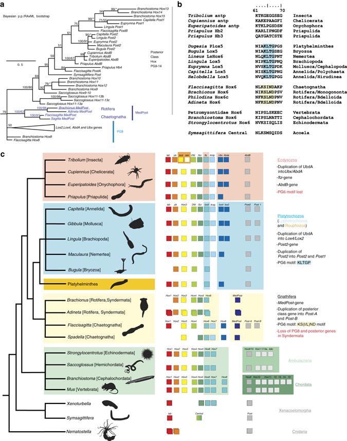
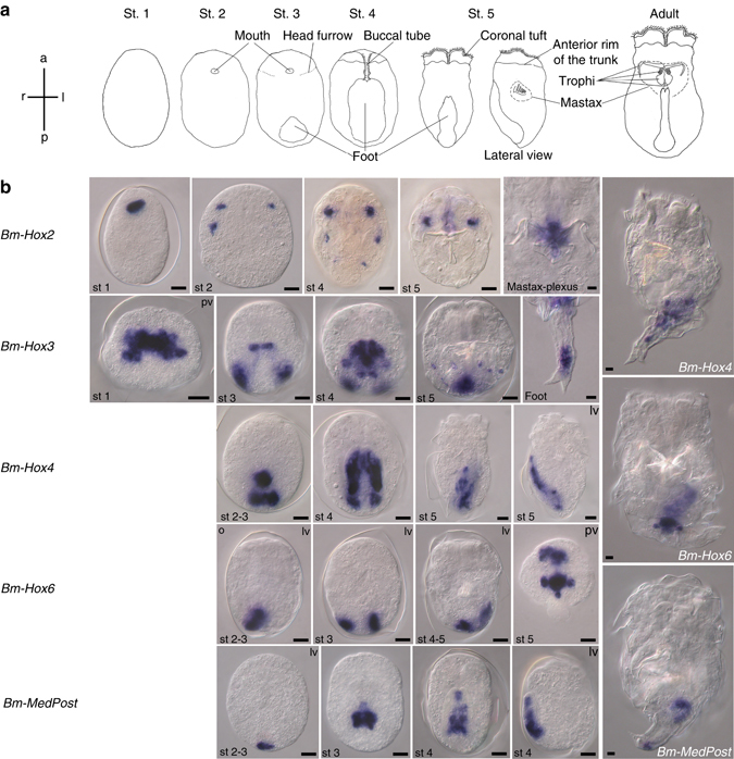
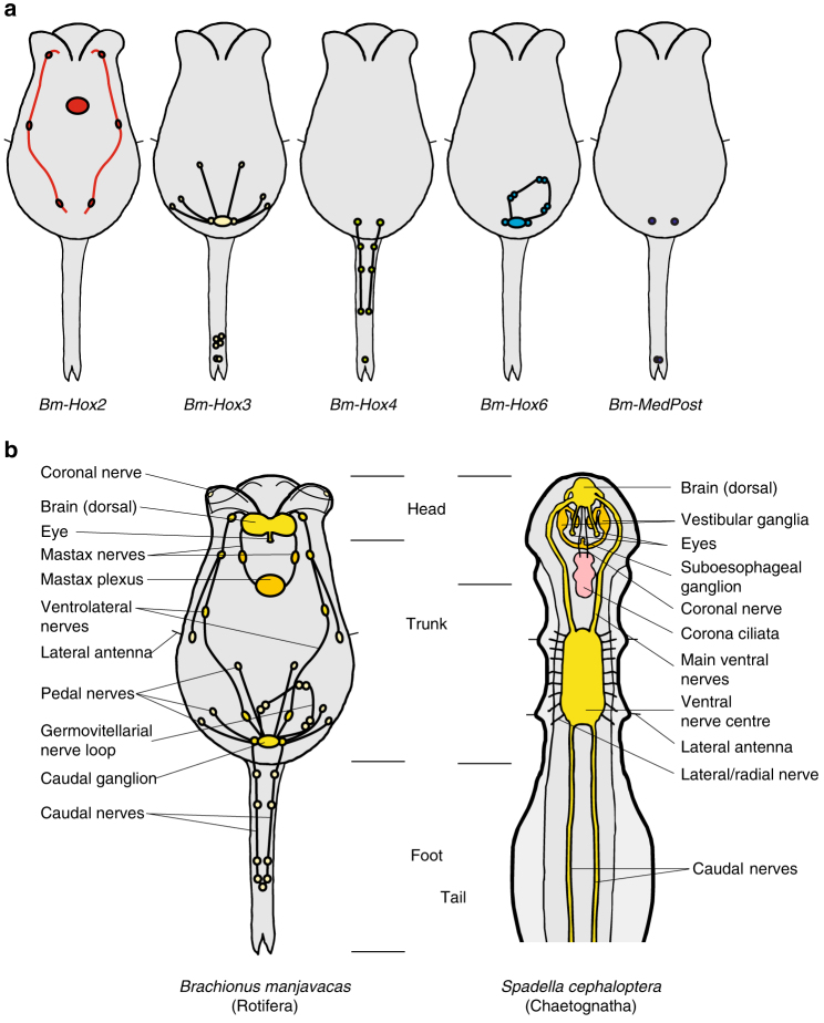
The phylum Rotifera consists of minuscule, nonsegmented animals with a unique body plan and an unresolved phylogenetic position. The presence of pharyngeal articulated jaws supports an inclusion in Gnathifera nested in the Spiralia. Comparison of Hox genes, involved in animal body plan patterning, can be used to infer phylogenetic relationships. Here, we report the expression of five Hox genes during embryogenesis of the rotifer Brachionus manjavacas and show how these genes define different functional components of the nervous system and not the usual bilaterian staggered expression along the anteroposterior axis. Sequence analysis revealed that the lox5-parapeptide, a key signature in lophotrochozoan and platyhelminthean Hox6/lox5 genes, is absent and replaced by different signatures in Rotifera and Chaetognatha, and that the MedPost gene, until now unique to Chaetognatha, is also present in rotifers. Collectively, our results support an inclusion of chaetognaths in gnathiferans and Gnathifera as sister group to the remaining spiralians.Rotifers are microscopic animals with an unusual, nonsegmented body plan consisting of a head, trunk and foot. Here, Fröbius and Funch investigate the role of Hox genes-which are widely used in animal body plan patterning-in rotifer embryogenesis and find non-canonical expression in the nervous system.


Conflict of interest statement

The authors declare no competing financial interests.

Figures

Fig. 1
Fig. 1
Hox gene data places rotifers and chaetognaths in Gnathifera within Spiralia. a Phylogenetic tree depicting the relationship of MedPost genes to PG8 and posterior class Hox genes. Tree topology is from Bayesian analysis. Bayesian posterior probabilities based on 400,000 trees from 40,000,000 generations and ML support values from 1000 iterations are shown above branches. Single values represent Bayesian posterior probabilities only. Asterisks denote ML support below 50%. b Alignment of ten amino acids of the carboxy flanking region to the homeodomain of PG6 genes. Sequences highlighted with yellow contain the new signature found in rotifers and chaetognaths. Blue highlighting marks the lox5-parapeptide of lophotrochozoan genes. Neither is found in Ecdysozoa, Ambulacraria, Chordata, or Xenacoelomorpha. c Summary of representative characteristics of the Hox cluster within different metazoan taxa. The tree to the left represents bilaterian phylogeny with Cnidaria as an outgroup. Boxes in the middle depict Hox gene contingents (color coded according to the assignment of the Hox genes to the different paralogous groups) isolated from representative species. The right hand column summarizes characteristic Hox gene evolution and duplication events along with presence of special Hox signatures resulting in Hox genes characterizing the respective groups
Fig. 2
Fig. 2
Expression of Hox genes during embryogenesis of Brachionus manjavacas. a Schematic of embryonic stages of Brachionus manjavacas with morphological characteristics used for staging. b Whole-mount in situ hybridization on amictic female embryos. Adults are only shown for genes with expression persisting into the adult stage. Anterior to the top. Mostly ventral views are shown. pv, posterior view, dorsal side up; lv, lateral view, ventral to the left. Scale bar, 10 µm
Fig. 3
Fig. 3
Body plans and nervous systems in Rotifera and Chaetognatha. a Diagram of Hox gene expression in the nervous system of Brachionus manjavacas. b Comparison of rotiferan and chaetognath body plans with respect to the structure of the nervous system. Both groups have a dorsal brain and additional nerve plexi: mastax nerves and ganglia in rotifers and vestibular, and esophageal ganglia in chaetognaths as well as a caudal ganglion in rotifers and a ventral nerve centre in chaetognaths with the latter possibly incorporating functional subsets, these are still separated from the caudal ganglion in rotifers, e.g., innervation of sensory lateral antennae

Similar articles

Cited by

References

    1. Lewis EB. A gene complex controlling segmentation in Drosophila. Nature. 1978;276:565–570. doi: 10.1038/276565a0. - DOI - PubMed
    1. Dessain S, Gross CT, Kuziora MA, McGinnis W. Antp-type homeodomains have distinct DNA binding specificities that correlate with their different regulatory functions in embryos. EMBO. J. 1992;11:991–1002. - PMC - PubMed
    1. Averof M, Akam M. Hox genes and the diversification of insect and crustacean body plans. Nature. 1995;376:420–423. doi: 10.1038/376420a0. - DOI - PubMed
    1. Fröbius AC, Matus DQ, Seaver EC. Genomic organization and expression demonstrate spatial and temporal Hox gene colinearity in the lophotrochozoan Capitella sp. I. PLoS. ONE. 2008;3:e4004. doi: 10.1371/journal.pone.0004004. - DOI - PMC - PubMed
    1. Kourakis MJ, Martindale MQ. Hox gene duplication and deployment in the annelid leech Helobdella. Evol. Dev. 2001;3:145–153. doi: 10.1046/j.1525-142x.2001.003003145.x. - DOI - PubMed

LinkOut - more resources