. 2017 Mar 28:3:17005.
doi: 10.1038/celldisc.2017.5. eCollection 2017.

Translation in astrocyte distal processes sets molecular heterogeneity at the gliovascular interface

Affiliations

Translation in astrocyte distal processes sets molecular heterogeneity at the gliovascular interface

Anne-Cécile Boulay et al. Cell Discov. .

Abstract

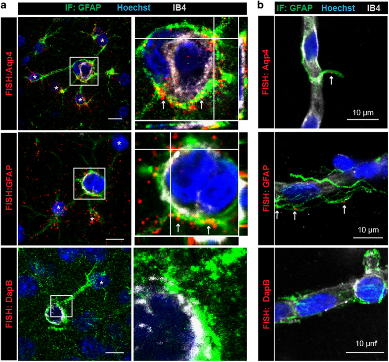
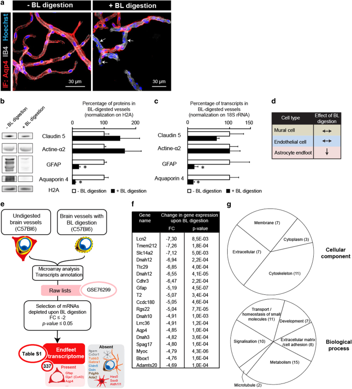
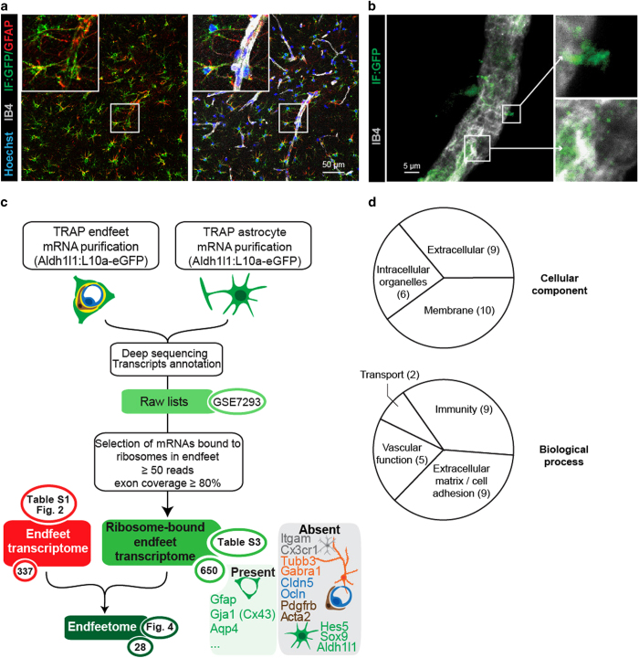
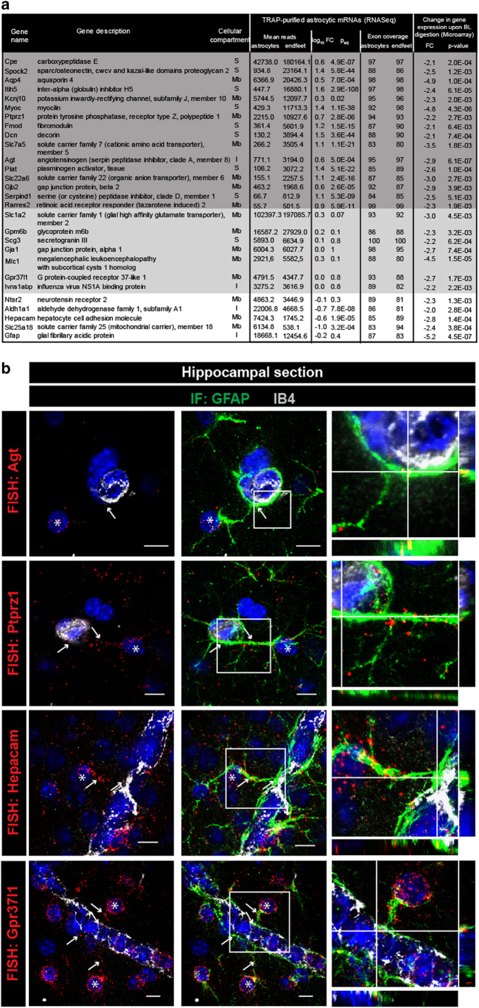
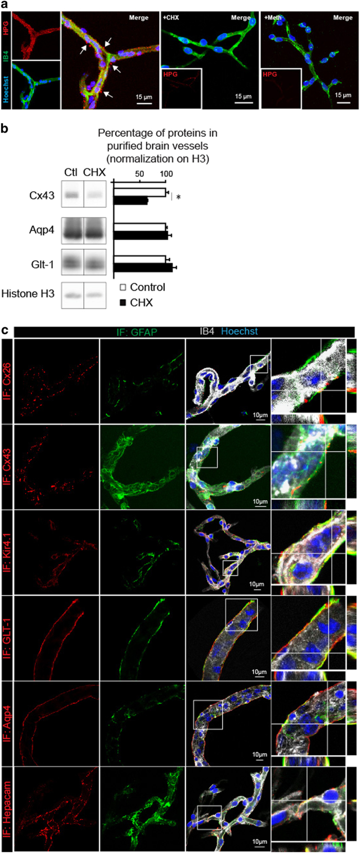
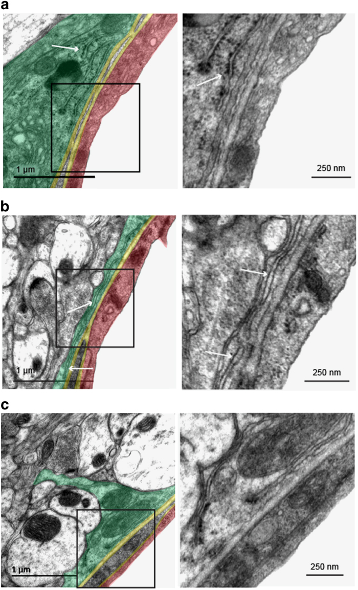
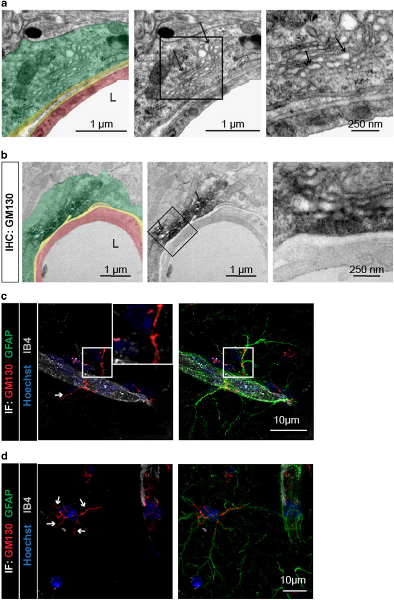
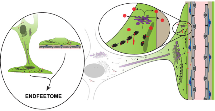
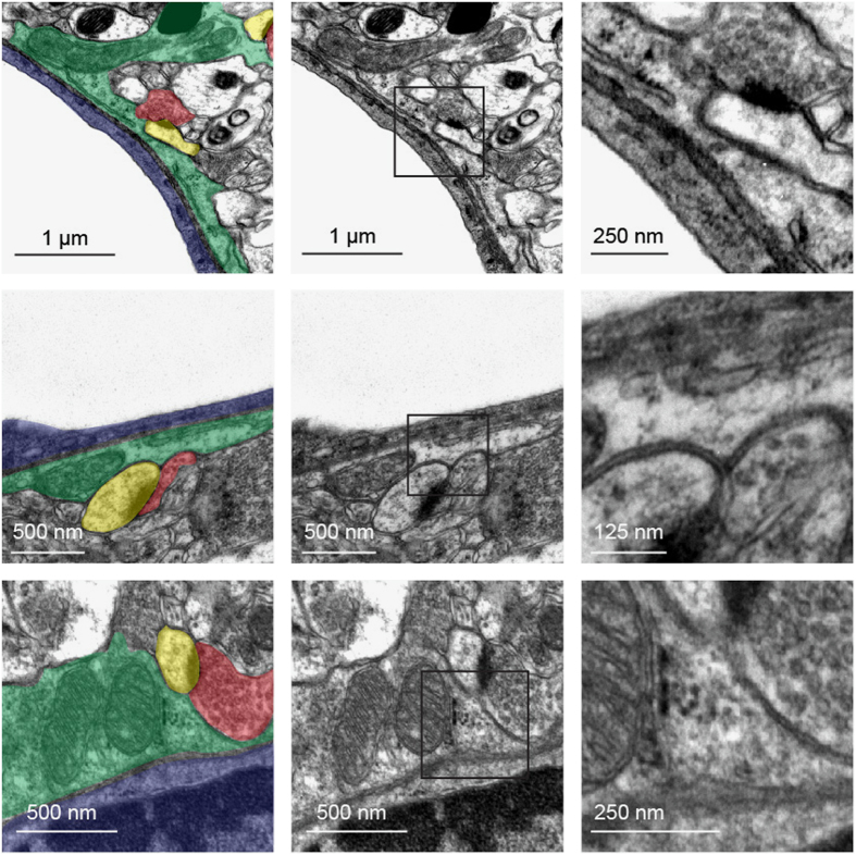
Astrocytes send out long processes that are terminated by endfeet at the vascular surface and regulate vascular functions as well as homeostasis at the vascular interface. To date, the astroglial mechanisms underlying these functions have been poorly addressed. Here we demonstrate that a subset of messenger RNAs is distributed in astrocyte endfeet. We identified, among this transcriptome, a pool of messenger RNAs bound to ribosomes, the endfeetome, that primarily encodes for secreted and membrane proteins. We detected nascent protein synthesis in astrocyte endfeet. Finally, we determined the presence of smooth and rough endoplasmic reticulum and the Golgi apparatus in astrocyte perivascular processes and endfeet, suggesting for local maturation of membrane and secreted proteins. These results demonstrate for the first time that protein synthesis occurs in astrocyte perivascular distal processes that may sustain their structural and functional polarization at the vascular interface.

Keywords: astrocyte; endfeet; gliovascular unit; local translation; mRNAs localization; translating ribosome immunoprecipitation.


Conflict of interest statement

The authors declare no conflict of interest.

Figures

Figure 1
Figure 1
mRNAs are present in astrocyte perivascular processes and endfeet. (a) Representative confocal images of astrocyte-specific mRNAs encoding Aqp4, GFAP and dapB (negative probe) detected by fluorescent in situ hybridization (FISH by RNAscope) on brain sections of 2-month-old C57BL6 mice. Astrocytes (here in the hippocampus) are immunostained for GFAP (green). GFAP is a cytoskeleton protein specific to astrocytes whose immunolabeling indicates the presence of astrocyte processes and endfeet. GFAP fibers do not fill the endfeet, which explains why astrocyte FISH signals are not necessarily co-localized with GFAP. Aqp4 or GFAP mRNAs are mostly detected in PVAPs and endfeet (red) (white arrows). Few Aqp4 mRNAs are detected at the level of IB4. They might belong to vascular cells, although Aqp4 is mostly expressed by astrocytes [17, 18, 20]. The astrocyte somata are indicated with an asterisk. Enlarged views of boxed areas show details of the FISH signals in PVAPs and endfeet. (b) Representative confocal images of astrocyte-specific mRNAs encoding Aqp4 and GFAP on 2-month-old C57BL6 mice-purified brain vessels. Perivascular Aqp4 or GFAP mRNAs are detected at the level of GFAP immunolabeled fibers attached to the surface of purified brain vessels (red) (white arrows). Enlarged views of boxed areas show details of the perivascular FISH signals. In a and b, the vessel surface is stained with IB4 (gray) and nuclei with Hoechst (blue). Orthogonal analysis of areas indicated by white lines show the detail of FISH signals at the level of GFAP fibers at the vessel surface.
Figure 2
Figure 2
Characterization of the endfeet transcriptome. (a) Representative confocal images of purified brain vessels with or without partial basal lamina (BL) digestion. The vessel surface is stained with IB4 (gray). Astrocyte perivascular membrane are immunolabeled for Aqp4 (red). The nuclei are stained with Hoechst (blue). Digestion of the BL partially removes astroglial perivascular membranes (white arrows). (b) Comparative Western blot analysis of Claudin5 (Cldn5), Smooth muscle actin (Acta2), GFAP and Aquaporin 4 (Aqp4) protein levels in brain vessels purified from 2-month-old mice with or without partial BL digestion. Histone2A (H2A) was used as the loading control. The percentage of expression in digested vessels compared with that in undigested vessels (set as 100%; n=3): Cldn5 149±62%, P=0.2; Acta2 164±53%, P=0.2; GFAP 20±4%, P=0.05; Aqp4 8±2%, P=0.05. Mann–Whitney two-tailed test, *P=0.05. (c) Comparative quantitative PCR analysis of Claudin5 (Cldn5), Smooth muscle actin (Acta2), GFAP and Aquaporin 4 (Aqp4) transcripts in brain vessels purified from 2-month-old mice with or without partial BL digestion. The percentage of expression in digested vessels compared with that in undigested vessels (set as 100%; n=3): Cldn5 70±5% P=0.2; Acta2 100±2% P=0.2; GFAP 9±2%, P=0.05; Aqp4 6±1%, P=0.05. Mann–Whitney two-tailed test, *P=0.05. (d) Recapitulative table of partial BL digestion effects on purified brain vessels and associated astrocyte endfeet. (e) Analysis flowchart of the endfeet transcriptome. Transcripts depleted upon BL digestion with a fold-change (FC) ⩽−2 and a P-value ⩽0.05 were selected. The endfeet transcriptome displays 337 mRNAs including the astroglial-specific Gja1 (Cx43), Aqp4 or GFAP. Neuronal-, microglial-, endothelial- and mural cell-specific mRNAs are absent, as well as the astroglial-specific Hes5, Sox9 and Aldh1l1 indicating that only a subset of astrocyte mRNAs are distributed in endfeet. (f) List of the 20 most depleted mRNAs upon BL digestion compared with the undigested vessels. (g) Gene Ontology analysis of the ‘cellular component’ and ‘biological process’ pathways significantly depleted in the brain vessels upon BL digestion. The amount of pathways for each category is indicated.
Figure 3
Figure 3
Analysis of the astrocyte endfeet ribosomal-bound transcriptome. (a, b) Representative confocal images of GFP-tagged ribosomes (green) in 2-month-old Aldh1l1:L10a-eGFP mice, (a) on brain section (here in the hippocampus) and (b) at the surface of purified brain vessels. Enlarged views of squared areas show details of perivascular astroglial ribosomes. The vessel surface is stained with IB4 (gray) and nuclei with Hoechst (blue). Astrocytes are immunostained for GFAP (red). (c) Flowchart for the combined analysis of the endfeet transcriptome and the endfeet and whole astrocyte TRAP ribosome-bound transcriptome. The selection criteria are detailed in the white box. Lists of transcripts and pathway analysis generated at each step are indicated in green circles, with the corresponding tables and the number of transcripts. The ribosome-bound endfeet transcriptome displays 650 mRNAs including the astroglial-specific Gja1 (Cx43), Aqp4 or GFAP. Neuronal-, microglial-, endothelial- and mural cell-specific mRNAs are absent, as well as the astroglial-specific Hes5, Sox9 and Aldh1l1 mRNAs indicating that only a subset of astrocyte mRNAs bound to ribosomes is present in endfeet. The combination of the endfeet transcriptome and the TRAP ribosome-bound transcriptomes defines the endfeetome, which displays 28 transcripts. (d) Gene Ontology analysis of the enriched ‘cellular component’ and ‘biological process’ pathways in the endfeet ribosome-bound transcriptome. Numbers indicate the amount of pathways for each category.
Figure 4
Figure 4
The endfeetome. (a) List of mRNAs. The endfeetome common to the endfeet transcriptome and the endfeet and whole astrocyte ribosome-bound (TRAP) transcriptomes. Endfeet mRNAs enriched in the whole astrocyte TRAP libraries compared to the endfeet TRAP libraries (RNAseq: log10 FC<0, Padj⩽0.05) are indicated in white. Endfeet mRNAs equally present in both types of TRAP libraries (RNAseq: Padj>0.05) are indicated in light gray. Endfeet mRNAs enriched in the endfeet TRAP libraries compared to whole astrocytes TRAP libraries (RNAseq: log10 FC>0, Padj⩽0.05) are indicated in dark gray. The cellular compartment of each corresponding protein is indicated (Mb, membrane; S, secreted; I, intracellular). (b) Representative confocal images of some endfeetome transcripts detected by FISH (red) in hippocampus slices. The vessel surface is stained with IB4 (gray) and nuclei with Hoechst (blue). The astrocytes are immunostained for GFAP. The astrocyte somata are indicated with an asterisk. The white arrows indicate extravascular FISH labeling on the vessels and at the level of GFAP positive filaments in PvAPs and endfeet. On the right, orthogonal analysis of areas indicated by white lines on the enlarged views shows the detail of FISH signals at the level of GFAP fibers at the vessel surface.
Figure 5
Figure 5
Protein translation occurs in astrocyte perivascular endfeet. (a) Representative confocal images of Click-iT HPG-labeled brains vessels purified from 2-month-old C57BL6 mice (red). The vessel surface is stained with IB4 (green) and nuclei by Hoechst (blue). Negative control experiments were performed using Cycloheximide (+CHX) or replacing HPG by methionine (+Meth). The white arrows indicate extravascular HPG labeling. (b) Cx43 turnover in endfeet is sustained by local protein translation. Western blot detection of Cx43, Aqp4 and Glt1 in protein extracts from brain vessel-associated astrocyte endfeet purified from 2-month-old C57BL6 mice submitted or not to Cycloheximide (+CHX). Protein level in untreated samples is set as 100% (n=3): Cx43 64±1%, *P=0.05; Aqp4 103±8%; Glt1 113±8%. Mann–Whitney two-tailed test. (c) Representative confocal images of immunofluorescent detection of endfeetome proteins in astrocyte endfeet (red) on 2-month-old C57BL6-purified brain vessels. The vessel surface is stained with IB4 (gray) and nuclei with Hoechst (blue). Astrocyte endfeet are immunolabeled for GFAP (gray). Enlarged views of boxed areas show details of immunofluorescence. Orthogonal analysis of areas indicated by white lines to show the detail of FISH signals at the level of GFAP fibers at the vessel surface.
Figure 6
Figure 6
Organization of endoplasmic reticulum in astrocyte endfeet. Representative transmission electron microscopy images showing endoplasmic reticulum (ER) in cortical astrocyte endfeet surrounding 5–20 μm diameter vessels of 2-month-old C57BL6 mice. On the left images, the different structures of the gliovascular unit are colored: an astrocyte endfoot (green), the basal lamina (yellow), an endothelial cell (red). Enlarged views of squared areas show details of the ribosomes and ER structures. (a) An astrocyte endfoot with rough ER (RER) (white arrow). (b) An astrocyte endfoot with smooth ER (SER) (white arrow). (c) An astrocyte endfoot with SER with areas containing few bound ribosomes (white arrow).
Figure 7
Figure 7
Organization of the Golgi apparatus in astrocyte perivascular processes and endfeet. (a, b) Representative transmission electron microscopy images of the Golgi apparatus (GA) in cortical astrocyte endfeet surrounding 5–20 μm diameter vessels of 2-month-old C57BL6 mice. Enlarged views of the squared areas show details of GA in the astrocyte endfeet. Black arrows indicate typical GA stacks with surrounding vesicles. On the left images, the different structures of the gliovascular unit are colored: an astrocyte endfoot (green), the basal lamina (yellow), an endothelial cell (red). (b) Immunolabeling for the cis-GA protein GM130 in an astrocyte endfoot. (c, d) Representative confocal images of cortical astrocytes immunolabeled for GFAP (green) and GM130 (red) on brain sections of 2-month-old C57BL6 mice. The vessels surface is stained with IB4 (gray) and nuclei with Hoechst (blue). The white arrows show the GA ramifications in the PvAPs and endfeet.
Figure 8
Figure 8
Astrocytes set protein synthesis in distal perivascular processes: graphical abstract. Astrocytes, the most numerous neuroglial cells in the central nervous system, are multipolar cells. They extend long processes terminated by endfeet (in green) at the surface of brain vessels (composed by mural cells in blue and endothelial cells in gray) and regulate vascular functions. In the present study, we demonstrate that some mRNAs (gray lines) are transported in astrocyte perivascular endfeet and bound to ribosomes (black dots) suggesting that they are translated on site. We also show that protein synthesis (red dots) occurs in endfeet. Finally, we show that endfeet are equipped with smooth and rough endoplasmic reticulum (gray) and the Golgi apparatus (purple), suggesting that the maturation of membrane and secreted proteins may occur locally. These results suggest that alternative routes for the translation, maturation and secretion of a specific pool of proteins are organized in the astrocyte perivascular endfeet. Proteins synthesized there might be either translated in the cytosol or in the RER. They can be further maturated in the local Golgi apparatus, inserted in the membrane or secreted in the perivascular space.
Figure 9
Figure 9
Presence of synapses abutting astrocyte endfeet. Representative transmission electron microscopy images of cortical astrocyte endfeet surrounding capillaries of a 2-month-old C57BL6 mouse. Enlarged views of the squared areas show details of the synapses abutting astrocyte endfeet. On the left image, astrocyte endfeet are colored in green, endothelial cells in blue, pre-synapses in red and post-synapses in yellow.

References

    1. Araque A, Carmignoto G, Haydon PG et al. Gliotransmitters travel in time and space. Neuron 2014; 81: 728–739. - PMC - PubMed
    1. Dallerac G, Chever O, Rouach N. How do astrocytes shape synaptic transmission? Insights from electrophysiology. Front Cell Neurosci 2013; 7: 159. - PMC - PubMed
    1. Clarke LE, Barres BA. Emerging roles of astrocytes in neural circuit development. Nat Rev Neurosci 2013; 14: 311–321. - PMC - PubMed
    1. Ghezali G, Dallerac G, Rouach N. Perisynaptic astroglial processes: dynamic processors of neuronal information. Brain Struct Funct 2015; 221: 2427–2442. - PubMed
    1. Mathiisen TM, Lehre KP, Danbolt NC, Ottersen OP. The perivascular astroglial sheath provides a complete covering of the brain microvessels: an electron microscopic 3D reconstruction. Glia 2010; 58: 1094–1103. - PubMed