. 2017 Mar 21:4:39.
doi: 10.3389/fvets.2017.00039. eCollection 2017.

Mesenchymal Stem Cell-Derived Exosomes: Immunomodulatory Evaluation in an Antigen-Induced Synovitis Porcine Model

Affiliations

Mesenchymal Stem Cell-Derived Exosomes: Immunomodulatory Evaluation in an Antigen-Induced Synovitis Porcine Model

Javier G Casado et al. Front Vet Sci. .

Abstract

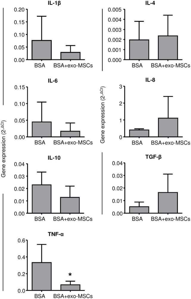
Synovitis is an inflammatory process associated with pain, disability, and discomfort, which is usually treated with anti-inflammatory drugs or biological agents. Mesenchymal stem cells (MSCs) have been also successfully used in the treatment of inflammatory-related diseases such as synovitis or arthritis. In the last years, the exosomes derived from MSCs have become a promising tool for the treatment of inflammatory-related diseases and their therapeutic effect is thought to be mediated (at least in part) by their immunomodulatory potential. In this work, we aimed to evaluate the anti-inflammatory effect of these exosomes in an antigen-induced synovitis animal model. To our knowledge, this is the first report where exosomes derived from MSCs have been evaluated in an animal model of synovitis. Our results demonstrated a decrease of synovial lymphocytes together with a downregulation of TNF-α transcripts in those exosome-treated joints. These results support the immunomodulatory effect of these exosomes and point out that they may represent a promising therapeutic option for the treatment of synovitis.

Keywords: exosomes; immunomodulation; inflammation; mesenchymal stem cells; synovitis.


Figures

Figure 1
Figure 1
Temporal scheme of the immunization protocol and monitoring. Subcutaneous BSA injections (black arrows), intra-articular injections of BSA or BSA co-administered with exo-mesenchymal stem cells (gray arrow), blood sampling (triangles), synovial fluid sampling (squares), and kinetic gait analysis (rhombus) are shown.
Figure 2
Figure 2
Humoral response in the antigen-induced animal model of synovitis. Plasma samples were weekly collected and anti-BSA IgG levels were quantified by ELISA immunoassay. The lower boundary of the box indicates the 25th percentile and the upper boundary the 75th percentile. Bars above and below the box indicate the 90th and 10th percentiles. The line within the box marks the median (n = 4).
Figure 3
Figure 3
Gene expression of cytokines in synovial fluid (SF). SF samples were collected 7 days after intra-articular injections and total RNA was isolated. The qRT-PCR products were quantified by the 2ΔCt method using β-2 microglobulin as a housekeeping gene. Graph represents the mean ± SD of three independently performed experiments (n = 4). *Statistically significant difference in a paired t-test (p ≤ 0.05).
Figure 4
Figure 4
Pressure platform (PP) gait analysis. Then, 7 days after intra-articular injections of BSA or BSA co-administered with exo-mesenchymal stem cells (MSCs), a PP gait analysis was performed to evaluate plantar pressure distributions. (A) A representative image of the gait analysis (LF, left forelimb; LH, left hind limb; RF, right forelimb; RH, right hind limb) is represented. (B) Impulses in forelimbs with intra-articular BSA co-administered with PBS or exo-MSCs (n = 4). The lower boundary of the box indicates the 25th percentile and the upper boundary the 75th percentile. Bars above and below the box indicate the 90th and 10th percentiles. The line within the box marks the median. Measurements compared in a paired t-test.

Similar articles

Cited by

References

    1. Scanzello CR, Goldring SR. The role of synovitis in osteoarthritis pathogenesis. Bone (2012) 51:249–57.10.1016/j.bone.2012.02.012 - DOI - PMC - PubMed
    1. Arend WP, Firestein GS. Pre-rheumatoid arthritis: predisposition and transition to clinical synovitis. Nat Rev Rheumatol (2012) 8:573–86.10.1038/nrrheum.2012.134 - DOI - PubMed
    1. McIlwraith CW. Use of synovial fluid and serum biomarkers in equine bone and joint disease: a review. Equine Vet J (2005) 37:473–82.10.2746/042516405774480102 - DOI - PubMed
    1. McIlwraith CW, Frisbie DD, Rodkey WG, Kisiday JD, Werpy NM, Kawcak CE, et al. Evaluation of intra-articular mesenchymal stem cells to augment healing of microfractured chondral defects. Arthroscopy (2011) 27:1552–61.10.1016/j.arthro.2011.06.002 - DOI - PubMed
    1. Kofoed H. Synovitis causes hypoxia and acidity in synovial fluid and subchondral bone. Injury (1986) 17:391–4.10.1016/0020-1383(86)90078-1 - DOI - PubMed